. 2018 May 4;46(8):4099-4113.
doi: 10.1093/nar/gky246.

Hrp48 and eIF3d contribute to msl-2 mRNA translational repression

Affiliations

Hrp48 and eIF3d contribute to msl-2 mRNA translational repression

Emilia Szostak et al. Nucleic Acids Res. .

Abstract

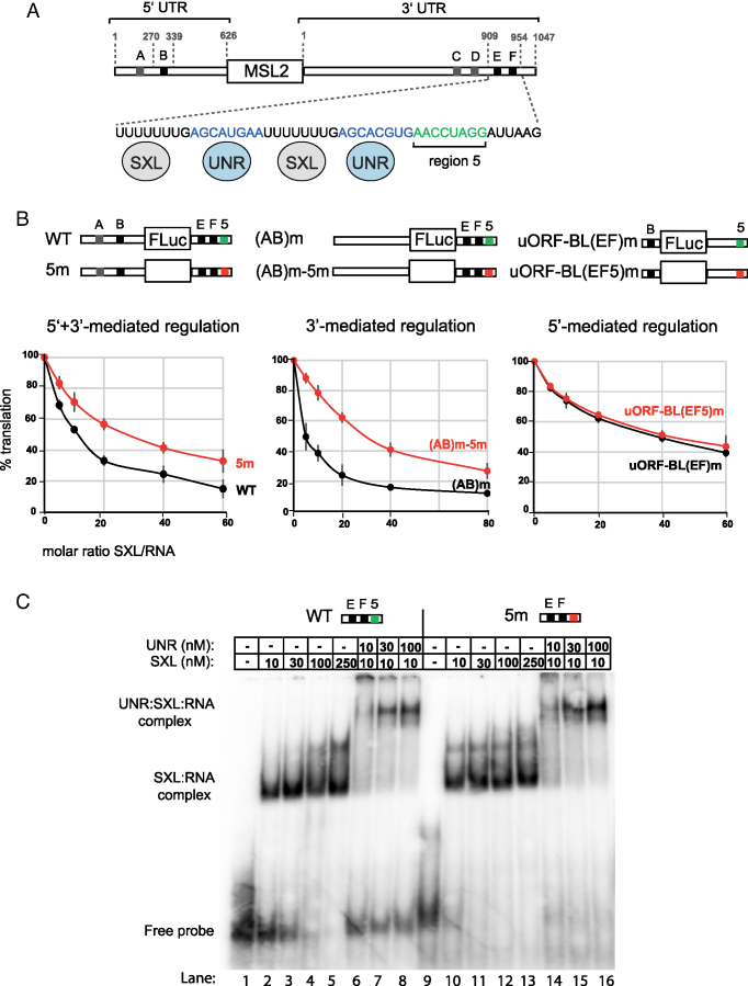
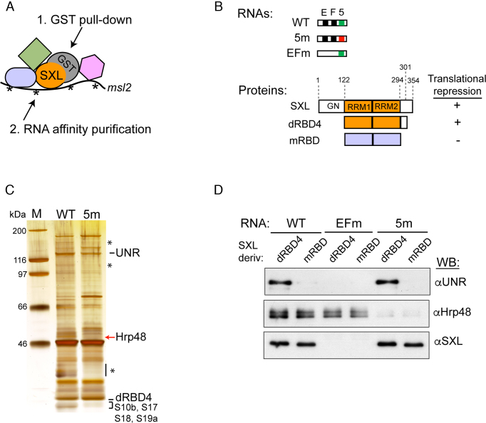
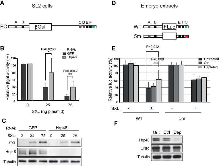
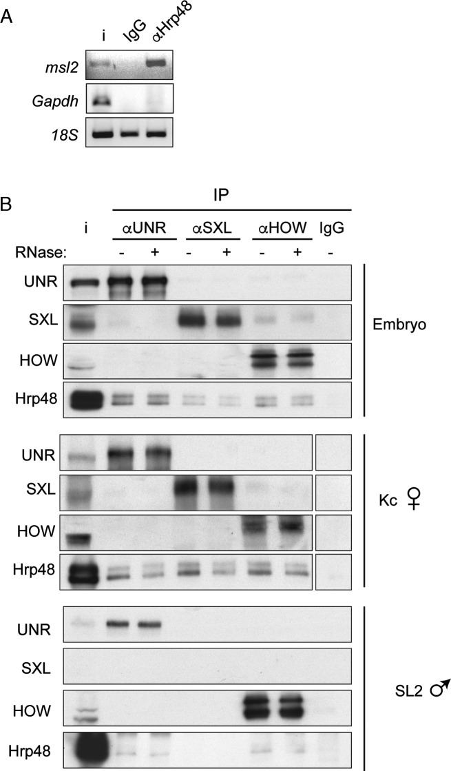
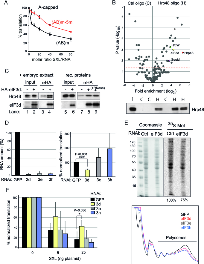
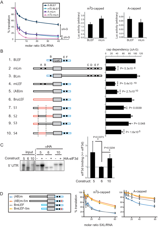
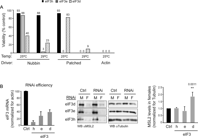
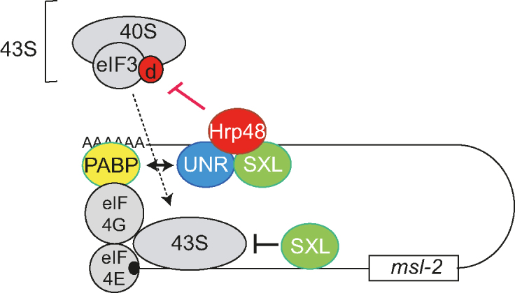
Translational repression of msl-2 mRNA in females of Drosophila melanogaster is an essential step in the regulation of X-chromosome dosage compensation. Repression is orchestrated by Sex-lethal (SXL), which binds to both untranslated regions (UTRs) of msl-2 and inhibits translation initiation by poorly understood mechanisms. Here we identify Hrp48 as a SXL co-factor. Hrp48 binds to the 3' UTR of msl-2 and is required for optimal repression by SXL. Hrp48 interacts with eIF3d, a subunit of the eIF3 translation initiation complex. Reporter and RNA chromatography assays showed that eIF3d binds to msl-2 5' UTR, and is required for efficient translation and translational repression of msl-2 mRNA. In line with these results, eIF3d depletion -but not depletion of other eIF3 subunits- de-represses msl-2 expression in female flies. These data are consistent with a model where Hrp48 inhibits msl-2 translation by targeting eIF3d. Our results uncover an important step in the mechanism of msl-2 translation regulation, and illustrate how general translation initiation factors can be co-opted by RNA binding proteins to achieve mRNA-specific control.


Figures

Figure 1.
Figure 1.
‘Region 5’ is important for translational repression of msl-2 mRNA independent of SXL and UNR binding. (A) Schematic representation of msl-2 mRNA. SXL binding sites are depicted with gray and black boxes (A–F); sites A, C, D (grey) are dispensable for translational repression while sites B, E and F (black) are required. Numbers indicate the length of the 5′ and 3′ UTRs (626 and 1047 nucleotides, respectively) and the position of the minimal sequences required for translational repression (nt 270–339 in the 5′ UTR and nt 909–954 in the 3′ UTR). A detail of the minimal functional 3′ UTR is shown, with the UNR binding sites in blue and the region 5 in green. SXL and UNR proteins are shown binding to their corresponding sites. (B) Region 5 is important for 3′-mediated regulation. In vitro translation assays were performed with a series of indicator constructs that support either 5′, 3′ or 5′+3′-mediated regulation, schematically represented above each graph. Constructs WT and 5m contain a 5′ UTR of 354 nt including sites A and B. Constructs (AB)m and (AB)m-5m contain a 5′ UTR of 626 nt lacking sites A and B. Constructs uORF-BL(EF)m and uORF-BL(EF5)m contain the minimal functional 5′ UTR. All constructs contain minimal 3′ UTR derivatives, as indicated. Region 5 was mutated to (CU)8 and is highlighted in red. In vitro translation assays were performed with increasing amounts of recombinant His-dRBD4. Renilla luciferase mRNA was co-translated as an internal control. Firefly luciferase was corrected for Renilla expression, and the data were plotted relative to the percentage of translation in the absence of SXL. Error bars represent the standard deviation of three experiments. (C) Gel mobility shift assays using the wild type minimal msl-2 3′ UTR (WT), or a derivative lacking region 5 (5m). Increasing amounts of GST-dRBD4 or UNR were added to the reaction, as indicated. The positions of the protein-RNA complexes and the free probe are indicated.
Figure 2.
Figure 2.
Hrp48 binds to region 5. (A) Schematic representation of the GRAB purification protocol. Recombinant GST-dRBD4 was incubated with biotinylated msl-2 RNA probes and Drosophila embryo extract in translation reaction conditions. A first purification step includes GST pull-down and elution with TEV protease, which separates the GST moiety. In the second purification step, the biotinylated RNA is pulled-down with streptavidin beads, and complexes are eluted with SDS buffer. (B) Schematic representation of the msl-2 RNA probes and the SXL derivatives used in this study. WT and 5m RNAs are as described in the legend of Figure 1C. EFm RNA lacks sites E and F, which have been substituted by CU repeats. D. melanogaster SXL is a 354 amino acid protein containing two RRM-type RNA binding domains and a glycine/ asparagine (GN)-rich amino-terminal region. The deletion derivative dRBD4 is fully competent for translational repression. mRBD contains the RNA-binding domains of the SXL homolog from Musca domestica, sharing 95% identity with Drosophila SXL but inactive in translational repression. (C) GRAB eluates obtained with WT and 5m RNAs were analyzed by PAGE and silver stained. Selected bands were cut and sent for identification by mass spectrometry. Asterisks denote bands that were not reproducibly absent in the 5m eluate. (D) Hrp48 binds to region 5 independently of SXL and UNR. RNA affinity chromatography was performed with WT, EFm and 5m RNAs, using the SXL derivatives described in part B. WB, Western blot.
Figure 3.
Figure 3.
Hrp48 contributes to msl-2 mRNA translational repression. (A) Schematic representation of the βGal reporter containing the full length 5′ and 3′ UTRs of msl-2 (FC) used in transfection assays. (B) Depletion of Hrp48 impairs SXL-mediated repression. Hrp48 was depleted from male SL2 cells, which were then transfected with FC, a control Renilla luciferase plasmid, and increasing amounts of a SXL-encoding plasmid. GFP RNAi was carried as negative control. βGal activity was normalized for Renilla expression and corrected for the levels of the reporter RNA. The data were plotted relative to the βGal activity in the absence of SXL. Error bars represent the standard deviation from seven independent experiments (Unpaired Student's t-test *P< 0.05, **P < 0.01). (C) The levels of Hrp48 and SXL were monitored by Western blot. Tubulin was used as loading control. (D) Schematic representation of msl-2 Luciferase reporters containing (WT) or lacking (5m) region 5. (E) Hrp48 functions through region 5. Hrp48 was depleted from embryo extracts after 15 rounds of incubation with an RNA oligomer containing two Hrp48 binding sites. Depletion with poly(C) was carried as control (Ctrl). The repression of the msl-2 Luciferase reporters was tested upon addition of 10 ng GST-dRBD4. Renilla luciferase was co-translated as internal control. Firefly luciferase activity was corrected for Renilla expression and plotted relative to the activity in the absence of SXL. Error bars represent the standard deviation from at least 11 replicates in 4 independent experiments. (F) The efficiency of depletion was assessed by western blot.
Figure 4.
Figure 4.
Hrp48 interacts with components of the msl-2 repressor complex. (A) Hrp48 interacts with endogenous msl-2 mRNA. Hrp48 was immunoprecipitated from Drosophila embryo extracts, and the presence of msl-2 and Gapdh mRNAs in the pellet was tested by semi-quantitative PCR. Immunoprecipitation with non-specific IgG was carried as negative control. 18S RNA is shown as a measure of background. (B) Hrp48 interacts with msl-2 repressors. SXL, UNR and HOW were immunoprecipitated from Drosophila embryo, Kc and SL2 cell extracts, and the presence of Hrp48 in the pellet was tested by Western blot. Samples were treated with RNase (+) or buffer (–). Non-specific IgG was carried as negative control. i, input.
Figure 5.
Figure 5.
Hrp48 binds to eIF3d, an initiation factor required for msl-2 mRNA regulation. (A) Region 5 is required for repression of A-capped mRNAs. In vitro translation reactions were performed as described in the legend of Figure 1B using A-capped (AB)m and (AB)m-5m mRNAs to measure 3′-mediated regulation. Error bars represent the standard error of three independent replicates. (B) Top, Volcano plot showing the mass spectrometry analysis of triplicate pull-downs of Drosophila embryo extracts with an oligomer containing Hrp48 binding sites. An unrelated oligomer (polyC) was used as control. Relevant proteins are marked with color. The red line indicates the significance threshold (Pval = 0.05). Bottom, Western blot of Hrp48 in the eluates to test the efficiency and specificity of pull-down. H, Hrp48 oligo; C, control oligo. (C) Hrp48 co-immunoprecipitates with eIF3d. Left, recombinant HA-tagged eIF3d was incubated with Drosophila embryo extract, captured with αHA beads, and the presence of Hrp48 in the pellet was tested by Western blot. Right, purified recombinant Hrp48 and HA-eIF3d were mixed in the absence (lanes 5–8) or presence of 100 units of RNase ONE (lane 9), captured with αHA beads, and the presence of Hrp48 in the pellet tested by Western blot. (D) eIF3d is necessary for msl-2 mRNA translation. eIF3d was depleted from SL2 cells and the efficiency of translation of the FC msl-2 reporter was measured. Depletion of two additional eIF3 subunits (eIF3e, eIF3h) and RNAi against GFP were carried as controls. The depletion efficiency was measured by RT-qPCR, and plotted relative to the amount of the corresponding eIF3 subunit in GFP RNAi cells (Left panel). To obtain the efficiency of FC translation, βGal activity was normalized for co-transfected Renilla expression and corrected for the levels of the reporter RNA. The data were plotted relative to the βGal activity in GFP RNAi cells (Right panel). Error bars represent the standard deviation from five experiments. (E) Depletion of eIF3d causes a mild defect in global translation. (Top) De novo protein synthesis was assessed by metabolic labeling with 35S-methionine. A Coomassie stained gel is shown as loading reference. Numbers represent quantification of the 35S signal corrected for loading. (Bottom) Polysome analysis of cells depleted of eIF3d, eIF3e or eIF3h. RNAi against GFP was carried as negative control. (F) eIF3d is required for efficient SXL-mediated repression. The ability of SXL to repress translation of the FC msl-2 reporter was measured in eIF3d depleted cells. A Renilla luciferase encoding plasmid was co-transfected as an internal control. RNAi against eIF3e, eIF3h and GFP were carried as controls. The data were processed as described for Figure 3B.
Figure 6.
Figure 6.
eIF3d binding to the 5′ UTR of msl-2 correlates with cap-independent repression. (A) Cap-dependency of repression of full length (mLm) and minimal (BLEF) msl-2 reporters. BLEF contains nucleotides 270–339 and 909–954 of the msl-2 5′ and 3′ UTRs, respectively (see also Figure 1A). Left, in vitro translation reactions were performed as described in the legend of Figure 1B using A-capped or m7G-capped mRNAs. The distance between the A and m7G repression lines (ΔA-G) was defined as the cap-dependency of the construct. Error bars represent the standard error of three independent experiments. Basal translation among m7G-capped (middle) or A-capped (right) construct was comparable. Notice the difference in absolute translation levels (ordinates) of m7G-capped versus A-capped constructs. (B) Analysis of cap-dependency of repression of msl-2 derivatives. Only the data obtained at SXL/RNA ratio = 40 are shown for simplicity. Error bars represent the standard error of at least three independent replicates. (C) eIF3d binds to msl-2 5′ UTR. Left, radiolabeled 5′ UTRs of constructs 5, 6 and 10 were incubated with Drosophila embryo extract in the presence (+) or absence (–) of HA-eIF3d and pulled-down with αHA beads. Immunoprecipitated material was resolved by SDS-PAGE. Right, quantification of four independent experiments. Error bars represent the standard deviation. (D) Effect of region 5 mutation on translational repression of constructs containing long versus short 5′ UTRs. Where it applies in this figure, significance was determined by unpaired Student's t-test (*P< 0.05, **P < 0.01, ***P < 0.001).
Figure 7.
Figure 7.
eIF3d is necessary for development and contributes to repression of msl-2 in female flies. (A) The eIF3 subunits d, e and h are required for development and display non-overlapping functions. The eIF3 subunits were depleted by expression of RNAi constructs under the nubbin, patched and actin-5c drivers, at 25 and 29°C. The percentage of embryos reaching pupae with respect to control was measured (exact percentage indicated on the top of each bar). Controls for eIF3d and e were Tubby siblings within the same cross. The control for eIF3h was a parallel w1118x RNAi cross. None of the pupae survived to adulthood, except for patched at 25°C. (B) eIF3d depletion de-represses msl-2 in vivo. eIF3d was depleted in salivary glands using the SgS3-GAL4 driver at 29°C. eIF3e and eIF3h depletions were carried as controls. The efficiency of depletion was assessed by RT-qPCR (left). MSL2 levels were monitored by Western blot (middle) and quantified (right). Error bars represent the standard deviation of three independent experiments. M, male; F, female; Ctrl, control crosses as in (A).
Figure 8.
Figure 8.
Model for translational repression of msl-2 mRNA. In this model, the findings of this manuscript are integrated with previous knowledge. SXL orchestrates a fail-safe mechanism of translational repression by binding to both the 5′ and 3′ UTRs of msl-2 mRNA. SXL bound to the 3′ UTR recruits UNR and interacts with Hrp48. Contacts of these factors with PABP and eIF3d contribute to inhibit 43S ribosomal complex recruitment. SXL bound to the 5′ UTR inhibits the scanning of those 43S complexes that presumably have escaped the 3′ UTR-mediated control. Findings in this work are highlighted in red.

References

    1. Sonenberg N., Hinnebusch A.G.. Regulation of translation initiation in Eukaryotes: Mechanisms and biological targets. Cell. 2009; 136:731–745. - PMC - PubMed
    1. Kong J., Lasko P.. Translational control in cellular and developmental processes. Nat. Rev. Genet. 2012; 13:383–394. - PubMed
    1. Szostak E., Gebauer F.. Translational control by 3′UTR binding proteins. Brief. Funct. Genomics. 2013; 12:58–65. - PMC - PubMed
    1. Blackinton J.G., Keene J.D.. Post-transcriptional RNA regulons affecting cell cycle and proliferation. Semin. Cell Dev. Biol. 2014; 34:44–54. - PMC - PubMed
    1. Graindorge A., Militti C., Gebauer F.. Post-transcriptional control of X-chromosome dosage compensation. Wiley Interdiscip. Rev. RNA. 2011; 2:534–545. - PubMed

Publication types

MeSH terms

LinkOut - more resources