. 2018 Jun 1;27(11):1927-1940.
doi: 10.1093/hmg/ddy101.

SNX14 mutations affect endoplasmic reticulum-associated neutral lipid metabolism in autosomal recessive spinocerebellar ataxia 20

Affiliations

SNX14 mutations affect endoplasmic reticulum-associated neutral lipid metabolism in autosomal recessive spinocerebellar ataxia 20

Dale Bryant et al. Hum Mol Genet. .

Abstract

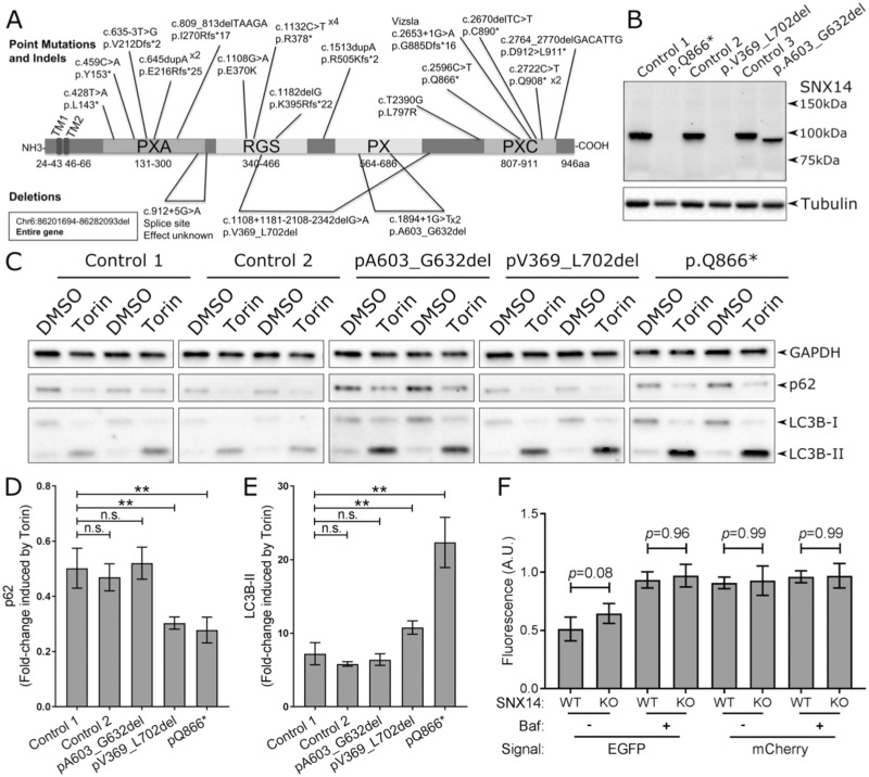
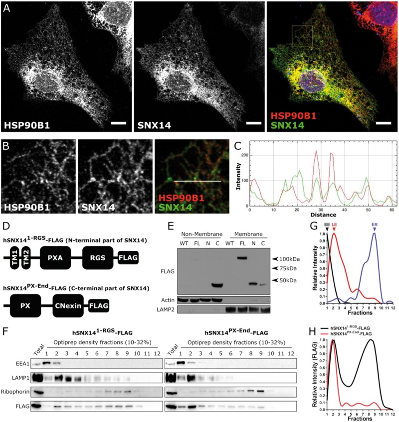
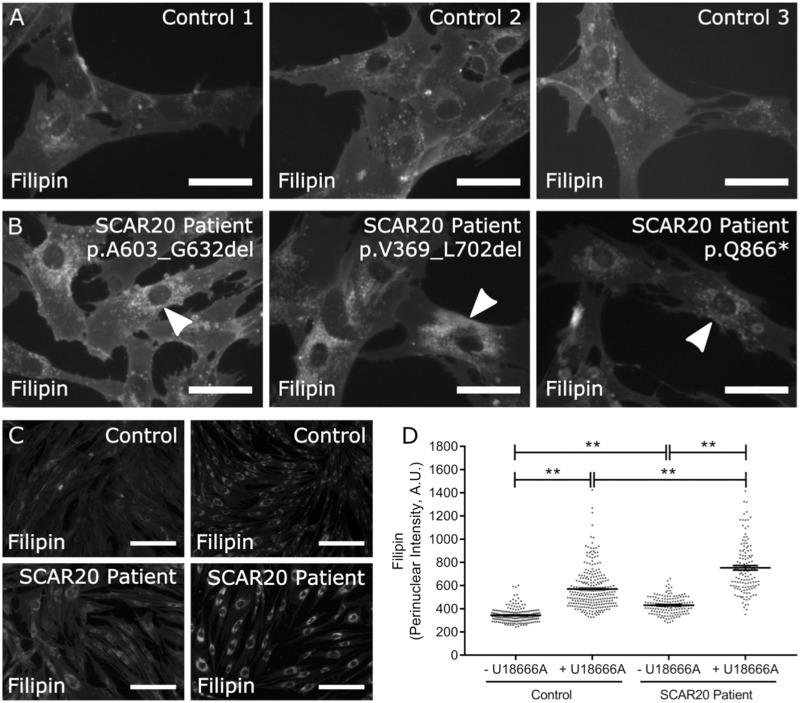
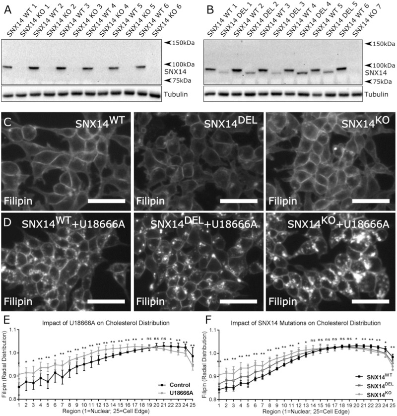
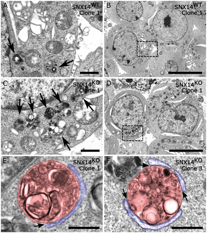
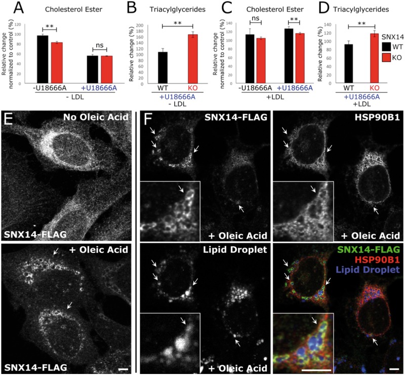
Mutations in SNX14 cause the autosomal recessive cerebellar ataxia 20 (SCAR20). Mutations generally result in loss of protein although several coding region deletions have also been reported. Patient-derived fibroblasts show disrupted autophagy, but the precise function of SNX14 is unknown. The yeast homolog, Mdm1, functions in endoplasmic reticulum (ER)-lysosome/vacuole inter-organelle tethering, but functional conservation in mammals is still required. Here, we show that loss of SNX14 alters but does not block autophagic flux. In addition, we find that SNX14 is an ER-associated protein that functions in neutral lipid homeostasis and inter-organelle crosstalk. SNX14 requires its N-terminal transmembrane helices for ER localization, while the Phox homology (PX) domain is dispensable for subcellular localization. Both SNX14-mutant fibroblasts and SNX14KO HEK293 cells accumulate aberrant cytoplasmic vacuoles, suggesting defects in endolysosomal homeostasis. However, ER-late endosome/lysosome contact sites are maintained in SNX14KO cells, indicating that it is not a prerequisite for ER-endolysosomal tethering. Further investigation of SNX14- deficiency indicates general defects in neutral lipid metabolism. SNX14KO cells display distinct perinuclear accumulation of filipin in LAMP1-positive lysosomal structures indicating cholesterol accumulation. Consistent with this, SNX14KO cells display a slight but detectable decrease in cholesterol ester levels, which is exacerbated with U18666A. Finally, SNX14 associates with ER-derived lipid droplets (LD) following oleate treatment, indicating a role in ER-LD crosstalk. We therefore identify an important role for SNX14 in neutral lipid homeostasis between the ER, lysosomes and LDs that may provide an early intervention target to alleviate the clinical symptoms of SCAR20.


Figures

Figure 1.
Figure 1.
SNX14 mutations enhance response to autophagy induction. (A) Mutations in the SNX14 gene reported to cause SCAR20. (B) SNX14 protein expression in control and SCAR20 patient dermal fibroblasts. Mutations in the SNX14 gene result in either no detectable SNX14 protein (c2596C > T; p.Gln866* and c.1108 + 1181_2108–2342del; p.Val369_Leu702del) or a truncated SNX14ΔPX protein (c1894 + 1G > T; p.A603_G632del). (C) Control and SCAR20 patient-derived fibroblasts were cultured for 6 h with or without autophagy inducing conditions (250 nm Torin1 or 0.1% DMSO) which affect levels of p62 and LC3B-II. (D) Fold-change of p62 expression in response to Torin1 treatment. (E) Fold-change of LC3B-II expression in response to Torin1 treatment. (D, E) n =6, error bars = SEM, **P 0.01, ns P 0.05, Student’s t-test. (F) Quantification of EGFP and mCherry fluorescence intensity in SNX14WT and SNX14KO HEK293 cells cultured under autophagy inducing conditions with or without inhibition of lysosome-autophagosome fusion with bafilomycin A (50 nm, 6 h) treatment. N =4–7 (Cells), error bars = STD, **P 0.01, ANOVA.
Figure 2.
Figure 2.
SNX14 is localized to the endoplasmic reticulum membrane. (A, B) Overexpressed SNX14 in U2OS cells display a similar subcellular distribution to ER associated HSP90B1 (Scale bar = 10 µm). (C) Line plot of the region designated in (B) showing the relationship between SNX14 (Green) and HSP90B1 (Red) intensity. (D) U2OS cell lines express either N- or C-terminal segments of SNX14-FLAG. (E) The N-terminal segment of SNX14-FLAG is predominantly responsible for membrane association. (F) Organelle fractionation reveals different distributions between N- and C-terminal SNX14 segments. (G) Relative intensity of early endosome (EE) associated EEA1, late endosome (LE) associated LAMP1 and endoplasmic reticulum (ER) associated Ribophorin of hSNX141-RGS-FLAG expressing U2OS cells. (H) Relative intensity profile of N- and C-terminal segments of SNX14 show different distribution profiles.
Figure 3.
Figure 3.
Accumulation of cholesterol in SCAR20 patients. (A, B) Filipin staining of dermal fibroblasts derived from (A) control and (B) SCAR20 patients show increased perinuclear intensity is indicated by arrowheads (Scale bar = 50 µm). (C) Filipin stained dermal fibroblasts derived from a control and SCAR20 patient (p.Q866*) show increased perinuclear distributions of filipin intensity following 24 h of U18666A treatment (Scale bar = 200 µm). (D) Quantification of filipin intensity in the perinuclear region of each cell. Perinuclear accumulation of cholesterol in SNX14 mutant fibroblasts is exacerbated by U18666A treatment. Each dot represents a single cell, n 143, bars = mean, error bars = SEM, **P 0.01, one-way ANOVA.
Figure 4.
Figure 4.
Perinuclear accumulation of cholesterol in SNX14 mutant cells. SNX14 protein in cell lysates from HEK293 clones generated from single cell sorting following CRISPR-Cas9 mediated targeting of the SNX14 gene. (A, B) Clones display either full length SNX14 protein (SNX WT 1–6), no SNX14 protein (SNX14 KO 1–7) or truncated SNX14 protein (SNX14 DEL 1–5). Cells were cultured (C) with or (D) without 23.5 µm U18666A for 24 h. SNX14 mutant cells showed greater accumulation of cholesterol with U18666A treatment. (E) U18666A increased perinuclear distribution of cholesterol. (F) SNX14 mutant clones treated with U18666A displayed an increased perinuclear distribution of cholesterol compared to SNX14WT clones. (C, D) Scale bar = 50 µm. (E, F) The mean radial distribution of filipin signal intensity plotted from nuclear to peripheral regions, N =6 (SNX14WT clones), N =5 (SNX14DEL clones), N =7 (SNX14KO clones), dots = mean, error bars = SD, **P 0.01, *P 0.05, n.s. P 0.05, Student’s t-test.
Figure 5.
Figure 5.
Loss of SNX14 results in greater accumulation of autophagic organelles. Electron microscopy images from (A, B) SNX14WT and (CF) SNX14KO HEK293 clones. Cell lines were cultured in with 23.5 µm U18666A for 24 h before processing. Arrows indicate autophagic structures containing undigested or partially digested material. (E, F) Examples of membrane contact sites (arrows) detected between autophagic organelles (pseudo coloured red) and the endoplasmic reticulum (pseudo coloured blue) in the absence of SNX14. (A, C) Scale bar = 1 µm; (B, D) Scale bar = 10 µm (E, F) Scale bar = 500 nm.
Figure 6.
Figure 6.
Loss of SNX14 disrupts endoplasmic reticulum-associated neutral lipid metabolism. (AD) Thin layer chromatography analysis of neutral lipids in SNX14WT and SNX14KO HEK293 cultured without (A, B) or with (C, D) exogenous cholesterol (Low-density lipoprotein; LDL). (A) Cholesterol esters (CE) decreased in SNX14KO cells. (B) Triacylglycerides (TAG) increased in SNX14KO cells on addition of U18666A. (C) CEs decreased when SNX14KO cells were treated with U18666A and LDL. (D) TAG levels increased in SNX14KO cells cultured with U18666A and LDL. N =2, error bars = SD, **P 0.01, ns P 0.05, Student’s t-test. (E) Immunofluorescent images of U2OS cells that were either untreated, or treated with 600 μm oleic acid for 8 h. SNX14-FLAG (anti-FLAG) was observed to accumulate in peri-nuclear ring-like formations following oleic acid treatment (arrows). (F) Co-fluorescent imaging of SNX14-FLAG (Green), ER marker HSP90B1 (Red) and LD stain monodansylpentane (Blue). Scale bar = 5 μm.

Similar articles

Cited by

References

    1. Thomas A.C., Williams H., Seto-Salvia N., Bacchelli C., Jenkins D., O'Sullivan M., Mengrelis K., Ishida M., Ocaka L., Chanudet E.. et al. (2014) Mutations in SNX14 cause a distinctive autosomal-recessive cerebellar ataxia and intellectual disability syndrome. Am. J. Hum. Genet., 95, 611–621. - PMC - PubMed
    1. Akizu N., Cantagrel V., Zaki M.S., Al-Gazali L., Wang X., Rosti R.O., Dikoglu E., Gelot A.B., Rosti B., Vaux K.K.. et al. (2015) Biallelic mutations in SNX14 cause a syndromic form of cerebellar atrophy and lysosome-autophagosome dysfunction. Nat. Genet., 47, 528–534. - PMC - PubMed
    1. Jazayeri R., Hu H., Fattahi Z., Musante L., Abedini S.S., Hosseini M., Wienker T.F., Ropers H.H., Najmabadi H., Kahrizi K. (2015) Exome sequencing and linkage analysis identified novel candidate genes in recessive intellectual disability associated with ataxia. Arch. Iranian Med., 18, 670–682. - PubMed
    1. Karaca E., Harel T., Pehlivan D., Jhangiani S.N., Gambin T., Coban Akdemir Z., Gonzaga-Jauregui C., Erdin S., Bayram Y., Campbell I.M.. et al. (2015) Genes that affect brain structure and function identified by rare variant analyses of Mendelian neurologic disease. Neuron, 88, 499–513. - PMC - PubMed
    1. Shukla A., Upadhyai P., Shah J., Neethukrishna K., Bielas S., Girisha K.M. (2017) Autosomal recessive spinocerebellar ataxia 20: report of a new patient and review of literature. Eur. J. Med. Genet., 60, 118–123. - PMC - PubMed

Publication types

MeSH terms

Supplementary concepts