. 2018 Jun 15;27(12):2113-2124.
doi: 10.1093/hmg/ddy118.

Metabolomic studies identify changes in transmethylation and polyamine metabolism in a brain-specific mouse model of tuberous sclerosis complex

Affiliations

Metabolomic studies identify changes in transmethylation and polyamine metabolism in a brain-specific mouse model of tuberous sclerosis complex

James McKenna 3rd et al. Hum Mol Genet. .

Abstract

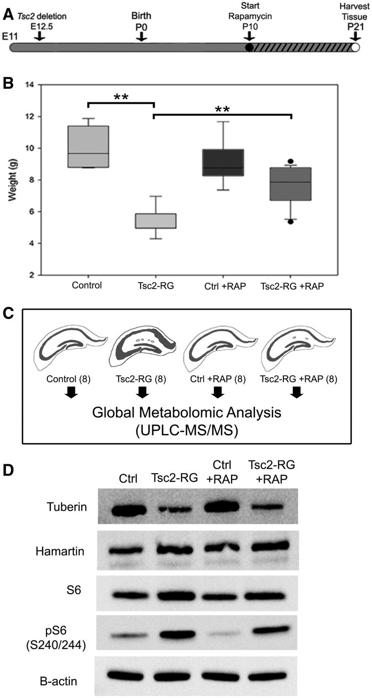
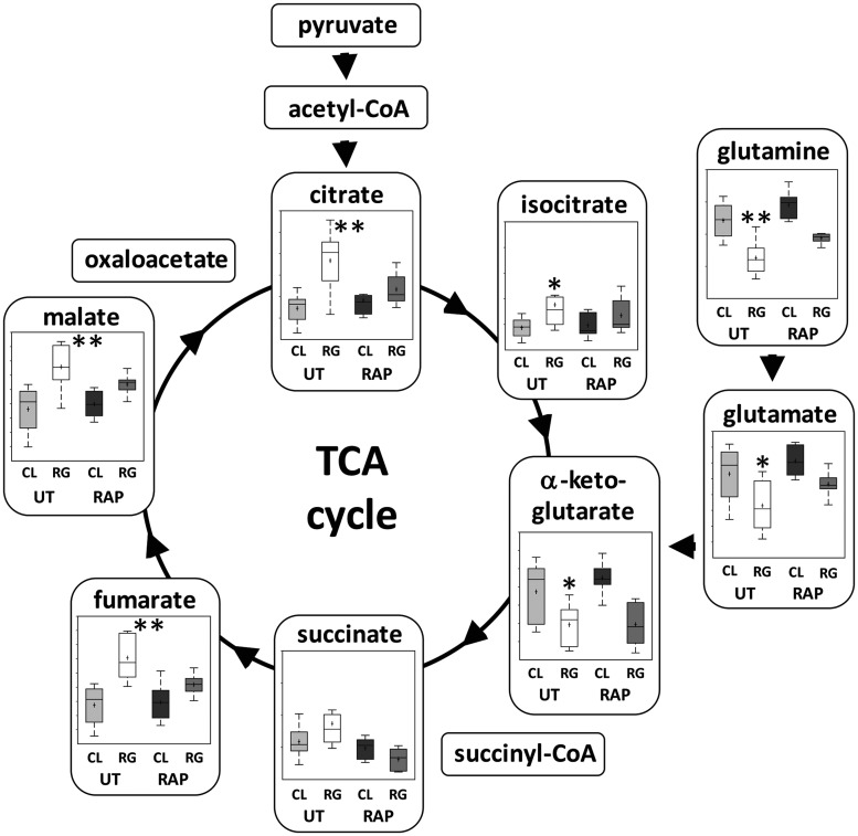
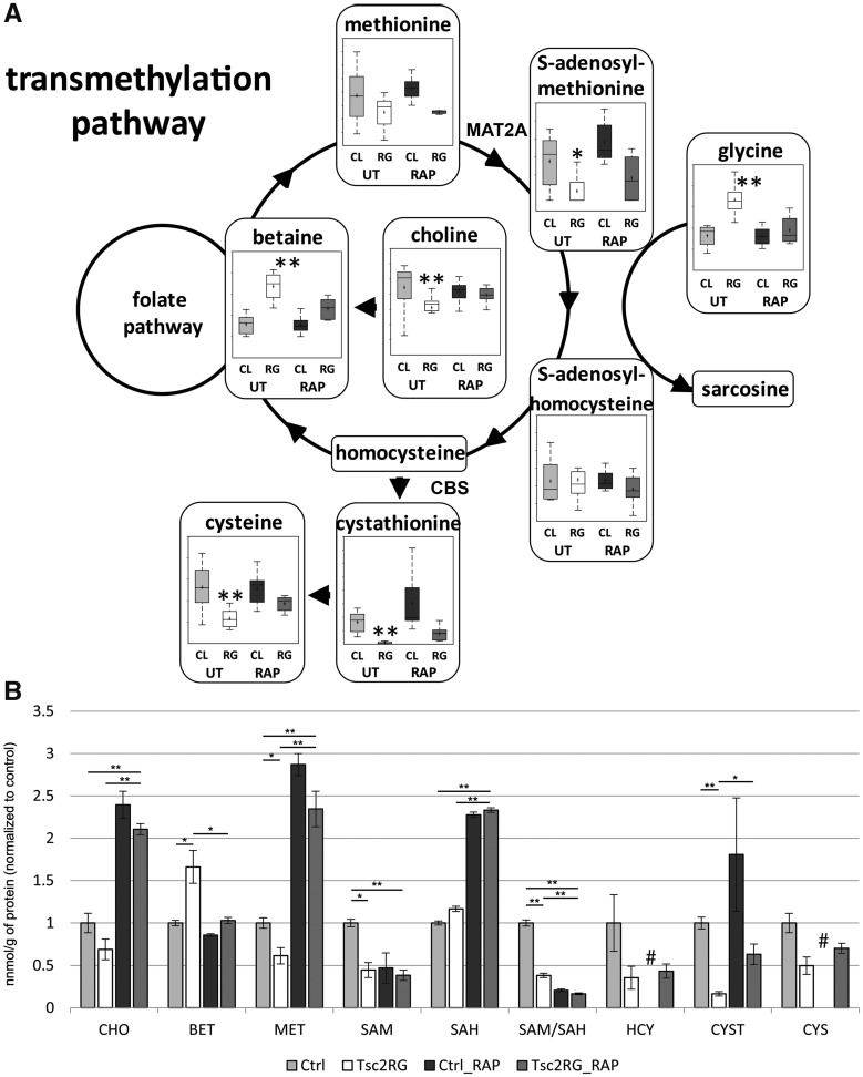
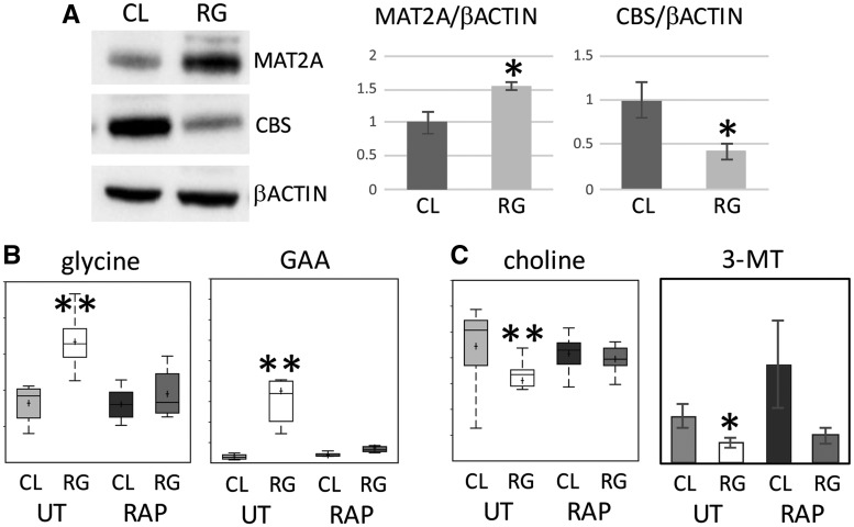
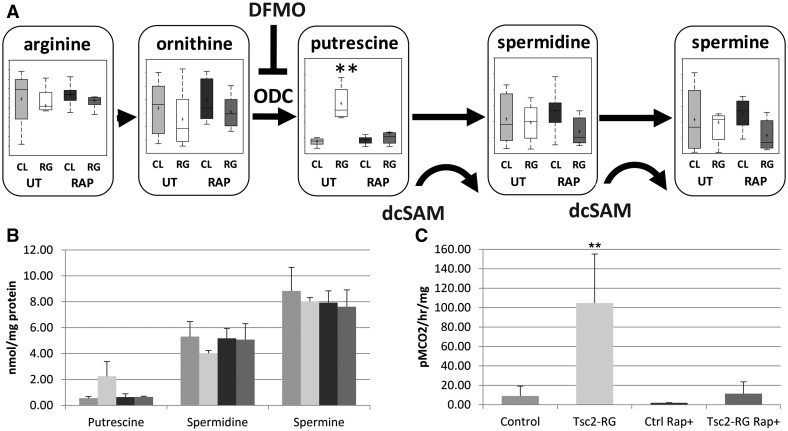
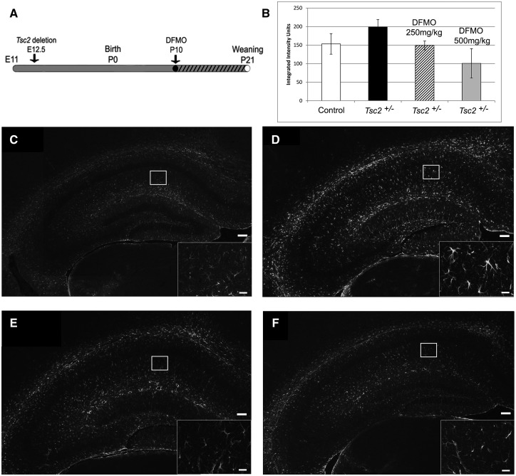
Tuberous sclerosis complex (TSC) is an autosomal dominant neurodevelopmental disorder and the quintessential disorder of mechanistic Target of Rapamycin Complex 1 (mTORC1) dysregulation. Loss of either causative gene, TSC1 or TSC2, leads to constitutive mTORC1 kinase activation and a pathologically anabolic state of macromolecular biosynthesis. Little is known about the organ-specific metabolic reprogramming that occurs in TSC-affected organs. Using a mouse model of TSC in which Tsc2 is disrupted in radial glial precursors and their neuronal and glial descendants, we performed an unbiased metabolomic analysis of hippocampi to identify Tsc2-dependent metabolic changes. Significant metabolic reprogramming was found in well-established pathways associated with mTORC1 activation, including redox homeostasis, glutamine/tricarboxylic acid cycle, pentose and nucleotide metabolism. Changes in two novel pathways were identified: transmethylation and polyamine metabolism. Changes in transmethylation included reduced methionine, cystathionine, S-adenosylmethionine (SAM-the major methyl donor), reduced SAM/S-adenosylhomocysteine ratio (cellular methylation potential), and elevated betaine, an alternative methyl donor. These changes were associated with alterations in SAM-dependent methylation pathways and expression of the enzymes methionine adenosyltransferase 2A and cystathionine beta synthase. We also found increased levels of the polyamine putrescine due to increased activity of ornithine decarboxylase, the rate-determining enzyme in polyamine synthesis. Treatment of Tsc2+/- mice with the ornithine decarboxylase inhibitor α-difluoromethylornithine, to reduce putrescine synthesis dose-dependently reduced hippocampal astrogliosis. These data establish roles for SAM-dependent methylation reactions and polyamine metabolism in TSC neuropathology. Importantly, both pathways are amenable to nutritional or pharmacologic therapy.


Figures

Figure 1.
Figure 1.
 Preparation and characterization of hippocampal samples for metabolomic profiling. (A) Time course of rapamycin treatment of control and Tsc2-RG mice. hGFAP-Cre-mediated disruption of the Tsc2 gene begins at embryonic day 12.5. Mice were treated with rapamycin by daily i.p. injection from P10 to P21 (denoted by hatched bar) and sacrificed for metabolomic profiling at P21. (B) Animal weights at P21. Note rapamycin treatment partially rescued impaired weight gain of Tsc2-RG mice. (C) Hippocampal tissue was isolated from eight animals per group and sent for metabolomic analysis. (D) Immunoblot analysis illustrating reduced tuberin protein and increased mTORC1 activity (pS6) in cortical lysates of Tsc2-RG mice compared with control lysates. Rapamycin treatment partially rescued mTORC1 activity in Tsc2-RG brains by P21. RAP, rapamycin. **P < 0.001 for pairwise comparisons.
Figure 2.
Figure 2.
 Metabolomic profile of hippocampi in untreated and rapamycin-treated control and Tsc2-RG mice. (A) Principal component analysis of the dataset attributed the most variability in metabolite expression to genotype (x-axis), with a weaker effect of treatment (y-axis). (B) Heat map of significantly (P < 0.05) altered metabolites between untreated control and Tsc2-RG mice. Metabolites are grouped by those upregulated in untreated Tsc2-RG and those downregulated in Tsc2-RG mice and are further organized by general biological function. The response of these metabolites to rapamycin (RAPA) treatment in control and Tsc2-RG mice is also indicated. Columns of signals represent data from individual subjects (eight per genotype/treatment group) with blue indicating downregulation and yellow upregulation. The intensity of the signal reflects the Z-score for that data point. Note that rapamycin treatment partially normalized levels of this set of metabolites in Tsc2-RG mice to those of untreated control animals, whereas rapamycin had minimal effect in control mice. (C) Schematic of altered amino acid synthesis in untreated Tsc2-RG versus control mice. Yellow and blue circles indicate amino acid derivatives upregulated and downregulated, respectively, in Tsc2-RG mice compared with controls. Thickness of the circle outline indicates the relative magnitude of change. (D) Pathway fold enrichment plot illustrating metabolic pathways significantly altered in Tsc2-RG mice (P-values indicated).
Figure 3.
Figure 3.
 Metabolomic profile of glutamine and TCA cycle metabolism in hippocampus of untreated and rapamycin-treated control and Tsc2-RG mice. (A) Box and whisker plots show levels of pathway metabolites in hippocampus of untreated (UT) and rapamycin (RAP) treated control (CL) and Tsc2-RG (RG) mice. Arrows indicate direction of metabolism. Acetyl-CoA, succinyl-CoA and oxaloacetate were not present in the dataset. Note the elevation of most TCA metabolites in untreated Tsc2-RG samples, with the exception of glutamine, glutamate and alpha-ketoglutarate, which are downregulated. Box plots: +, mean value; center line of box, median value; top line of box, limit of upper quartile; bottom line of box, limit of lower quartile; top of whisker, maximum of distribution; bottom of whisker, minimum of distribution; ordinate represents scaled intensity. *P < 0.05, **P < 0.001 for pairwise comparison of untreated control and Tsc2-RG values.
Figure 4.
Figure 4.
 Metabolomic profile of transmethylation and part of transulfuration pathways in untreated and rapamycin-treated control and Tsc2-RG mice. (A) Box and whisker plots show levels of pathway metabolites in hippocampus of untreated (UT) and rapamycin (RAP)-treated control (CL) and Tsc2-RG (RG) mice. Arrows indicate direction of metabolism. Sarcosine, homocysteine and folate pathway metabolites were not present in the dataset. Box plots: +, mean value; center line of box, median value; top line of box, limit of upper quartile; bottom line of box, limit of lower quartile; top of whisker, maximum of distribution; bottom of whisker, minimum of distribution. MAT2A, methionine S-adenosyltransferase 2A, CBS, cystathionine beta synthase. *P < 0.05, **P < 0.001 for pairwise comparison of untreated control and Tsc2-RG values. (B) Transmethylation pathway metabolites in cortical samples from control and Tsc2-RG mice as measured by LC-MS/MS. These values were normalized to untreated control values. CHO, choline; BET, betaine; MET, methionine; SAM, S-adenosylmethionine; SAH, S-adenosylhomocysteine; HCY, homocysteine; CYST, cystathionine; CYS, cysteine. Significant pairwise comparisons are indicated by * P < 0.05, **P < 0.01. # indicates absence of rapamycin-treated control samples from HCY and CYS metabolite datasets.
Figure 5.
Figure 5.
Dysregulation of enzymes and SAM-dependent enzymatic reactions in Tsc2-RG mice. (A) Immunoblot showing increased expression of MAT2A and reduced expression of CBS proteins in cortices of Tsc2-RG (RG) mice compared with control. Immunoblot band intensities for MAT2A and CBS relative to β-actin are shown normalized to control (CL) values. (B) Accumulation of precursors of SAM-dependent enzymatic reactions in Tsc2-RG hippocampus. Box and whisker plots showing increased levels of glycine and GAA in Tsc2-RG hippocampus. (C) Decreased levels of products of SAM-dependent enzymatic reactions in Tsc2-RG brains. Box and whisker plot showing decreased levels of choline and histogram showing reduced 3-MT in Tsc2-RG hippocampus and cortex, respectively. UT, untreated; RAP, rapamycin treated. *P < 0.05, **P < 0.001 for pairwise comparison of untreated control and Tsc2-RG values.
Figure 6.
Figure 6.
 Metabolomic profile of polyamine metabolism in untreated and rapamycin-treated control and Tsc2-RG mice. (A) Pathway diagram depicting dysregulation of polyamine synthesis, mainly the accumulation of putrescine, in Tsc2-RG hippocampus. Box and whisker plots show levels of pathway intermediate metabolites in hippocampus of untreated (UT) and rapamycin (RAP)-treated control (CL) and Tsc2-RG (RG) mice. dcSAM (decarboxylated SAM) acts as an aminopropyl donor in the conversion of putrescine to spermidine and spermidine to spermine. ODC, ornithine decarboxylase; DFMO, α-difluoromethylornithine; an irreversible ODC inhibitor. Box plots: +, mean value; center line of box, median value; top line of box, limit of upper quartile; bottom line of box, limit of lower quartile; top of whisker, maximum of distribution; bottom of whisker, minimum of distribution; **P < 0.001 for pairwise comparison of untreated control and Tsc2-RG values. (B) Elevated putrescine levels in cortical samples from Tsc2-RG mice by HPLC. Spermidine and spermine levels are unaffected. (C) Elevated ODC activity in cortical samples from Tsc2-RG mice.
Figure 7.
Figure 7.
 Inhibition of ODC with DFMO in Tsc2+/− mice decreases astrogliosis in the hippocampal CA1 region in a dose-dependent manner. (A) Time course of DFMO treatment of Tsc2+/− mice. Mice were treated with DFMO (250 or 500 mg/kg) by daily i.p. injection from P10 to P21 (denoted by hatched bar) and sacrificed for GFAP immunofluorescence (IF) at P21. (B) Quantification of hippocampal GFAP IF intensity from control mice and untreated and DFMO-treated Tsc2+/− mice. (C) Representative IF of control, (D) untreated Tsc2+/−, (E) 250 mg/kg DFMO-treated Tsc2+/− and (F) 500 mg/kg DFMO-treated Tsc2+/- hippocampi using anti-GFAP antibody to detect astrogliosis (scale bar = 100 µm). Astrocytes in the CA1 region (inset, scale bar = 20 µm). *P < 0.05.

Similar articles

Cited by

References

    1. Crino P.B., Nathanson K.L., Henske E.P. (2006) The tuberous sclerosis complex. N. Engl. J. Med., 355, 1345–1356. - PubMed
    1. Consortium. (1993) Identification and characterization of the tuberous sclerosis gene on chromosome 16. Cell, 75, 1305–1315. - PubMed
    1. van Slegtenhorst M., de Hoogt R., Hermans C., Nellist M., Janssen B., Verhoef S., Lindhout D., van den Ouweland A., Halley D., Young J.. et al. (1997) Identification of the tuberous sclerosis gene TSC1 on chromosome 9q34. Science, 277, 805–808. - PubMed
    1. Menon S., Dibble C., Talbott G., Hoxhaj G., Valvezan A., Takahashi H., Cantley L.C., Manning B.D. (2014) Spatial control of the TSC complex integrates insulin and nutrient regulation of mTORC1 at the lysosome. Cell, 156, 771–785. - PMC - PubMed
    1. Sabatini D.M. (2017) Twenty-five years of mTOR: uncovering the link from nutrients to growth. Proc. Natl. Acad. Sci. U. S. A., 114, 11818–11825. - PMC - PubMed

Publication types

MeSH terms