. 2018 Apr 10;16(1):14.
doi: 10.1186/s12964-018-0226-1.

Matrix metalloproteinase-9 (MMP9) is involved in the TNF-α-induced fusion of human M13SV1-Cre breast epithelial cells and human MDA-MB-435-pFDR1 cancer cells

Affiliations

Matrix metalloproteinase-9 (MMP9) is involved in the TNF-α-induced fusion of human M13SV1-Cre breast epithelial cells and human MDA-MB-435-pFDR1 cancer cells

Julian Weiler et al. Cell Commun Signal. .

Abstract

Background: In addition to physiological events such as fertilisation, placentation, osteoclastogenesis, or tissue regeneration/wound healing, cell fusion is involved in pathophysiological conditions such as cancer. Cell fusion, which applies to both the proteins and conditions that induce the merging of two or more cells, is not a fully understood process. Inflammation/pro-inflammatory cytokines might be a positive trigger for cell fusion. Using a Cre-LoxP-based cell fusion assay we demonstrated that the fusion between human M13SV1-Cre breast epithelial cells and human MDA-MB-435-pFDR1 cancer cells was induced by the pro-inflammatory cytokine tumour necrosis factor-α (TNF-α).

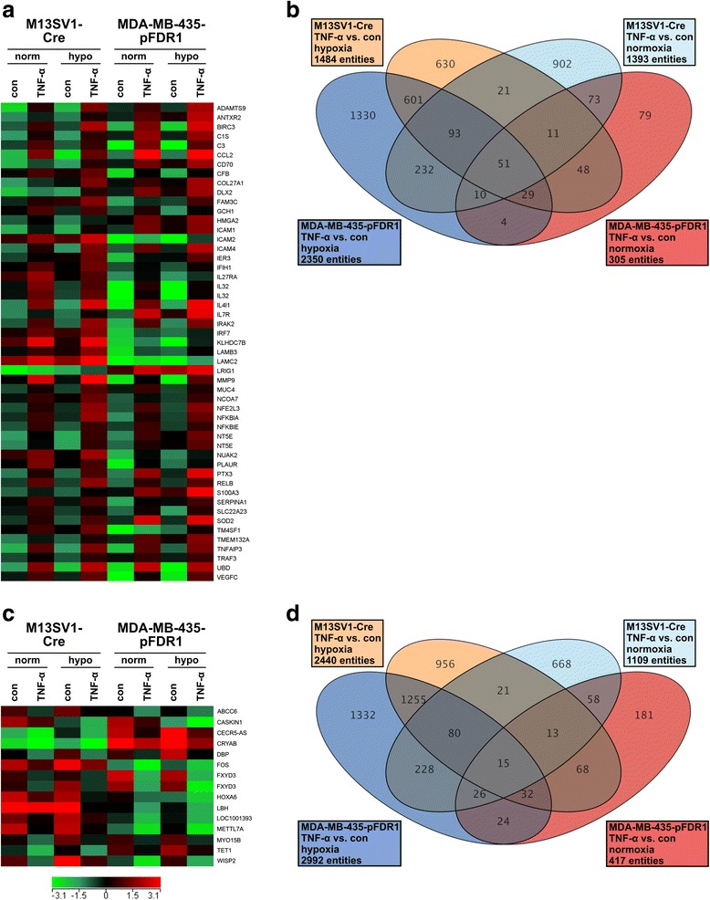
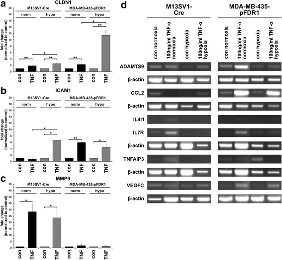
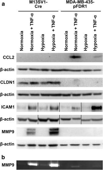
Methods: The gene expression profile of the cells in the presence of TNF-α and under normoxic and hypoxic conditions was analysed by cDNA microarray analysis. cDNA microarray data were verified by qPCR, PCR, Western blot and zymography. Quantification of cell fusion events was determined by flow cytometry. Proteins of interest were either blocked or knocked-down using a specific inhibitor, siRNA or a blocking antibody.

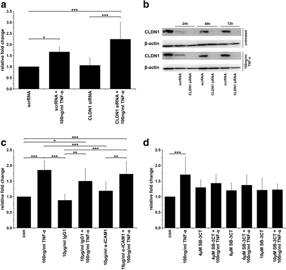
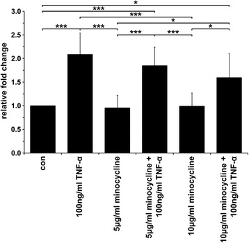
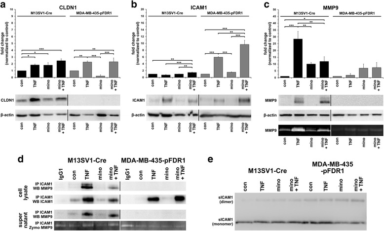
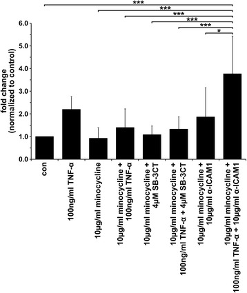
Results: The data showed an up-regulation of various genes, including claudin-1 (CLDN1), ICAM1, CCL2 and MMP9 in M13SV1-Cre and/or MDA-MB-435-pFDR1 cells. Inhibition of these proteins using a blocking ICAM1 antibody, CLDN1 siRNA or an MMP9 inhibitor showed that only the blockage of MMP9 was correlated with a decreased fusion rate of the cells. Likewise, the tetracycline-based antibiotic minocycline, which exhibits anti-inflammatory properties, was also effective in both inhibiting the TNF-α-induced MMP9 expression in M13SV1-Cre cells and blocking the TNF-α-induced fusion frequency of human M13SV1-Cre breast epithelial cells and human MDA-MB-435-pFDR1 cancer cells.

Conclusions: The matrix metalloproteinase-9 (MMP9) is most likely involved in the TNF-α-mediated fusion of human M13SV1-Cre breast epithelial cells and human MDA-MB-435-pFDR1 cancer cells. Likewise, our data indicate that the tetracycline-based antibiotic minocycline might exhibit anti-fusogenic properties because it inhibits a cell fusion-related mechanism.

Keywords: Breast cancer; Cell fusion; MMP9; Minocycline; TNF-α.


Conflict of interest statement

Ethics approval and consent to participate

Not applicable.

Consent for publication

Not applicable.

Competing interests

The authors declare that they have no competing interests.

Publisher’s Note

Springer Nature remains neutral with regard to jurisdictional claims in published maps and institutional affiliations.

Figures

Fig. 1
Fig. 1
Differentially regulated genes in TNF-α-treated M13SV1-Cre and MDA-MB-435-pFDR1 cells under normoxic and hypoxic conditions. The up-regulated and down-regulated genes in TNF-α-treated cells compared with untreated cells that matched the filter criteria (flags: not detected flags and compromised spots were removed) and fold changes (2-fold up and down). a A heat map analysis of TNF-α up-regulated genes, b a Venn diagram of TNF-α up-regulated genes, c a heat map analysis of TNF-α down-regulated genes, d a Venn diagram of TNF-α down-regulated genes
Fig. 2
Fig. 2
Validation of microarray data by qPCR and conventional PCR. a qPCR CLDN1, b qPCR ICAM1, c qPCR MMP9, d conventional PCR. MMP9 was significantly up-regulated in TNF-α-treated (100 ng/ml) M13SV1-Cre cells, whereas significantly elevated CLDN1 and ICAM1 expression levels were detected in TNF-α-treated (100 ng/ml) MDA-MB-435-pFDR1 cells. Likewise, increased CCL2 and VEGFC expression levels were detected in TNF-α-treated (100 ng/ml) MDA-MB-435-pFDR1 cells. Data are presented as the mean ± SD of at least three independent experiments (a) or the representative data of three independent experiments (b). Statistical analysis: unpaired, two-tailed Student’s t-test: * = p < 0.05; ** = p < 0.01
Fig. 3
Fig. 3
Validation of qPCR and conventional RT-PCR data by Western blot and zymography. a Western blot analysis: CCL2 protein expression was only detected in TNF-α-treated (100 ng/ml) MDA-MB-435-pFDR1 cells, whereas CLDN1 expression was absent from these cells but was clearly detectable in M13SV1-Cre cells. Increased ICAM1 expression was observed in TNF-α-treated cells, whereas increased MMP9 expression was only found in TNF-α-stimulated M13SV1-Cre cells. Two discrete bands were detected for MMP9 in Western blot analysis; the upper band represents inactive MMP9 and the lower band represents active MMP9. ICAM1 Western blot data were rearranged because ICAM1 samples were originally loaded in a different order. b Zymography: markedly increased MMP9 expression levels and activity were detected in TNF-α-treated M13SV1-Cre cells, whereas even in TNF-α-stimulated MDA-MB-435 cells slightly enhanced MMP9 expression levels could be identified. The data shown are representative of at least three independent experiments
Fig. 4
Fig. 4
TNF-α-induced cell fusion could be blocked by the inhibition of MMP9. a The knockdown of CLDN1 expression with specific siRNA was not correlated with a decreased TNF-α-induced (100 ng/ml) fusion rate. b siRNA-mediated CLDN1 expression levels of M13SV1-Cre cells were stably down-regulated over 72 h. c Inhibition of ICAM1 activity using a blocking antibody did not impair the TNF-α-induced fusion between M13SV1-Cre and MDA-MB-435-pFDR1 cells. d The TNF-α-induced fusion of M13SV1-Cre and MDA-MB-435-pFDR1 cells was impaired by the MMP9 inhibitor SB-3CT in a dose-dependent manner. The mean ± SD of at least three independent experiments is shown. Statistical analysis: ANOVA F-test and Scheffé post-hoc test: * = p < 0.05; ** = p < 0.01; *** = p < 0.001
Fig. 5
Fig. 5
TNF-α-induced fusion was inhibited by the tetracycline-based antibiotic minocycline. TNF-α-induced (100 ng/ml) cell fusion was impaired by minocycline in a dose-dependent manner. The data shown are the mean ± SD of three independent experiments. Statistical analysis: ANOVA F-test and Scheffé post-hoc test: * = p < 0.05; ** = p < 0.01; *** = p < 0.001
Fig. 6
Fig. 6
TNF-α-induced MMP9 expression in M13SV1-Cre cells was blocked by minocycline. M13SV1-Cre and MDA-MB-435-pFDR1 cells were treated for 3 days with 100 ng/ml TNF-α, 10 μg/ml minocycline and a combination of both. The expression levels of a) CLDN1, b) ICAM1 and c) MMP9 were determined by qPCR, Western blot analysis and zymography (only MMP9). Significantly decreased MMP9 mRNA levels were detected in TNF-α and minocycline-treated M13SV1-Cre cells as compared with TNF-α-treated cells and were validated by zymography. d) Co-immunoprecipitation data show that MMP9 binds to ICAM1. ICAM1 expression and co-immunoprecipitated MMP9 were markedly decreased in the lysates of TNF-α + minocycline-treated M13SV1-Cre cells compared with the lysates from TNF-α-stimulated cells. e) No differences in sICAM1 levels were detected in M13SV1-Cre and MDA-MB-435-pFDR1 cells that were treated with TNF-α, minocycline and a combination of both. Shown are the mean ± SD or representative Western blot data for at least three independent experiments. Statistical analysis: ANOVA F-test and Scheffé post-hoc test: * = p < 0.05; ** = p < 0.01; *** = p < 0.001
Fig. 7
Fig. 7
The co-administration of minocycline, SB-3CT and the ICAM1 blocking antibody, yielded different results. No additive inhibitory effect of minocycline and SB-3CT regarding the TNF-α-induced fusion of M13SV1-Cre and MDA-MB-435-pFDR1 cells was observed. In contrast, the incubation of cells in the presence of minocycline, ICAM1 blocking antibody and TNF-α yielded a significantly increased fusion rate. The data shown are the mean ± SD of at least three independent experiments. Statistical analysis: ANOVA F-test and Scheffé post-hoc test: * = p < 0.05; ** = p < 0.01; *** = p < 0.001

Similar articles

Cited by

References

    1. Aguilar PS, Baylies MK, Fleissner A, Helming L, Inoue N, Podbilewicz B, Wang H, Wong M. Genetic basis of cell-cell fusion mechanisms. Trends Genet. 2013;29(7):427–437. doi: 10.1016/j.tig.2013.01.011. - DOI - PMC - PubMed
    1. Dittmar T, Zanker KS. Cell fusion in health and disease. Volume II, vol 2. Adv Exp med biol. Dordrecht, The Netherlands: Springer; 2011. - PubMed
    1. Dittmar T, Zänker KS. Cell fusion in health and disease: volume I, vol 1. Adv Exp med biol. Dordrecht, The Netherlands: Springer; 2011. - PubMed
    1. Zhou X, Platt JL. Molecular and cellular mechanisms of mammalian cell fusion. Adv Exp Med Biol. 2011;713:33–64. doi: 10.1007/978-94-007-0763-4_4. - DOI - PubMed
    1. Huppertz B, Bartz C, Kokozidou M. Trophoblast fusion: fusogenic proteins, syncytins and ADAMs, and other prerequisites for syncytial fusion. Micron. 2006;37(6):509–517. doi: 10.1016/j.micron.2005.12.011. - DOI - PubMed

Publication types

MeSH terms

LinkOut - more resources