. 2018 Jun;209(2):551-565.
doi: 10.1534/genetics.118.300909. Epub 2018 Apr 10.

Differential Expression of Histone H3.3 Genes and Their Role in Modulating Temperature Stress Response in Caenorhabditis elegans

Affiliations

Differential Expression of Histone H3.3 Genes and Their Role in Modulating Temperature Stress Response in Caenorhabditis elegans

Kamila Delaney et al. Genetics. 2018 Jun.

Abstract

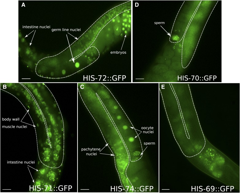
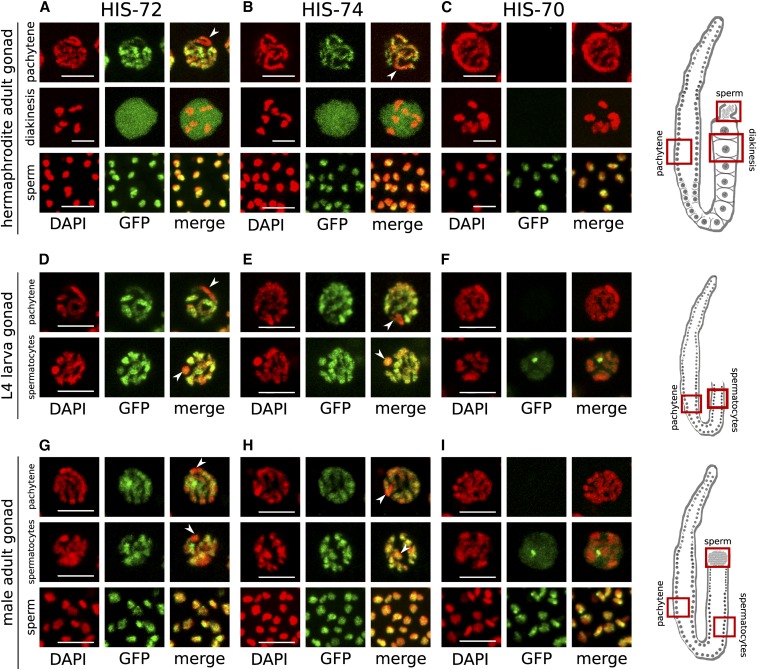
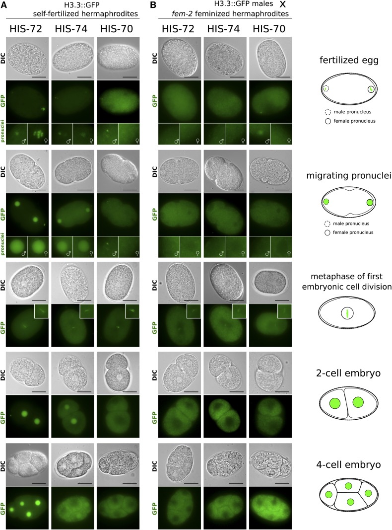
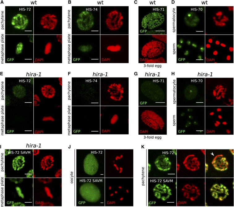
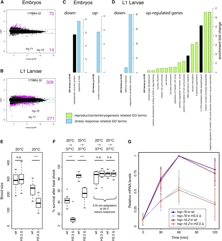
Replication-independent variant histones replace canonical histones in nucleosomes and act as important regulators of chromatin function. H3.3 is a major variant of histone H3 that is remarkably conserved across taxa and is distinguished from canonical H3 by just four key amino acids. Most genomes contain two or more genes expressing H3.3, and complete loss of the protein usually causes sterility or embryonic lethality. Here, we investigate the developmental expression patterns of the five Caenorhabditis elegans H3.3 homologs and identify two previously uncharacterized homologs to be restricted to the germ line. Despite these specific expression patterns, we find that neither loss of individual H3.3 homologs nor the knockout of all five H3.3-coding genes causes sterility or lethality. However, we demonstrate an essential role for the conserved histone chaperone HIRA in the nucleosomal loading of all H3.3 variants. This requirement can be bypassed by mutation of the H3.3-specific residues to those found in H3. While even removal of all H3.3 homologs does not result in lethality, it leads to reduced fertility and viability in response to high-temperature stress. Thus, our results show that H3.3 is nonessential in C. elegans but is critical for ensuring adequate response to stress.

Keywords: C. elegans; H3.3; HIRA; Histone variants; germ line; stress response.


Figures

Figure 1
Figure 1
H3.3 homologs are differentially expressed. Signals of GFP fusions with H3.3 homologs in representative parts of adult hermaphrodites are shown. (A) HIS-72 is expressed ubiquitously in both the soma and germ line. (B) HIS-71 is expressed in somatic cells, but absent from the germ line. (C) HIS-74 expression is restricted to the germ line at all stages of germ line development, including mature oocytes and sperm. (D) HIS-70 is restricted to the male germ line and only detectable in mature sperm in adult hermaphrodites. (E) HIS-69 is not detectable in any cells. The germ line is outlined by a dashed white line. Note the autofluorescence of the intestine in all panels. Bars represent 20 µm.
Figure 2
Figure 2
HIS-72 and HIS-74 are expressed in all germ cells, whereas HIS-70 is male germ line-specific. (A–C) Hermaphrodite adult stage. HIS-72 (A) and HIS-74 (B) expression shown in pachytene, oocytes, and sperm. In pachytene nuclei, the GFP signal is chromatin associated, but appears depleted from parts of the chromatin (arrows). In oocytes, strong nucleoplasmic signals obscure the chromatin-associated signal. (C) HIS-70 is only detectable in sperm. (D–F) Hermaphrodite L4 larval stage. HIS-72 (D) and HIS-74 (E) expression is present in all germ cells, including pachytene and spermatocyte stages. (F) HIS-70 is only detectable in spermatocytes, but absent from pachytene. (G–I) Male adult stage. HIS-72 (G) and HIS-74 (H) expression is visible in all germ cells, including pachytene stage and in spermatocytes and sperm. (I) HIS-70 is only detectable in spermatocytes and sperm. Bars represent 5 µm. Cartoon images of germ lines highlight the regions shown in (A–I).
Figure 3
Figure 3
Dynamics of H3.3 proteins in early embryos. Representative live-cell images of early stages in embryonic development are shown. Stages include eggs after fertilization, migrating pronuclei, the metaphase of first cell division, the two-cell embryo, and the four-cell embryo, as outlined by cartoons on the right. (A) Eggs of self-fertilizing hermaphrodites carrying H3.3::GFP fusions. HIS-72 (left panels) is present in both male and female pronuclei upon fertilization and remains chromatin associated during metaphase of the first mitotic division. It is subsequently present in all cells. HIS-74 (center panels) is present in both male and female pronuclei upon fertilization and remains chromatin associated during metaphase of the first mitotic division. It is present in both nuclei at the two-cell stage, but subsequently becomes diluted. It is sometimes still faintly visible in two nuclei at the four-cell stage and then becomes undetectable. HIS-70 (right panels) is faintly detectable in the male but not the female pronucleus, remains chromatin associated during the first mitotic division, and subsequently becomes undetectable. (B) Eggs of feminized fem-2 hermaphrodites fertilized by males carrying H3.3::GFP fusions. HIS-72 and HIS-74 are not detectable in the pronuclei, while HIS-70 is visible in the male pronucleus. All three GFP fusions appear faintly chromatin associated during the first mitotic division and become undetectable after that. In GFP images representing fertilized eggs and migrating pronuclei, female pronuclei are highlighted by enlarged images with female symbols and male pronuclei are highlighted by enlarged images with male symbols. Bars represent 20 µm.
Figure 4
Figure 4
Chromatin association of all H3.3 homologs depends on HIRA-1. (A–D) Chromatin association of H3.3 homologs in a wild-type (wt) background. (A) HIS-72 in pachytene and mitotic metaphase nuclei, (B) HIS-74 in pachytene and mitotic metaphase nuclei, (C) HIS-71 in threefold embryo, and (D) HIS-70 in spermatocyte nucleus and mature sperm. (E–H) H3.3 homolog signal is lost in hira-1 deletion background at the same stages as in (A–D). (E) HIS-72, (F) HIS-74, (G) HIS-71, and (H) HIS-70. Residual signal remains at HIS-70 foci. See text for details. (I–K) Chromatin localization of HIS-72 in hira-1 deletion background is restored upon mutation of the H3.3-specific motif in HIS-72 to the canonical H3-specific motif (AAIG to SAVM). (I) HIS-72 SAVM in pachytene and mitotic metaphase nuclei. (J and K) Chromatin association of HIS-72 (top panels) and HIS-72 SAVM (bottom panels) appears different. HIS-72 SAVM is more strongly chromatin associated in oocyte nuclei, with chromosomes being visible despite the strong nucleoplasmic background (J), and there is no obvious depletion of GFP signal in any part of the chromatin (K). Bars represent 2 µm in all panels except (C and G), where they represent 10 µm.
Figure 5
Figure 5
H3.3 modulates fertility and temperature stress response. (A and B) MA plots of RNA sequencing results showing genes up- and downregulated in H3.3 null mutant (H3.3 ∆) worms compared to wild-type (wt) worms. (A) Embryos. (B) L1 larvae. Log2 fold changes (FC) are plotted against log2 counts per million (CPM). The values are averages from two biological replicates. The blue lines show the FC ± 2 cutoff. The most significant genes—his-71, his-72, and Y75B8A.33—are highlighted. (C and D) Gene ontology (GO) enrichment analysis for genes differentially expressed in H3.3 ∆ animals compared to wt animals at embryonic (C) and L1 larval stages (D). Enrichment fold change of significant GO terms is plotted separately for up- and downregulated genes. (E) Brood size analysis. Wt and H3.3 ∆ strain brood sizes at 20 and 25°. Brood sizes of individual worms were counted over a period of 4 days after the first egg was laid (N = 20). *** indicates P ≤ 0.001. n.s., not significant. (F) Resistance to heat shock. Percentage of surviving wt and H3.3 ∆ worms after 100 min heat shock at 37°, without and with 3.5 hr adaptation at 30° (N = 8). *** indicates P ≤ 0.001. n.s., not significant. Worms were either maintained at 20 or 25° prior to heat shock. (G) Relative mRNA levels of hsp-70 (blue) and hsp-16.2 (red) upon heat shock in wt (dark colors) and H3.3 ∆ (light colors) worms. Expression levels were determined by quantitative PCR, and the maximum expression levels set to 1.

Similar articles

Cited by

References

    1. Adam S., Polo S. E., Almouzni G., 2013. Transcription recovery after DNA damage requires chromatin priming by the H3.3 histone chaperone HIRA. Cell 155: 94–106. 10.1016/j.cell.2013.08.029 - DOI - PubMed
    1. Akiyama T., Suzuki O., Matsuda J., Aoki F., 2011. Dynamic replacement of histone H3 variants reprograms epigenetic marks in early mouse embryos. PLoS Genet. 7: e1002279 10.1371/journal.pgen.1002279 - DOI - PMC - PubMed
    1. Arico J. K., Katz D. J., van der Vlag J., Kelly W. G., 2011. Epigenetic patterns maintained in early Caenorhabditis elegans embryos can be established by gene activity in the parental germ cells. PLoS Genet. 7: e1001391 10.1371/journal.pgen.1001391 - DOI - PMC - PubMed
    1. Arribere J. A., Bell R. T., Fu B. X., Artiles K. L., Hartman P. S., et al. , 2014. Efficient marker-free recovery of custom genetic modifications with CRISPR/Cas9 in Caenorhabditis elegans. Genetics 198: 837–846. 10.1534/genetics.114.169730 - DOI - PMC - PubMed
    1. Ausió J., 1999. Histone H1 and evolution of sperm nuclear basic proteins. J. Biol. Chem. 274: 31115–31118. 10.1074/jbc.274.44.31115 - DOI - PubMed

Publication types

MeSH terms