. 2018 Jun 22;123(1):57-72.
doi: 10.1161/CIRCRESAHA.117.311828. Epub 2018 Apr 10.

Cardiac c-Kit Biology Revealed by Inducible Transgenesis

Affiliations

Cardiac c-Kit Biology Revealed by Inducible Transgenesis

Natalie A Gude et al. Circ Res. .

Abstract

Rationale: Biological significance of c-Kit as a cardiac stem cell marker and role(s) of c-Kit+ cells in myocardial development or response to pathological injury remain unresolved because of varied and discrepant findings. Alternative experimental models are required to contextualize and reconcile discordant published observations of cardiac c-Kit myocardial biology and provide meaningful insights regarding clinical relevance of c-Kit signaling for translational cell therapy.

Objective: The main objectives of this study are as follows: demonstrating c-Kit myocardial biology through combined studies of both human and murine cardiac cells; advancing understanding of c-Kit myocardial biology through creation and characterization of a novel, inducible transgenic c-Kit reporter mouse model that overcomes limitations inherent to knock-in reporter models; and providing perspective to reconcile disparate viewpoints on c-Kit biology in the myocardium.

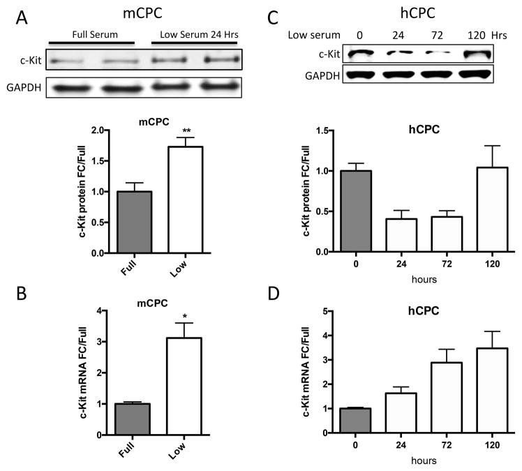
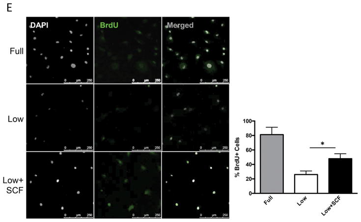
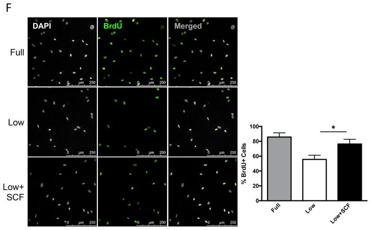
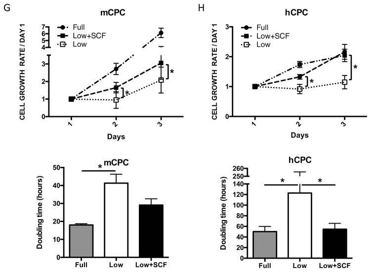
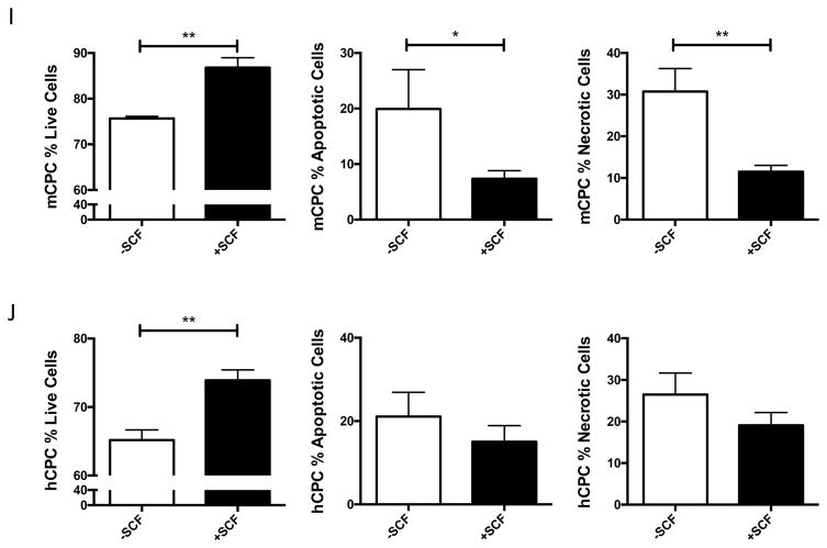
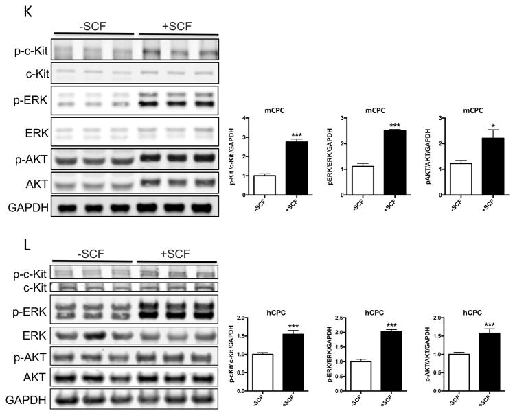
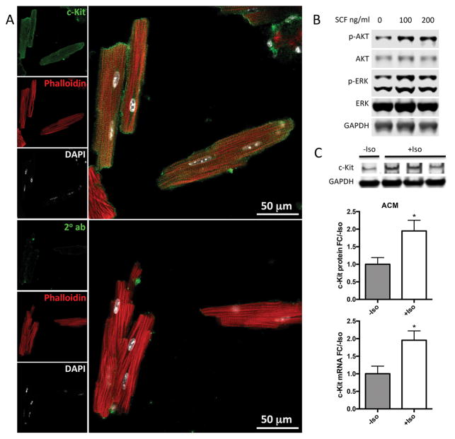
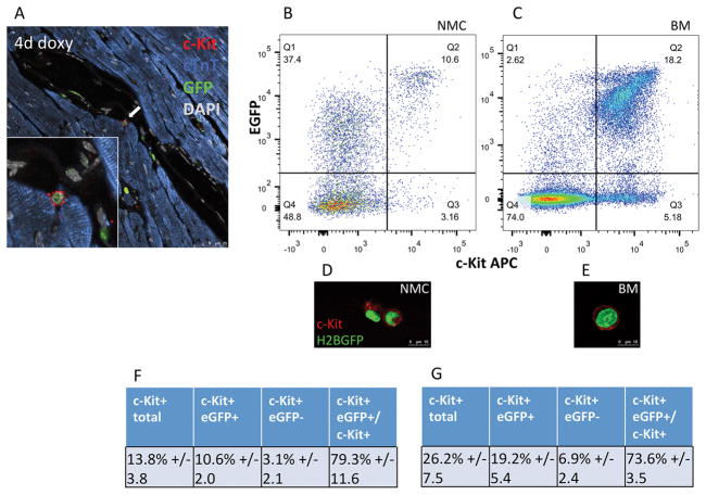
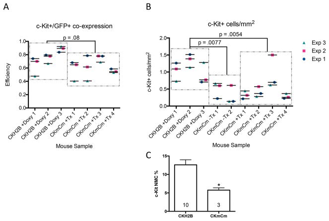
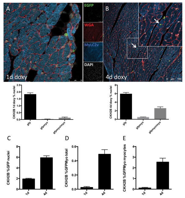
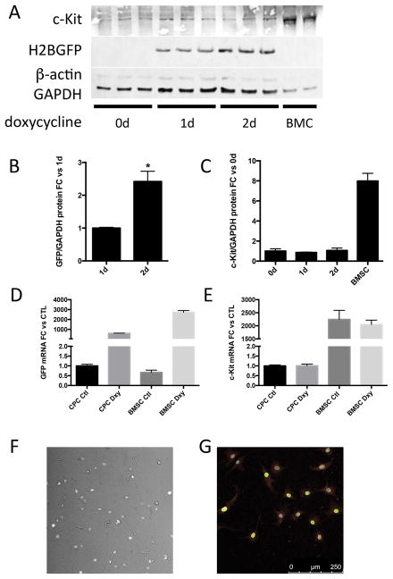
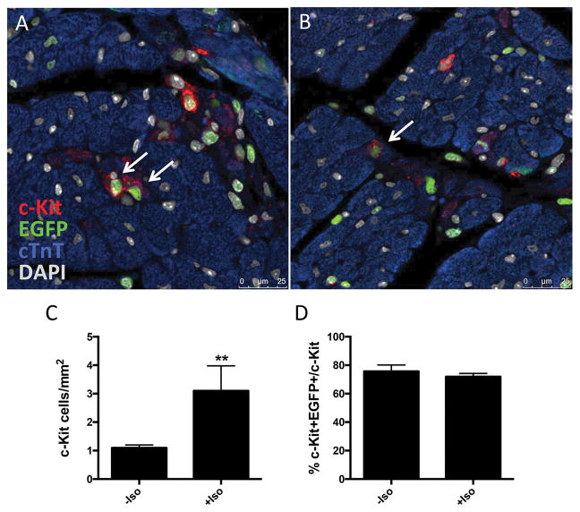
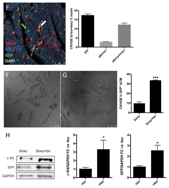
Methods and results: In vitro studies confirm a critical role for c-Kit signaling in both cardiomyocytes and cardiac stem cells. Activation of c-Kit receptor promotes cell survival and proliferation in stem cells and cardiomyocytes of either human or murine origin. For creation of the mouse model, the cloned mouse c-Kit promoter drives Histone2B-EGFP (enhanced green fluorescent protein; H2BEGFP) expression in a doxycycline-inducible transgenic reporter line. The combination of c-Kit transgenesis coupled to H2BEGFP readout provides sensitive, specific, inducible, and persistent tracking of c-Kit promoter activation. Tagging efficiency for EGFP+/c-Kit+ cells is similar between our transgenic versus a c-Kit knock-in mouse line, but frequency of c-Kit+ cells in cardiac tissue from the knock-in model is 55% lower than that from our transgenic line. The c-Kit transgenic reporter model reveals intimate association of c-Kit expression with adult myocardial biology. Both cardiac stem cells and a subpopulation of cardiomyocytes express c-Kit in uninjured adult heart, upregulating c-Kit expression in response to pathological stress.

Conclusions: c-Kit myocardial biology is more complex and varied than previously appreciated or documented, demonstrating validity in multiple points of coexisting yet heretofore seemingly irreconcilable published findings.

Keywords: c-Kit protein; myocardium; myocytes, cardiac; signal transduction; stem cell.


Figures

Figure 1
Figure 1. c-Kit signaling promotes proliferation and survival in CPCs
c-Kit protein and RNA were quantified in mouse and human CPCs cultured in full or low serum media. c-Kit protein levels increase twofold in mCPCs cultured for 24 hours in low serum versus full growth media as measured by immunoblotting (A). c-Kit RNA levels in mCPCs exposed to low serum increase threefold as measured by qPCR (B). hCPC culture for five days in low serum exhibit an initial decrease in c-Kit protein levels which return to full serum levels by five days (C), while c-Kit mRNA increases steadily over time (D). Decreased proliferation in mCPCs and hCPCs cultured in low serum is partially rescued by SCF treatment as indicated by BrdU incorporation (green=BrdU, E,F) and daily cell counts (G,H). Doubling time of CPCs calculated from growth rates increases following serum depletion but is restored with addition of SCF (G,H). Viability is higher, while apoptosis and necrosis are lower in SCF treated CPCs as measured by AnnexinV/PI flow cytometry analysis (I,J). Specific activation of c-Kit in mCPCs and hCPCs is confirmed by increased phosphorylation of c-Kit, ERK and AKT following SCF treatment (K,L). n=3 for all experiments. p<0.05 denotes statistical significance. Statistical significance between two groups was determined by unpaired t-test for all data except J, which was analyzed by paired t-test. *=p<0.05, **=p<0.01, ***=p<0.001.
Figure 2
Figure 2. Cardiac myocytes express active c-Kit and upregulate c-Kit expression in response to stress in vitro
Adult mouse cardiac myocytes (ACM) were isolated, fixed and immunolabeled for c-Kit (green) colocalized with phalloidin (red) and DAPI (white) (A). Activation of c-Kit by SCF was confirmed by immunoblotting for phospho-ERK and phospho-AKT (B). ACM were treated with isoproterenol to induce cellular stress. c-Kit mRNA and protein levels were measured by qPCR and immunoblotting, respectively. c-Kit protein and mRNA levels increased significantly in isoproterenol treated ACMs as measured by immunoblotting and qPCR, respectively (C). n=3 experiments for A,B. *=p<0.05 as measured by t-test.
Figure 3
Figure 3. H2BEGFP reporter expression is induced in c-Kit+ cells in vivo
Cardiac c-Kit+ cells from CKH2B mice express H2BEGFP reporter (white arrow) following administration of doxycycline, as visualized by immunolabeling (A, red=c-Kit, green=EGFP, blue=cardiac troponin T(cTnT), gray=DAPI). Coexpression of c-Kit and H2BEGFP is detected in fresh isolates of nonmyocyte cardiac cells (NMC, B) and whole bone marrow (BM, C) from doxycycline treated CKH2B mice as measured by flow cytometry, and illustrated by corresponding representative confocal images (red=c-Kit, green=H2BEGFP). Tabulation of flow cytometry analysis of c-Kit+ and H2BEGFP in CKH2B NMC (F) and BM (G). n=4 animals.
Figure 4
Figure 4. Similar c-Kit+/EGFP+ co-localization but decreased total cardiac c-Kit+ cells per area in CKmCm knock-in mice versus CKH2B transgenic mice
Paraffin sections were immunolabeled for c-Kit and EGFP to quantitate reporter coexpression in cardiac c-Kit+ cells in situ. EGFP co-localization with c-Kit+ cells in hearts of CKmCm and CKH2B mice is comparable (p=0.0838)(A). Significantly fewer c-Kit+ cells per area were present in CKmCm+Tx and CKmCm-Tx mice compared to doxycycline treated CKH2B mice (p=0.0054 and p=0.0077, respectively). No statistical differences were found between CKmCm-Tx and CKmCm+Tx mice (0.9058)(B). CKH2B hearts n=3, CKmCm-Tx hearts n=2, CKmCm+Tx hearts n=4. Significance determined by mixed linear modeling as described in the Statistics section and the Supplemental Statistical Analysis. p<0.05 denotes significance. (C). Comparative flow cytometry analysis of nonmyocytes isolated from CKmCm+Tx versus CKH2B+doxycycline hearts reveals significantly lower percentage of c-Kit+ NMCs in CKmCm hearts. n=10 CKH2B, n=3 CKmCm, statistical significance determined by two-tailed t-test, *=p<0.05.
Figure 5
Figure 5. H2BEGFP reporter accumulates in cardiac myocytes in vivo
EGFP is detected and quantitated in nuclei of immunolabeled CKH2B hearts treated for one day (A) or four days (B) with doxycycline (red=wheat germ agglutinin, green=H2BEGFP, blue=myosin light chain 2v (MYLC2V), gray=DAPI). Quantitation of the increase in EGFP+ cells per total nuclei (C), EGFP+ myocytes per total nuclei (D) and EGFP+ myocytes per total myocytes (E) represented in histograms. n=3 hearts, minimum 10 fields of view per heart.
Figure 6
Figure 6. H2BEGFP reporter is induced in CKH2B c-Kit+ CPCs in vitro
CKH2B CPCs express c-Kit and accumulate H2BEGFP after treatment with doxycycline (A). BMC=c-KitrtTA whole bone marrow. Quantitation of H2BEGFP protein levels in A relative to 1 day of doxycycline treatment (B) and of c-Kit relative to untreated CPCs (C). Quantitation of mRNA levels of EGFP (D) and c-Kit (E) relative to untreated CPCs. H2BEGFP fluorescence in c-Kit+ CKH2B CPCs following doxycycline induction in live cells (F) and fixed cells immunolabeled for c-Kit (G, red=c-Kit, green=EGFP, gray=DAPI).
Figure 7
Figure 7. Cardiac c-Kit+ expression increases following diffuse injury in vivo
H2BEGFP expression in cardiac c-Kit+ cells visualized by immunostaining and confocal microscopy (A,B), red=c-Kit, green=EGFP, blue=cTnT, gray=DAPI, white arrows indicate c-Kit+/EGFP+ cells. Quantitation of c-Kit+ immunolabeled cells per area of tissue (C), and percentage of c-Kit+ cells expressing H2BEGFP reporter in uninjured versus injured CKH2B hearts (D). H2BEGFP expression in adult cardiac myocytes in hearts from doxycycline and isoproterenol treated CKH2B revealed and quantitated by immunostaining (E) (red=WGA, green=EGFP, blue=MyLC2V, gray=DAPI, white arrow indicates H2BEGFP in nonmyocyte, green arrow indicates H2BEGFP in cardiomyocyte). ACM isolated from CKH2B mice treated with doxycycline only (F) or doxycycline and isoproterenol (G) for three days as imaged and quantitated by confocal microscopy. Protein levels of c-Kit and EGFP quantitated in ACM isolated from Doxy or Doxy+Iso treated CKH2B hearts (H). Bright field=ACM, green=H2BEGFP fluorescence. n=3 mice (F–H), n=4 isolations (I).

Comment in

References

    1. Orlic D, Kajstura J, Chimenti S, Limana F, Jakoniuk I, Quaini F, Nadal-Ginard B, Bodine DM, Leri A, Anversa P. Mobilized bone marrow cells repair the infarcted heart, improving function and survival. Proceedings of the National Academy of Sciences of the United States of America. 2001;98:10344–10349. - PMC - PubMed
    1. Beltrami AP, Barlucchi L, Torella D, Baker M, Limana F, Chimenti S, Kasahara H, Rota M, Musso E, Urbanek K, Leri A, Kajstura J, Nadal-Ginard B, Anversa P. Adult cardiac stem cells are multipotent and support myocardial regeneration. Cell. 2003;114:763–776. - PubMed
    1. Fransioli J, Bailey B, Gude NA, Cottage CT, Muraski JA, Emmanuel G, Wu W, Alvarez R, Rubio M, Ottolenghi S, Schaefer E, Sussman MA. Evolution of the c-kit-positive cell response to pathological challenge in the myocardium. Stem cells. 2008;26:1315–1324. - PMC - PubMed
    1. Tallini YN, Greene KS, Craven M, Spealman A, Breitbach M, Smith J, Fisher PJ, Steffey M, Hesse M, Doran RM, Woods A, Singh B, Yen A, Fleischmann BK, Kotlikoff MI. c-kit expression identifies cardiovascular precursors in the neonatal heart. Proceedings of the National Academy of Sciences of the United States of America. 2009;106:1808–1813. - PMC - PubMed
    1. Miyamoto S, Kawaguchi N, Ellison GM, Matsuoka R, Shin’oka T, Kurosawa H. Characterization of long-term cultured c-kit+ cardiac stem cells derived from adult rat hearts. Stem Cells and Development. 2010;19:105–116. - PubMed

Publication types