. 2018 May;49(5):1241-1247.
doi: 10.1161/STROKEAHA.117.020544. Epub 2018 Apr 10.

Beneficial Role of Neutrophils Through Function of Lactoferrin After Intracerebral Hemorrhage

Affiliations

Beneficial Role of Neutrophils Through Function of Lactoferrin After Intracerebral Hemorrhage

Xiurong Zhao et al. Stroke. 2018 May.

Abstract

Background and purpose: Intracerebral hemorrhage (ICH) is a devastating disease with a 30-day mortality of ~50%. There are no effective therapies for ICH. ICH results in brain damage in 2 major ways: through the mechanical forces of extravasated blood and then through toxicity of the intraparenchymal blood components including hemoglobin/iron. LTF (lactoferrin) is an iron-binding protein, uniquely abundant in polymorphonuclear neutrophils (PMNs). After ICH, circulating blood PMNs enter the ICH-afflicted brain where they release LTF. By virtue of sequestrating iron, LTF may contribute to hematoma detoxification.

Methods: ICH in mice was produced using intrastriatal autologous blood injection. PMNs were depleted with intraperitoneal administration of anti-Ly-6G antibody. Treatment of mouse brain cell cultures with lysed RBC or iron was used as in vitro model of ICH.

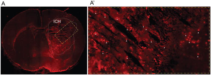
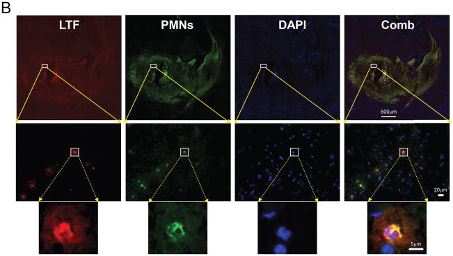
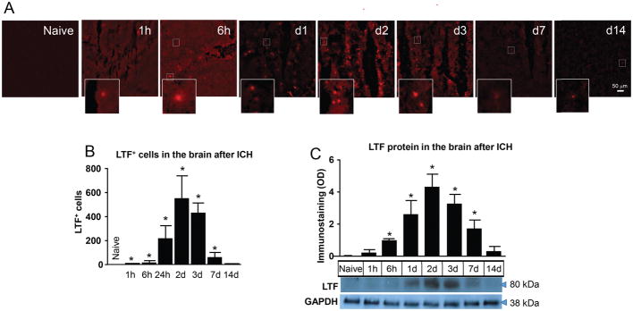
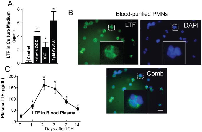
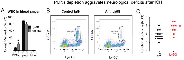
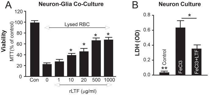
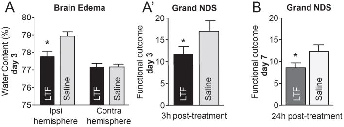
Results: LTF mRNA was undetectable in the mouse brain, even after ICH. Unlike mRNA, LTF protein increased in ICH-affected hemispheres by 6 hours, peaked at 24 to 72 hours, and remained elevated for at least a week after ICH. At the single cell level, LTF was detected in PMNs in the hematoma-affected brain at all time points after ICH. We also found elevated LTF in the plasma after ICH, with a temporal profile similar to LTF changes in the brain. Importantly, mrLTF (recombinant mouse LTF) reduced the cytotoxicity of lysed RBC and FeCl3 to brain cells in culture. Ultimately, in an ICH model, systemic administration of mrLTF (at 3, 24, and 48 hours after ICH) reduced brain edema and ameliorated neurological deficits caused by ICH. mrLTF retained the benefit in reducing behavioral deficit even with 24-hour treatment delay. Interestingly, systemic depletion of PMNs at 24 hours after ICH worsened neurological deficits, suggesting that PMN infiltration into the brain at later stages after ICH could be a beneficial response.

Conclusions: LTF delivered to the ICH-affected brain by infiltrating PMNs may assist in hematoma detoxification and represent a powerful potential target for the treatment of ICH.

Keywords: brain edema; inflammation; lactoferrin; neutrophil; transferrin.


Figures

Fig-1
Fig-1. LTF+ PMNs in brain after ICH in mouse
(A)-LTF immunofluorescence (red) in brain coronal section at 48h after ICH. A′ is a highlighted area in A. The arrowheads in A′ indicate the LTF+-cells. (B)-Double immunofluorescence of LTF-(red) and neutrophil-(green) in the brain at 24h after ICH. The nuclei are stained with DAPI-(blue). A combination-(Comb) of LTF and PMN staining shows the 100% co-localization.
Fig-2
Fig-2. ICH transiently increases LTF in the ICH-affected brain
(A)-Representative images of LTF immunofluorescence in striatum of naïve mouse and mice at 1h-14d after ICH in the hematoma-affected sub-cortical striatum. The scale bar=50 μm. (B)-Bar graph quantitating presence of LTF+-cells in brain at 1–14days after ICH. Data are expressed as mean±SEM (n=5). *P≤0.05, vs. naïve group. (C)-Representative LTF and GAPDH Western blot in naïve striatum and in hematoma-affected striatum at 1h-14d after ICH. Data are expressed as mean±SEM (n=3). *P≤0.05, vs. naïve group.
Fig-3
Fig-3. LTF in Peripheral Blood PMNs
(A)- Quantification (using ELISA) of LTF released into the culture medium by purified blood PMN exposed to 15min oxygen-glucose-deprivation, lysed-RBC, or calcium inophore (A23187; 1μM). The data are mean±SEM (n=3). *p≤0.05, vs. control. (B)-LTF immunofluorescence-(green) in purified mouse blood PMNs. The LTF+-cell has a lobular nucleus, which is typical morphology of a mature neutrophil. Bar=20μm. (C)-X-Y Plot of blood plasma LTF levels at indicated time point after ICH. The data are mean±SEM (n=5). *p≤0.05, vs. d0/prior to ICH.
Fig-4
Fig-4. PMNs-depletion aggravates Neurological deficits
To deplete PMNs, mice were injected with anti-Ly-6G, a neutrophil neutralizing antibody or isotype IgG at 500μg/mouse at 24h and 48h after ICH (i.p.)(n=10 mice/group). The Ly-6G-mediated PMNs depletion at d3 resulted in 50–70% reduction of PMNs while having negligible effect on lymphocytes (Lymph) or monocytes (mono), as determined on tail blood smears using microscopic morphological features (A); and about 90% depletion as measured with flow cytometry using CD45+/CD11b+/Ly6C-Intermediate/SSC-High as markers for gating on cardiac puncture-collected blood (B). The neurological deficit scores (NDS) were quantified with Postural Flexing and Forward Placing at 3d after ICH (C). The data are mean±SEM. *p≤0.05, vs. control.
Fig-5
Fig-5. mrLTF protects brain cells from ICH-like Injury
(A)-MTT/(viability index) in the mouse neuron-glia co-culture at 24h after exposure to lysed RBC in presence of 0–100μg/ml of mrLTF. The data are mean±SEM (n=3). *p≤0.05, vs. control without rLTF (“0”). The “Con” is the naïve control. (B)-LDH in cortical neuron culture at 6h after exposure to 10μM FeCl3, with or without 20μg/ml of mrLTF. The data are mean±SEM (n=3). *p≤0.05; **p≤0.01 vs. other two groups.
Fig-6
Fig-6. mrLTF protects mouse from ICH-injury
(A)-Brain edema (water content in the ipsilateral- and contralateral-striatum) on d3 after ICH. The data are mean±SEM (n=7). *p≤0.05, vs. saline control. (A)-Grand NDS (A combination score of sensory-motor behavioral tests) on d3 after ICH. The data are mean±SEM (n=12/group). *p≤0.05, vs. saline control. (B)-Grand NDS on d7 after ICH in animals treated with mrLTF starting 24h after ICH. (n=5/group) *p≤0.05, vs. saline control

References

    1. Mayer SA, Rincon F. Treatment of intracerebral haemorrhage. Lancet neurology. 2005;4:662–672. - PubMed
    1. Qureshi AI, Mendelow AD, Hanley DF. Intracerebral haemorrhage. Lancet. 2009;373:1632–1644. - PMC - PubMed
    1. Wagner KR, Sharp FR, Ardizzone TD, Lu A, Clark JF. Heme and iron metabolism: Role in cerebral hemorrhage. Journal of cerebral blood flow and metabolism: official journal of the International Society of Cerebral Blood Flow and Metabolism. 2003;23:629–652. - PubMed
    1. Nakamura T, Keep RF, Hua Y, Hoff JT, Xi G. Oxidative DNA injury after experimental intracerebral hemorrhage. Brain research. 2005;1039:30–36. - PubMed
    1. Nunez MT, Urrutia P, Mena N, Aguirre P, Tapia V, Salazar J. Iron toxicity in neurodegeneration. Biometals. 2012;25:761–776. - PubMed

Publication types