. 2018 Apr 10;9(2):e00626-17.
doi: 10.1128/mBio.00626-17.

Extracellular Electron Transfer Powers Enterococcus faecalis Biofilm Metabolism

Affiliations

Extracellular Electron Transfer Powers Enterococcus faecalis Biofilm Metabolism

Damien Keogh et al. mBio. .

Erratum in

Abstract

Enterococci are important human commensals and significant opportunistic pathogens. Biofilm-related enterococcal infections, such as endocarditis, urinary tract infections, wound and surgical site infections, and medical device-associated infections, often become chronic upon the formation of biofilm. The biofilm matrix establishes properties that distinguish this state from free-living bacterial cells and increase tolerance to antimicrobial interventions. The metabolic versatility of the enterococci is reflected in the diversity and complexity of environments and communities in which they thrive. Understanding metabolic factors governing colonization and persistence in different host niches can reveal factors influencing the transition to biofilm pathogenicity. Here, we report a form of iron-dependent metabolism for Enterococcus faecalis where, in the absence of heme, extracellular electron transfer (EET) and increased ATP production augment biofilm growth. We observe alterations in biofilm matrix depth and composition during iron-augmented biofilm growth. We show that the ldh gene encoding l-lactate dehydrogenase is required for iron-augmented energy production and biofilm formation and promotes EET.IMPORTANCE Bacterial metabolic versatility can often influence the outcome of host-pathogen interactions, yet causes of metabolic shifts are difficult to resolve. The bacterial biofilm matrix provides the structural and functional support that distinguishes this state from free-living bacterial cells. Here, we show that the biofilm matrix can immobilize iron, providing access to this growth-promoting resource which is otherwise inaccessible in the planktonic state. Our data show that in the absence of heme, Enterococcus faecalis l-lactate dehydrogenase promotes EET and uses matrix-associated iron to carry out EET. Therefore, the presence of iron within the biofilm matrix leads to enhanced biofilm growth.

Keywords: Enterococcus faecalis; biofilm; extracellular electron transfer; iron; metabolism.


Figures

FIG 1
FIG 1
E. faecalis flow cell biofilms in iron-supplemented medium. (a) CLSM images at 8, 12, and 18 h for normal (10% TSBG) and supplemented (10% TSBG plus 0.2 mM FeCl3) media. Selected optical sections at the indicated depths are followed by representative lateral views in yz at the right of each image set. Bar, 100 μm. (b) 3D reconstructions of high-magnification CLSM stacks of E. faecalis flow cell biofilms grown in normal or supplemented medium for 18 h at 37°C or 120 h at 22°C. Biofilm depth is color coded as indicated on z-depth scale (0 to 24 μm), and the lateral box dimensions are 85 by 85 µm (20 µm between ticks).
FIG 2
FIG 2
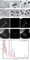
Electron micrographs of the E. faecalis biofilm matrix with iron supplementation. Representative images from TEM of E. faecalis biofilm from flow cell in normal 10% TSBG (a, b, and c) or 10% TSBG supplemented with 0.2 mM FeCl3 (d, e, and f). In panels a to f, bars represent 2 μm (black) and 0.5 μm (white), and red arrows highlight examples of electron-dense particles. The biofilm matrix from biofilms grown in normal medium (g, h, and i) or with iron supplementation (j, k, and l) was examined by HAADF STEM and EDS mapping at ×300,000 magnification: HAADF STEM (g and j), iron EDS map (h and k), and merged EDS-STEM images (i and l) are shown at ×300,000 magnification. Bars in panels g to l represent 1 µm. The EDS spectra for iron, corresponding to the images, are shown in panel m.
FIG 3
FIG 3
E. faecalis biofilm growth under metal supplementation. (a) Time course of E. faecalis biofilm growth in TSBG supplemented with FeCl3. (b) E. faecalis biofilm growth at 120 h in TSBG supplemented with metals as indicated. (c) ICP-MS analysis of E. faecalis grown in TSBG (normal) and TSBG supplemented with 2 mM FeCl3 (supplemented). (a and b) Data at each time point or metal supplement represent an independent experiment, with the data merged for representation. (a to c) n = 3 biological replicates. Statistical significance was determined by two-way analysis of variance with Tukey’s test for multiple comparisons. n = 3 with four technical replicates. *, P ≤ 0.05; **, P ≤ 0.01; ***, P ≤ 0.001; ****, P ≤ 0.0001. (a) Statistical analysis was calculated with time set as a repeated measure. (a, b, and c) Error bars represent standard deviations from the mean.
FIG 4
FIG 4
Extracellular electron transfer in E. faecalis biofilm. (a) Chronocoulometry current (Q) measurement, expressed in millicoulombs, of E. faecalis biofilm on a screen-printed electrode over 40 h in TSBG supplemented with 2 mM FeCl3 (supplemented). Abiotic controls and medium controls are indicated. Statistical significance was determined by one-way analysis of variance with Tukey’s test for multiple comparisons. n = 3 biological replicates. Error bars represent standard deviations from the mean. ****, P ≤ 0.0001. (b) Chronocoulometry of E. faecalis biofilm in iron-supplemented medium with a chelator spike (4 mM 2,2′-dipyridyl) at 7.5 h. Representative data from four independent experiments are shown, where the trend is consistent among all experiments. Statistical significance was determined by a paired two-tailed t test; error bars (light green or light red shading) represent standard deviations from the mean. ****, P ≤ 0.0001. (c) Chronocoulometry current (Q) measurement, expressed in millicoulombs, of E. faecalis wild-type, ldh1 mutant, and complemented ldh1 mutant biofilm on a screen-printed electrode over 40 h in TSBG supplemented with 2 mM FeCl3. Statistical significance was determined by one-way analysis of variance with Tukey’s test for multiple comparisons. n = 3 biological replicates. Error bars represent standard deviations from the mean. ****, P ≤ 0.0001. (d) ATP quantification within biofilm grown in TSBG and TSBG supplemented with 2 mM FeCl3 for 24 h. Nisin was included at 5 µg/ml, and erythromycin was included at 300 µg/ml for strains carrying a plasmid. Statistical significance was determined by one-way analysis of variance with Sidak’s test for multiple comparisons. n = 2 biological replicates. Error bars represent standard errors of the means. **, P ≤ 0.01; ns, not significant. ATP production was significantly increased for E. faecalis when grown in iron-supplemented medium in each of two individual experiments (P < 0.05) (data not shown).
FIG 5
FIG 5
Model for fermentation and EET-dependent respiration metabolism in E. faecalis biofilm. Schematic model for E. faecalis biofilm metabolism describing EET through DET and MET mechanisms. ATP is generated by proton flow through membrane-integrated ATP synthases. Glycolysis and fermentation are required prior to EET-dependent respiration. This generates the fermentation end products required as the substrates for dehydrogenases acting as electron donors. DET occurs in the absence of heme, where extracellular iron can be reduced and thereby serve as an iron sink for electrons of the respiratory electron transport chain. MET occurs in the absence of heme, when iron is transported intracellularly and reduced, thereafter being exported and serving as an electron mediator/shuttle.

References

    1. Hufnagel M, Liese C, Loescher C, Kunze M, Proempeler H, Berner R, Krueger M. 2007. Enterococcal colonization of infants in a neonatal intensive care unit: associated predictors, risk factors and seasonal patterns. BMC Infect Dis 7:107. doi: 10.1186/1471-2334-7-107. - DOI - PMC - PubMed
    1. Sekirov I, Russell SL, Antunes LC, Finlay BB. 2010. Gut microbiota in health and disease. Physiol Rev 90:859–904. doi: 10.1152/physrev.00045.2009. - DOI - PubMed
    1. Flemming HC, Wingender J, Szewzyk U, Steinberg P, Rice SA, Kjelleberg S. 2016. Biofilms: an emergent form of bacterial life. Nat Rev Microbiol 14:563–575. doi: 10.1038/nrmicro.2016.94. - DOI - PubMed
    1. Paganelli FL, Willems RJ, Leavis HL. 2012. Optimizing future treatment of enterococcal infections: attacking the biofilm? Trends Microbiol 20:40–49. doi: 10.1016/j.tim.2011.11.001. - DOI - PubMed
    1. Winstedt L, Frankenberg L, Hederstedt L, von Wachenfeldt C. 2000. Enterococcus faecalis V583 contains a cytochrome bd-type respiratory oxidase. J Bacteriol 182:3863–3866. doi: 10.1128/JB.182.13.3863-3866.2000. - DOI - PMC - PubMed

Publication types

MeSH terms