. 2018 Apr 10;8(1):5777.
doi: 10.1038/s41598-018-24217-1.

Satiation state-dependent dopaminergic control of foraging in Drosophila

Affiliations

Satiation state-dependent dopaminergic control of foraging in Drosophila

Dan Landayan et al. Sci Rep. .

Abstract

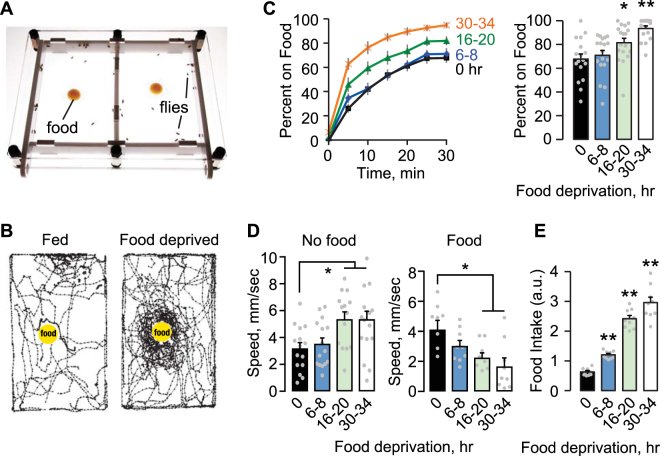
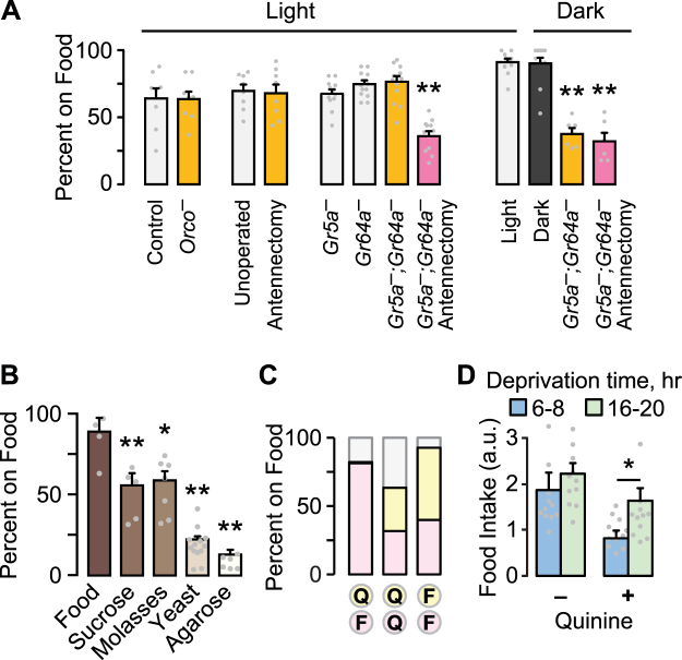
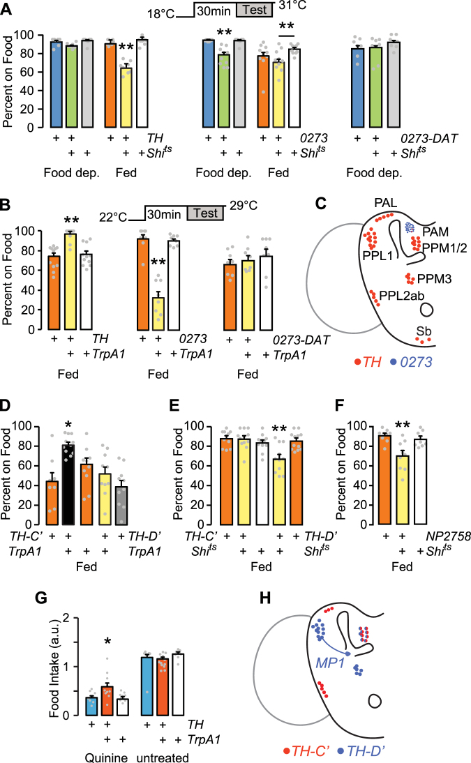
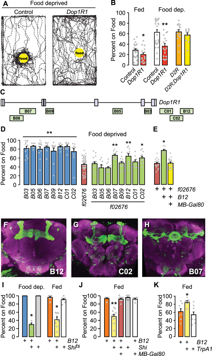
Hunger evokes stereotypic behaviors that favor the discovery of nutrients. The neural pathways that coordinate internal and external cues to motivate foraging behaviors are only partly known. Drosophila that are food deprived increase locomotor activity, are more efficient in locating a discrete source of nutrition, and are willing to overcome adversity to obtain food. We developed a simple open field assay that allows flies to freely perform multiple steps of the foraging sequence, and we show that two distinct dopaminergic neural circuits regulate measures of foraging behaviors. One group, the PAM neurons, functions in food deprived flies while the other functions in well fed flies, and both promote foraging. These satiation state-dependent circuits converge on dopamine D1 receptor-expressing Kenyon cells of the mushroom body, where neural activity promotes foraging independent of satiation state. These findings provide evidence for active foraging in well-fed flies that is separable from hunger-driven foraging.


Conflict of interest statement

The authors declare no competing interests.

Figures

Figure 1
Figure 1
Food deprivation effect on foraging behavior. (A) Two-sided chamber for foraging assays. Flies and 100 ul of cornmeal molasses food on a Parafilm square placed in each chamber via sliding side doors. The chamber is lit from below. Fly locomotion is recorded from above. (B) 10 sec locomotor traces of 20 flies (fed and 20 hr food deprived) each filmed soon after addition of food (yellow dot). Tracking traces were generated with DIAS software. (C) Left: The percent of flies on food over time for a food deprivation time course. Right, food occupancy averaged at 25–30 min. P < 0.0001, ANOVA/Bonferroni comparison to 0 hr. n = 17–18 groups. (D) Locomotor speed. Left, speed at 20 min of acclimation, without food. Right, speed averaged over 0–10 min after food introduction. P = 0.0091 no food, P = 0.0066 food, ANOVA/Bonferroni compared to 0 hr. n = 9–15 groups. (E) Intake with increasing food deprivation time. P < 0.0001, ANOVA/Bonferroni comparison to 0 hr. n = 9 groups. *P < 0.05, **P < 0.01.
Figure 2
Figure 2
Environmental and sensory information in foraging. (A) Food occupancy following sensory ablations in 16–20 hr food deprived flies. Antennectomy is surgical removal of the third antennal segment. Orco flies lack the Orco olfactory coreceptor; Gr5a and Gr64a are taste receptor mutants. P < 0.0001 for both Light and Dark, ANOVA/Bonferroni compared to control, n = 8–12 groups. Light/dark tests were performed in an incubator, where unknown environmental factors increased food occupancy overall. (B) Occupancy of 16–20 hr food deprived flies to agarose with the indicated food component. P < 0.0001, ANOVA/Bonferroni comparison to Food. n = 4–5 groups. (C) Food occupancy for flies given the choice between two closely apposed sources of food (yellow and pink): unadulterated food (F) and 10 mM quinine food (Q). n = 5 groups. (D) When presented with a single food source, flies consumed greater quantities of quinine food (3 mM) when food-deprived for 16–20 hr (long) versus 6–8 hr (short). P = 0.0251, Mann Whitney test, n = 12. *P < 0.05, **P < 0.01. See also Figure S1.
Figure 3
Figure 3
Satiation state-dependent effects of dopamine neuron activity on foraging. (A) Acute inactivation of dopamine neurons with Shibirets (Shits), food occupancy in fed and 16–20 hr food-deprived flies. P = 0.0012 ANOVA/Tukey’s, n = 8–11 groups with TH-Gal4. P = 0.0001 Kruskal-Wallis/Dunn’s, n = 8–10 groups food deprived; P = 0.0139 ANOVA/Tukey’s, n = 8–9 groups fed, with 0273-Gal4. 0273-DAT: 0273-Gal4 with R58E02-Gal80 to specifically block GAL4 activity in the PAM cluster dopamine neurons. n = 6 groups. (B) Acute activation of dopamine neurons in fed flies, food occupancy. P = 0.0002, ANOVA/Tukey’s, n = 8–11 groups with TH-Gal4. P = 0.0002, Kruskal-Wallis/Dunn’s, n = 8 groups with 0273-Gal4. 0273-DAT: n = 8 groups. (C) Dopamine neuron clusters in the adult brain that express TH-Gal4 and 0273-Gal4. (D) Acute activation of subsets of TH-Gal4 neurons, food occupancy in fed flies. P = 0.0002, ANOVA/Tukey’s, n = 8–11 groups. (E) Acute inactivation of subsets of TH-Gal4 neurons, food occupancy in fed flies. P < 0.0001, ANOVA/Tukey’s, n = 11–12 groups. (F) Acute inactivation of neurons with NP2758-Gal4. P = 0.001, ANOVA/Tukey’s, n = 8–9 groups. (G) Food intake in 4–6 hr food-deprived flies. P = 0.0053, ANOVA/Tukey’s, n = 15–19 groups. (H) Dopamine neurons that express TH-C’-Gal4 and TH-D’-Gal4. MP1: PPL1-γ1pedc neuron labeled by TH-D’-Gal4 and NP2758-Gal4. *P < 0.05, **P < 0.01. See also Figure S2.
Figure 4
Figure 4
Dopamine receptor-expressing neurons in the mushroom body control foraging. (A) Locomotor traces of food-deprived flies 5 min after addition of food. Dop1R1 mutant f02676 vs. the Berlin genetic background control strain. (B) Food occupancy for the indicated genotypes that were fed or food deprived. t-test P = 0.0492 fed (n = 16–20 groups), P = 0.001 food deprived (n = 16–20 groups). D2R: the loss-of-function mutation f06521. (C) Location of Dop1R1 enhancer fragments. (D) Genetic rescue of Dop1R1 mutant food occupancy in 16–20 hr food deprived animals. Dop1R1-Gal4 strains (blue) were made heterozygous in f02676 homozygotes (rescuing configuration, green). P < 0.0001 ANOVA/Bonferroni’s comparison to f02676, n = 8–16 groups. (E) Inclusion of MB-Gal80, preventing GAL4 activity in the mushroom bodies blocks B12 rescue. P < 0.0001 ANOVA/Tukey’s, n = 10–19 groups. (FH) Expression pattern of Dop1R1-Gal4 strains (CD8-GFP, green), and bruchpilot (magenta) to show the synaptic neuropil. (I) Acute silencing of B12 Dop1R1-Gal4 neurons with Shits, food occupancy, food deprived and fed. Food deprived: P < 0.0001 Kruskal-Wallis/Dunn’s, n = 4 groups. Fed: P = 0.0002 Kruskal-Wallis/Dunn’s, n = 7–8 groups. (J) Addition of MB-Gal80 in B12 Dop1R1-Gal4 > Shits fed flies, food occupancy. P < 0.0001 Kruskal-Wallis/Dunn’s, n = 6–10 groups. (K) Activation of B12 Dop1R1-Gal4 neurons in fed flies increased food occupancy. P = 0.0054, ANOVA/Tukey’s, n = 7–9 groups. *P < 0.05, **P < 0.01. See also Figure S3.

References

    1. Benoit SC, Tracy AL. Behavioral controls of food intake. Peptides. 2008;29:139–47. doi: 10.1016/j.peptides.2007.10.019. - DOI - PMC - PubMed
    1. Craig W. Appetites and Aversions as Constituents of Instincts. Proc Natl Acad Sci. 1917;3:685–8. doi: 10.1073/pnas.3.12.685. - DOI - PMC - PubMed
    1. DiLeone RJ, Taylor JR, Picciotto MR. The drive to eat: comparisons and distinctions between mechanisms of food reward and drug addiction. Nat Neurosci. 2012;15:1330–5. doi: 10.1038/nn.3202. - DOI - PMC - PubMed
    1. Kenny PJ. Common cellular and molecular mechanisms in obesity and drug addiction. Nat Rev Neurosci. 2011;12:638–51. doi: 10.1038/nrn3105. - DOI - PubMed
    1. Jeong YT, et al. An odorant-binding protein required for suppression of sweet taste by bitter chemicals. Neuron. 2013;79:725–37. doi: 10.1016/j.neuron.2013.06.025. - DOI - PMC - PubMed

Publication types