. 2018 Apr 10;8(1):5760.
doi: 10.1038/s41598-018-24148-x.

Study of mucin turnover in the small intestine by in vivo labeling

Affiliations

Study of mucin turnover in the small intestine by in vivo labeling

Hannah Schneider et al. Sci Rep. .

Abstract

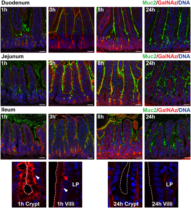
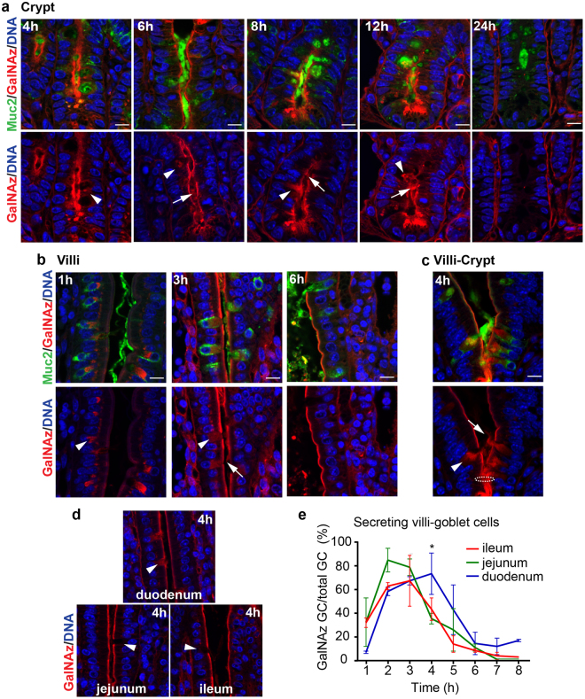
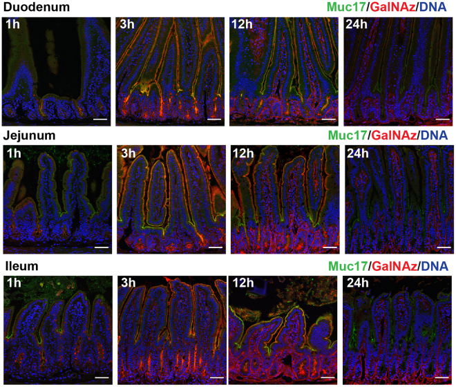
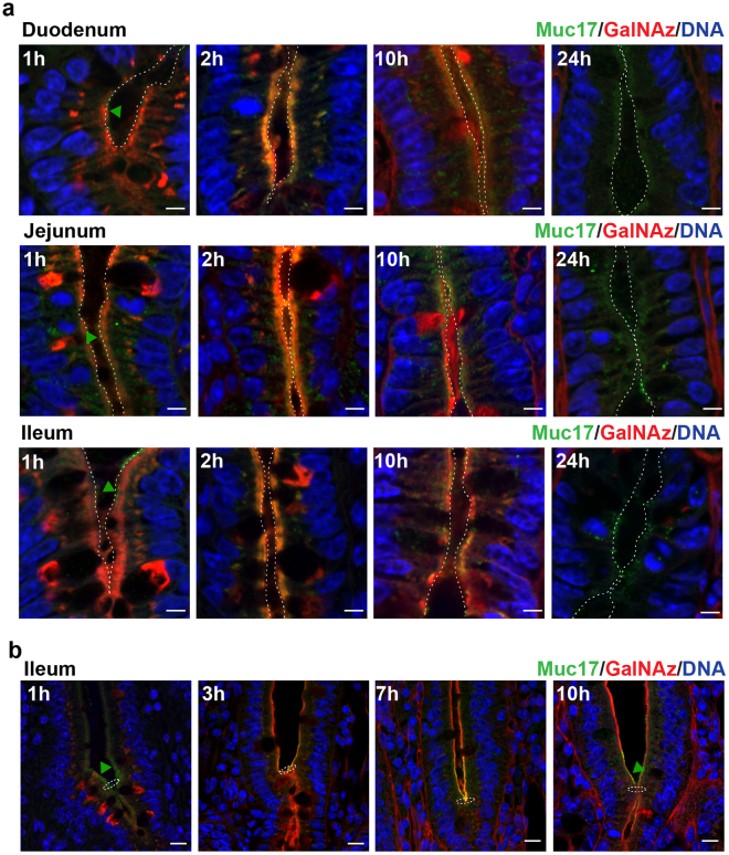
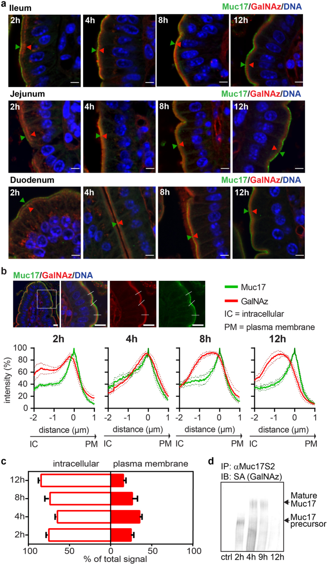
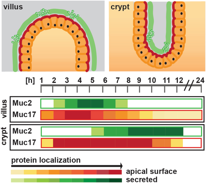
Mucins are highly glycosylated proteins which protect the epithelium. In the small intestine, the goblet cell-secreted Muc2 mucin constitutes the main component of the loose mucus layer that traps luminal material. The transmembrane mucin Muc17 forms part of the carbohydrate-rich glycocalyx covering intestinal epithelial cells. Our study aimed at investigating the turnover of these mucins in the small intestine by using in vivo labeling of O-glycans with N-azidoacetylgalactosamine. Mice were injected intraperitoneally and sacrificed every hour up to 12 hours and at 24 hours. Samples were fixed with preservation of the mucus layer and stained for Muc2 and Muc17. Turnover of Muc2 was slower in goblet cells of the crypts compared to goblet cells along the villi. Muc17 showed stable expression over time at the plasma membrane on villi tips, in crypts and at crypt openings. In conclusion, we have identified different subtypes of goblet cells based on their rate of mucin biosynthesis and secretion. In order to protect the intestinal epithelium from chemical and bacterial hazards, fast and frequent renewal of the secreted mucus layer in the villi area is combined with massive secretion of stored Muc2 from goblet cells in the upper crypt.


Conflict of interest statement

The authors declare no competing interests.

Figures

Figure 1
Figure 1
GalNAz labeling of Muc2 is used to analyze small intestinal mucus turnover. Incorporated GalNAz detected by TAMRA (red), immunostaining of Muc2 (green) and DNA stain using Hoechst (blue). Intracellular red staining was first observed at 1 h after injection. Secretion of labeled mucus was seen after 3 h. TAMRA staining was still clearly visible at 8 h but was mostly absent at 24 h (full sets are shown in Supplementary Figs S1–S3). Magnifications of crypt and villi epithelium are shown below after 1 h of labeling (arrows indicate Golgi staining) and after 24 h (no staining in epithelium). Dotted lines indicate the cell surface. LP = lamina propria. Scale bars are 50 µm.
Figure 2
Figure 2
Ileal Muc2 secretion is faster at the villi than in the crypts. GalNAz-TAMRA (red) in comparison to immunostained Muc2 (green) in ileal sections. DNA detected with Hoechst in blue. (a) GalNAz-positive mucin granules in crypt GCs appear at 4 h after labeling. Stained Muc2 was secreted after 6 h and 12 h. (b) After 1 h, labeled Muc2 was observed in the Golgi of villi GCs. TAMRA-stained Muc2 was secreted at 3 h but was no longer observed after 6 h. (c) Fast producing and secreting GCs were observed just above the crypt opening (4 h). Intracellular labeled Muc2 is shown by arrowheads, secreted labeled mucus by arrows. Dotted circle indicates the crypt opening. Scale bars are 10 µm. (d) Muc2 secretion at 4 h at the different imntestinal locations. Arrowhead indicate GCs. (e) Quantification of GalNAz-Muc2 secreting GCs at the villi in relation to the total number of GCs. Most GCs were emptied after 2 h in ileum and jejunum with slower secretion in the duodenum. Significant differences were seen at 4 h. Data is presented as mean in % (*p > 0.05, n = 3 per time point).
Figure 3
Figure 3
GalNAz-labeled Muc17 is expressed along the villi. GalNAz labeling of Muc17 was executed by intraperitoneal injection in mice, followed by TAMRA detection in methacarn fixed small intestinal sections. Muc17 was detected by immunostaining (green) along the villi and at the crypt openings, showing a characteristic patchy appearance. Hoechst DNA stain is shown in blue. GalNAz-labeled Muc17 was present at the villi at 3 h and 12 h throughout the small intestine, with no staining detectable after 24 h. Red signal in the crypts was already observed 1 h after GalNAz injection and intensifies at 3 h and 12 h (full sets are shown in Supplementary Figs 6–8). Scale bars are 50 µm.
Figure 4
Figure 4
Muc17 is located at the apical surface of the crypt and above the crypt opening. (a) GalNAz-TAMRA (red), anti-Muc17 (green) and DNA (blue) staining of crypts in small intestinal sections. At 1 h, GalNAz staining was detected intracellularly and at the apical membrane. Green arrowheads point to immunostained Muc17 localized at the plasma membrane. 2 h post injection the GalNaz signal co-localized with the Muc17 immunostaining. A partial co-localization was still seen after 10 h, with no red staining after 24 h. Dotted lines indicate the cell surface. Scale bars are 5 µm. (b) In the ileum, GalNAz staining was present at 1 h on the apical epithelium above the crypt opening. Overlap of the GalNAz signal with Muc17 immunostaining at the apical plasma membrane can be detected at 3 h to 7 h. 10 h post injection GalNAz was still visible at the plasma membrane but no longer overlapped with the Muc17 immunostaining. The presence of immunostained Muc17 at the plasma membrane is pointed out by green arrowheads. Dotted circles indicate the crypt opening. Scale bars are 10 µm.
Figure 5
Figure 5
Muc17 is stably expressed apically in epithelial cells on the villi tip. (a) Close up view of upper villi epithelium with continuous Muc17 expression at the plasma membrane (anti-Muc17, green; DNA, blue). Intracellular GalNAz-labeled Muc17 (red) was seen at 2 h and overlapped with anti-Muc17 staining at the plasma membrane at 4 h and 8 h. After 12 h, GalNAz-Muc17 was again detected intracellularly where it localized close to the plasma membrane (a full set for ileum is shown in Supplementary Fig. S9). Arrowheads demonstrate the variation in localization of GalNAz-labeled Muc17 (red) in reference to immunostained Muc17 (green) at the plasma membrane. Scale bars are 5 µm. (b) Line profile quantification comparing the GalNAz signal with anti-Muc17 staining at villi tips. A representative image of an ileal section is shown, with the boxed in area displayed to the right and lines representing analyzed segments. Intensity profiles summarize quantification for all small intestinal regions with SEM (n = 5–6, individual line profiles are shown in Supplementary Fig. S10). Scale bars are 10 µm. (c) Representation of GalNAz-labeled intracellular and plasma membrane-localized Muc17 2 h to 12 h after GalNAz injection. (d) Muc17 was immunoprecipitated from small intestinal scrapings and analyzed by immunoblotting. GalNAz-biotin-labeled Muc17 was detected using streptavidin (SA), unlabeled samples served as negative control. A large molecular weight band of mature Muc17 was detected at 4 h and 9 h, with a smaller band visible at 2 h, likely representing a less glycosylated precursor.
Figure 6
Figure 6
Overview of Muc2 and Muc17 turnover in the small intestine. Schematic representation of Muc2 secretion and Muc17 plasma membrane localization in small intestinal crypts and villi. Both processes occur rapidly at the villi surface, but the Muc2 secretion was more transient than the Muc17 surface localization. In the crypt, Muc2 secretion occurs later and slightly slower, whereas Muc17 plasma membrane localization was equally fast, but extends over a much longer time.

References

    1. Bevins CL, Salzman NH. Paneth cells, antimicrobial peptides and maintenance of intestinal homeostasis. Nat. Rev. Microbiol. 2011;9:356–368. doi: 10.1038/nrmicro2546. - DOI - PubMed
    1. Allen A, Flemstrom G. Gastroduodenal mucus bicarbonate barrier: protection against acid and pepsin. Am. J. Physiol Cell Physiol. 2005;288:C1–19. doi: 10.1152/ajpcell.00102.2004. - DOI - PubMed
    1. Ermund A, Schutte A, Johansson ME, Gustafsson JK, Hansson GC. Studies of mucus in mouse stomach, small intestine, and colon. I. Gastrointestinal mucus layers have different properties depending on location as well as over the Peyer’s patches. Am. J. Physiol Gastrointest. Liver Physiol. 2013;305:G341–G347. doi: 10.1152/ajpgi.00046.2013. - DOI - PMC - PubMed
    1. Rodriguez-Pineiro AM, et al. Studies of mucus in mouse stomach, small intestine, and colon. II. Gastrointestinal mucus proteome reveals Muc2 and Muc5ac accompanied by a set of core proteins. Am. J. Physiol Gastrointest. Liver Physiol. 2013;305:G348–G356. doi: 10.1152/ajpgi.00047.2013. - DOI - PMC - PubMed
    1. Ambort, D., Johansson, M. E., Gustafsson, J. K., Ermund, A. & Hansson, G. C. Perspectives on mucus properties and formation–lessons from the biochemical world. Cold Spring Harb. Perspect. Med. 2 (2012). - PMC - PubMed

Publication types