Review
. 2018 May;15(5):309-323.
doi: 10.1038/nrgastro.2018.27. Epub 2018 Apr 11.

Expert consensus document: Advances in the evaluation of anorectal function

Affiliations
Review

Expert consensus document: Advances in the evaluation of anorectal function

Emma V Carrington et al. Nat Rev Gastroenterol Hepatol. 2018 May.

Abstract

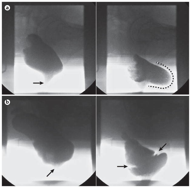
Faecal incontinence and evacuation disorders are common, impair quality of life and incur substantial economic costs worldwide. As symptoms alone are poor predictors of underlying pathophysiology and aetiology, diagnostic tests of anorectal function could facilitate patient management in those cases that are refractory to conservative therapies. In the past decade, several major technological advances have improved our understanding of anorectal structure, coordination and sensorimotor function. This Consensus Statement provides the reader with an appraisal of the current indications, study performance characteristics, clinical utility, strengths and limitations of the most widely available tests of anorectal structure (ultrasonography and MRI) and function (anorectal manometry, neurophysiological investigations, rectal distension techniques and tests of evacuation, including defecography). Additionally, this article provides our consensus on the clinical relevance of these tests.


Conflict of interest statement

COMPETING INTERESTS

E.V.C. has received honoraria for teaching from MMS/Laborie.

S.M.S. has received honoraria for teaching from MMS/Laborie.

J.R.-T. has received research funding from Newton Foundation-CONACYT, Sanfer and Asofarma Laboratories, speaker fees from Covidien/Medtronics, Takeda, Allergan, Astra Zeneca, Sanofi and Sanfer.

F.M. has served as consultant for Medtronic, Laborie.

A.B. has licensed intellectual property in a portable anorectal manometry device to Medspira Inc.

M.F. has received research funding from Covidien/Medtronic, speaker fees from Covidien/Medtronic, Sandhill, MMS/Laborie, Reckitt Benckiser, and Mui Scientific.

S.S.R. has served on advisory boards for Forest labs, Synergy Pharmaceuticals, Vibrant Ltd, Intone, and has received research grants from Forest labs, Synergy, InTone, and Medtronic. H.H. and A.M. declare no competing interests.

Figures

Figure 1
Figure 1
Schematic of standardized protocol for high-resolution anorectal manometry.
Figure 2
Figure 2
Representative high-resolution anorectal manometry traces and resultant line traces to assess anorectal function. a | Sphincter hypotonia in a patient with fecal incontinence, visualized in the color contour plot as a band of pale green (~ 20–25mmHg) set between normal blue (~5mmHg) rectal (superiorly) and atmospheric (inferiorly) pressures. b | Dyssynergia (paradoxical anal sphincter contraction during push visualized in the color contour plot as a band of purple (~ 150–175mmHg) within the anal canal and band of yellow (~50mmHg) in a patient with evacuation disorder.
Figure 3
Figure 3
Representative endoanal ultrasonography images. a | The mid anal canal in healthy volunteer, demonstrating an intact internal anal sphincter (IAS) (arrow) appearing hypoechoic, and an intact external anal sphincter (EAS) (arrowhead) appearing hyperechoic. b Mid anal canal in a patient with fecal incontinence, demonstrating an IAS defect between the 1 and 5 o’clock positions (between the arrows). c | Mid anal canal in a patient with fecal incontinence demonstrating an EAS defect, evident as an area of hypoechoic discontinuity between the 12 and 2 o’clock positions (extent of defect between dashed lines). d | Mid anal canal in a patient with fecal incontinence demonstrating IAS atrophy (global thinning of the smooth muscle ring, which is of mixed echogenicity difficult to distinguish from surrounding structures, arrow). The EAS is intact.
Figure 4
Figure 4
Schematic of rectal barostat setup. a | Typical rectal barostat setup. b | Barostat conditioning distension protocol.
Figure 5
Figure 5
Representative barium defecography images. a | A significant rectocele; the left panel shows a lateral view of the rectum at rest, opacified by barium neostool with the anal canal closed (arrow). The right panel clearly demonstrates a large retaining rectocele at end evacuation (extent of anterior bulging highlighted by dashed line) b) Obstructing full thickness intussusception; the left panel shows a lateral view of the rectum at rest, with the anal canal closed (arrow). The right panel shows an image at mid evacuation with clear invagination of the mid rectum (between arrows) secondary to a full thickness rectal intussusception; this is causing occlusion of the distal rectal lumen with retention of neostool proximal to this.

References

    1. Palit S, Lunniss PJ, Scott SM. The physiology of human defecation. Dig Dis Sci. 2012;57:1445–64. - PubMed
    1. Perry S, Shaw C, McGrother C, et al. Prevalence of faecal incontinence in adults aged 40 years or more living in the community. Gut. 2002;50:480–4. - PMC - PubMed
    1. Whitehead WE, Borrud L, Goode PS, et al. Fecal incontinence in US adults: epidemiology and risk factors. Gastroenterology. 2009;137:512–7. 517e1–2. - PMC - PubMed
    1. Johanson JF, Lafferty J. Epidemiology of fecal incontinence: the silent affliction. Am J Gastroenterol. 1996;91:33–6. - PubMed
    1. Higgins PD, Johanson JF. Epidemiology of constipation in North America: a systematic review. Am J Gastroenterol. 2004;99:750–9. - PubMed

Publication types

MeSH terms