

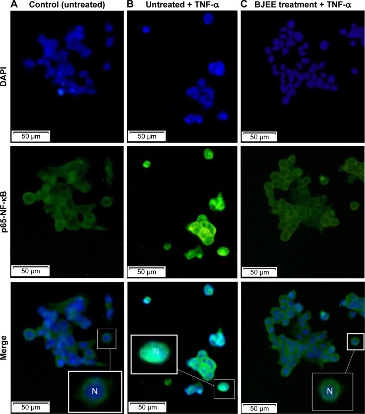
The apoptotic effects of Brucea javanica fruit extract against HT29 cells associated with p53 upregulation and inhibition of NF-κB translocation
- PMID: 29636600
- PMCID: PMC5881282
- DOI: 10.2147/DDDT.S155115
The apoptotic effects of Brucea javanica fruit extract against HT29 cells associated with p53 upregulation and inhibition of NF-κB translocation
Abstract
Background: Brucea javanica (L.) Merr. is a plant from the genus Brucea, which is used in local traditional medicine to treat various diseases. Recent studies revealed an impressive anticancer efficiency of B. javanica extract in different types of cancer cells.
Purpose: In this study, we have investigated the cytotoxic effects of the B. javanica hexane, ethanolic extracts against colon cancer cells. HT29 colon cells were selected as an in vitro cancer model to evaluate the anticancer activity of B. javanica ethanolic extract (BJEE) and the possible mechanisms of action that induced apoptosis.
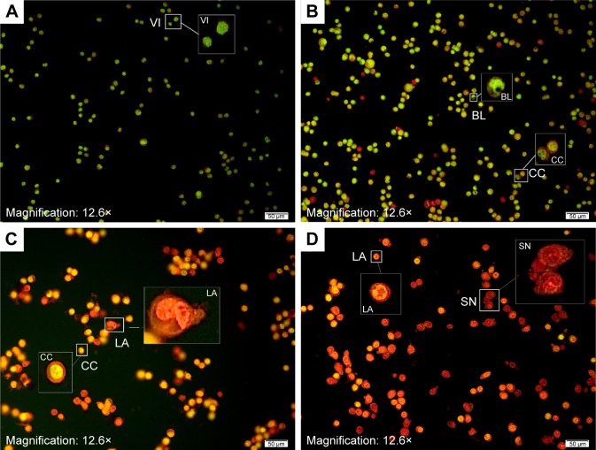
Methods: 3-(4,5-dimethylthiazol-2-yl)-2, 5,-diphenyltetrazolium bromide (MTT), lactate dehydrogenase, acridine orange/propidium iodide, and annexin-V-fluorescein isothiocyanate assays were performed to determine the antiproliferative and apoptosis validation of BJEE on cancer cells. Measurement of reactive oxygen species (ROS) production, caspase activities, nucleus factor-κB activity, and gene expression experiments was done to investigate the potential mechanisms of action in the apoptotic process.
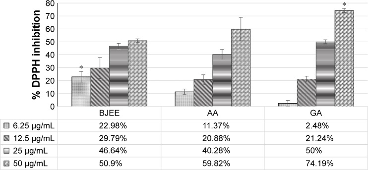
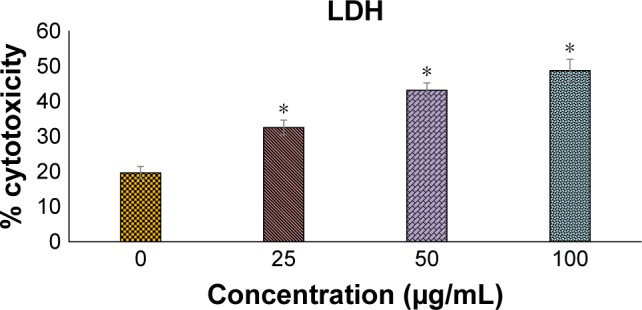
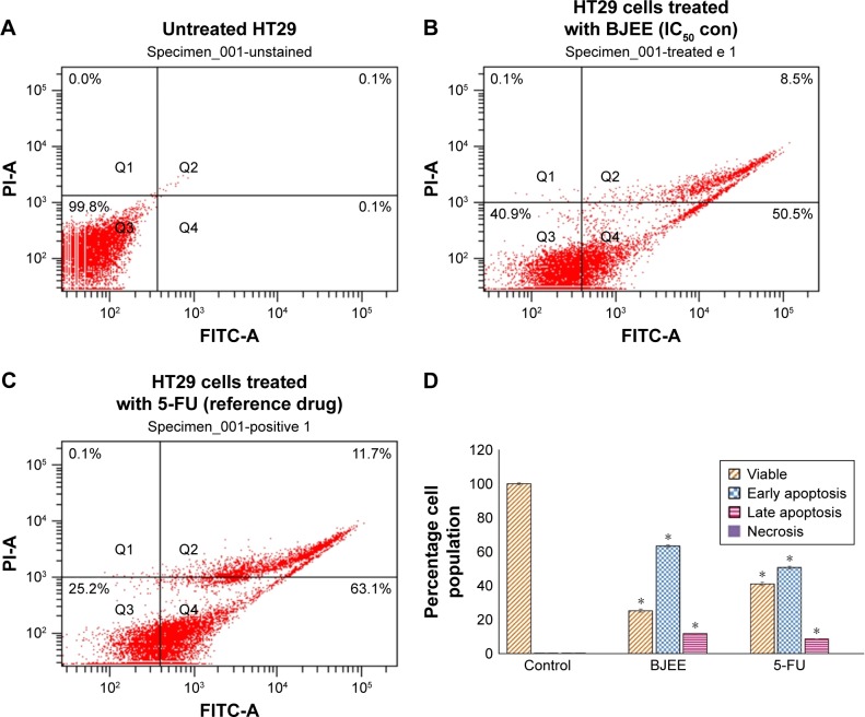
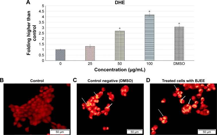
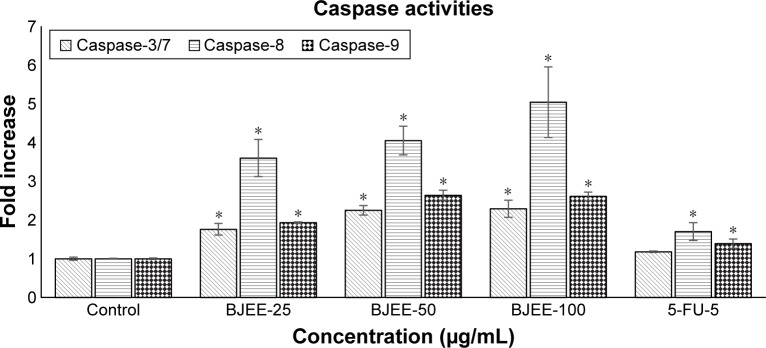
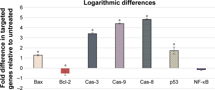
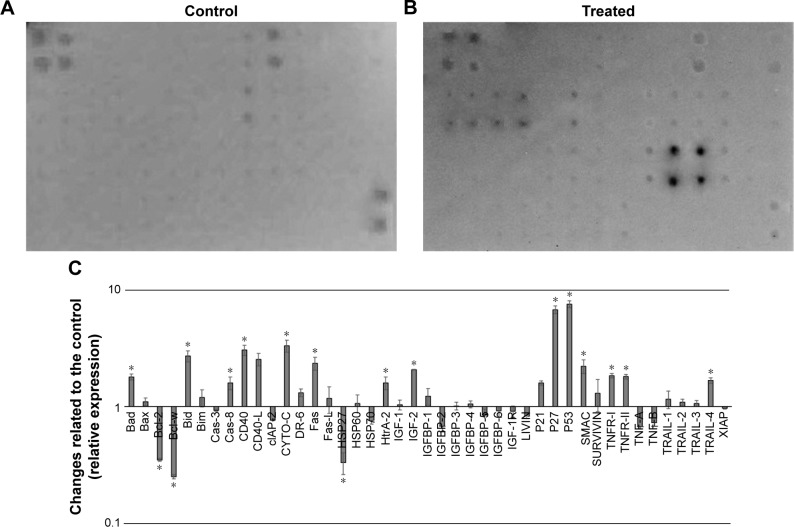
Results: The results obtained from this study illustrated the significant antiproliferative effect of BJEE on colorectal cancer cells, with a concentration value that inhibits 50% of the cell growth of 25±3.1 µg/mL after 72 h of treatment. MTT assay demonstrated that the BJEE is selectively toxic to cancer cells, and BJEE induced cell apoptosis via activation of caspase-8 along with modulation of apoptosis-related proteins such as Fas, CD40, tumor necrosis factor-related apoptosis-inducing ligands, and tumor necrosis factor receptors, which confirmed the contribution of extrinsic pathway. Meanwhile, increased ROS production in treated cells subsequently activated caspase-9 production, which triggered the intrinsic pathways. In addition, overexpression of cytochrome-c, Bax, and Bad proteins along with suppression of Bcl-2 illustrated that mitochondrial-dependent pathway also contributed to BJEE-induced cell death. Consistent with the findings from this study, BJEE-induced cancer cell death proceeds via extrinsic and intrinsic mitochondrial-dependent and -independent events.
Conclusion: From the evidence obtained from this study, it is concluded that the BJEE is a promising natural extract to combat colorectal cancer cells (HT29 cells) via induction of apoptosis through activation of extrinsic and intrinsic pathways.
Keywords: Brucea javanica; HT29; apoptosis; apoptosis protein array; cancer; mitochondrial pathway.
Conflict of interest statement
Disclosure The authors report no conflicts of interest in this work.
Figures
References
-
- David AR, Zimmerman MR. Cancer: an old disease, a new disease or something in between? Nat Rev Cancer. 2010;10(10):728–733. - PubMed
-
- Parkin DM, Pisani P, Ferlay J. Global cancer statistics. CA Cancer J Clin. 1999;49(1):33–64. - PubMed
-
- Schrag D, Cramer LD, Bach PB, Begg CB. Age and adjuvant chemotherapy use after surgery for stage III colon cancer. J Natl Cancer Inst. 2001;93(11):850–857. - PubMed
MeSH terms
Substances
LinkOut - more resources
Full Text Sources
Other Literature Sources
Research Materials
Miscellaneous
