Review
. 2018 Jan 31:2018:3537471.
doi: 10.1155/2018/3537471. eCollection 2018.

Regulation of Tumor Progression by Programmed Necrosis

Affiliations
Review

Regulation of Tumor Progression by Programmed Necrosis

Su Yeon Lee et al. Oxid Med Cell Longev. .

Abstract

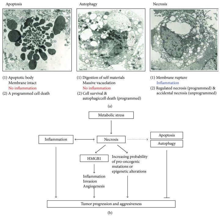
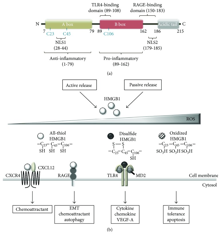
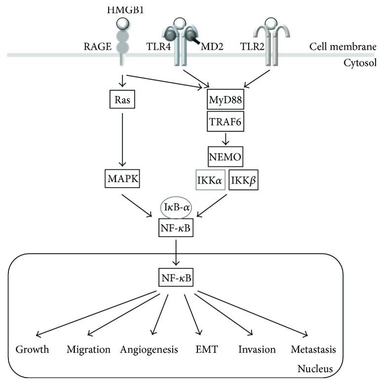
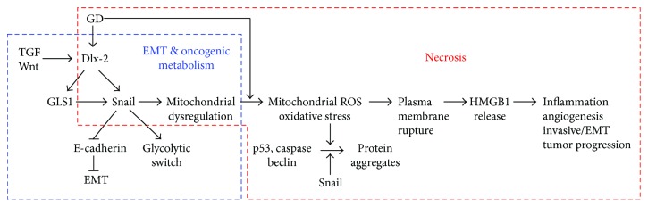
Rapidly growing malignant tumors frequently encounter hypoxia and nutrient (e.g., glucose) deprivation, which occurs because of insufficient blood supply. This results in necrotic cell death in the core region of solid tumors. Necrotic cells release their cellular cytoplasmic contents into the extracellular space, such as high mobility group box 1 (HMGB1), which is a nonhistone nuclear protein, but acts as a proinflammatory and tumor-promoting cytokine when released by necrotic cells. These released molecules recruit immune and inflammatory cells, which exert tumor-promoting activity by inducing angiogenesis, proliferation, and invasion. Development of a necrotic core in cancer patients is also associated with poor prognosis. Conventionally, necrosis has been thought of as an unregulated process, unlike programmed cell death processes like apoptosis and autophagy. Recently, necrosis has been recognized as a programmed cell death, encompassing processes such as oncosis, necroptosis, and others. Metabolic stress-induced necrosis and its regulatory mechanisms have been poorly investigated until recently. Snail and Dlx-2, EMT-inducing transcription factors, are responsible for metabolic stress-induced necrosis in tumors. Snail and Dlx-2 contribute to tumor progression by promoting necrosis and inducing EMT and oncogenic metabolism. Oncogenic metabolism has been shown to play a role(s) in initiating necrosis. Here, we discuss the molecular mechanisms underlying metabolic stress-induced programmed necrosis that promote tumor progression and aggressiveness.


Figures

Figure 1
Figure 1
Metabolic stress-induced necrosis is closely associated with tumor progression and aggressiveness. (a) Cell death is generally classified into three categories: apoptosis, autophagy, and necrosis, which are characterized by the distinct biochemical and morphological changes listed. (b) Diagram depicting how necrosis promotes tumor progression and aggressiveness by releasing the proinflammatory and angiogenic cytokine HMGB1, which can contribute to malignancy through increasing the probability of proto-oncogenic mutations or epigenetic alterations.
Figure 2
Figure 2
The structure and redox state of HMGB1. (a) Structure of HMGB1. HMGB1 is a-215 amino acid protein, composed of two DNA-binding HMG boxes (a and b) and an acidic C-terminal tail. It contains two nuclear localization signals (NLS) and three conserved redox-sensitive cysteine residues: C23 and C45 in box A, which can form an intramolecular disulfide bond, and C106 in box B. Amino acids 89–108 and 150–183 of HMGB1 are responsible for binding to TLR4 and RAGE, respectively. The A box acts in anti-inflammatory effects, whereas the B box domain plays an important role in pro-inflammatory effects. (b) Redox states of HMGB1 regulate its receptor-binding and extracellular activity. Fully reduced all-thiol HMGB1 (at-HMGB1) has chemoattractant activity. at-HMGB1 forms a heterocomplex with CXCL12 and binds CXCR4, promoting the recruitment of inflammatory cells to damaged tissues. at-HMGB1 binding to RAGE supports its chemoattractant activity via increasing CXCL12 secretion. Disulfide HMGB1 (ds-HMGB1) has sole cytokine activity. ds-HMGB1 induces the release of proinflammatory cytokines via TLR4-mediated signaling. All-oxidized HMGB1 has no cytokine or chemotaxis activity, thereby inducing immune tolerance.
Figure 3
Figure 3
Molecular mechanisms of HMGB1-induced tumor progression and metastasis. HMGB1 is ubiquitous in the tumor microenvironment and functions through activating NF-κB signaling pathways. Extracellular HMGB1 binds to several receptors, including RAGE, TLR2, and TLR4, and activates downstream signaling pathways, such as MAP kinases and myeloid differentiation primary response protein 88- (MyD88-) dependent NF-κB pathways. NF-κB increases expression of its target genes (such as IL-6, IL-8, and Snail) to regulate cancer growth, angiogenesis, EMT, invasion, and metastasis.
Figure 4
Figure 4
Snail and Dlx-2 regulate metabolic stress-induced necrosis in tumors by inducing EMT, mitochondrial dysregulation, and oncogenic metabolism. GD-induced Snail and Dlx-2 may cause mitochondrial dysfunction, facilitating ROS production in response to GD. Increased ROS can induce insoluble protein aggregates containing p53, caspase, and beclin and cause necrosis through triggering the plasma membrane rupture and HMGB1 release. In addition, metabolic stress-induced necrosis is driven by increased ROS, which is stimulated by Snail and Dlx-2, which mediates EMT for tumor invasion in the absence of metabolic stress. The Dlx-2/GLS1/glutamine metabolic axis can regulate TGF-β/Wnt-induced, Snail-dependent EMT, and glycolytic switch. Metabolic stress-induced Snail and Dlx-2 expression contributes to tumor progression by promoting necrosis as well as by inducing EMT and oncogenic metabolism.

References

    1. Tomes L., Emberley E., Niu Y., et al. Necrosis and hypoxia in invasive breast carcinoma. Breast Cancer Research and Treatment. 2003;81(1):61–69. doi: 10.1023/A:1025476722493. - DOI - PubMed
    1. Kunz M., Ibrahim S. M. Molecular responses to hypoxia in tumor cells. Molecular Cancer. 2003;2(1):p. 23. doi: 10.1186/1476-4598-2-23. - DOI - PMC - PubMed
    1. Gatenby R. A., Gillies R. J. Why do cancers have high aerobic glycolysis? Nature Reviews Cancer. 2004;4(11):891–899. doi: 10.1038/nrc1478. - DOI - PubMed
    1. Jin S., DiPaola R. S., Mathew R., White E. Metabolic catastrophe as a means to cancer cell death. Journal of Cell Science. 2007;120(3):379–383. doi: 10.1242/jcs.03349. - DOI - PMC - PubMed
    1. Jin S., White E. Role of autophagy in cancer: management of metabolic stress. Autophagy. 2007;3(1):28–31. doi: 10.4161/auto.3269. - DOI - PMC - PubMed