. 2018 Mar 15;10(3):966-974.
eCollection 2018.

COX-2 contributes to LPS-induced Stat3 activation and IL-6 production in microglial cells

Affiliations

COX-2 contributes to LPS-induced Stat3 activation and IL-6 production in microglial cells

Jie Zhu et al. Am J Transl Res. .

Abstract

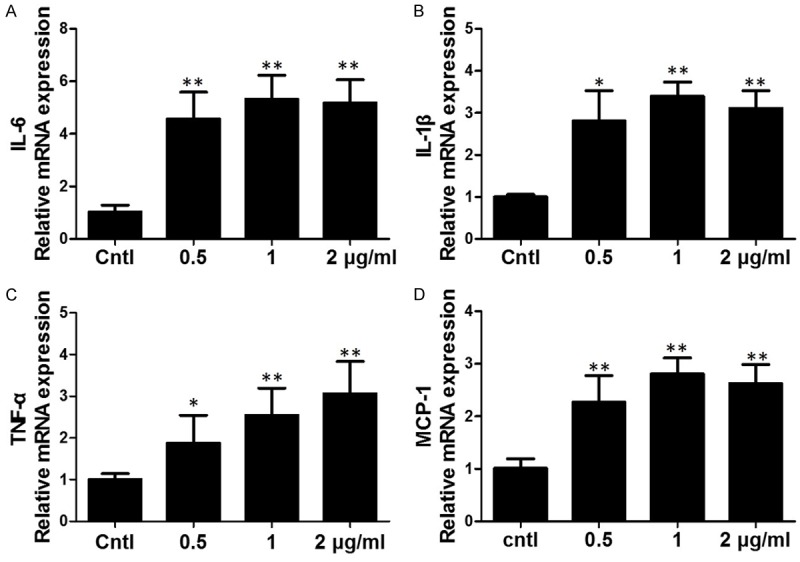
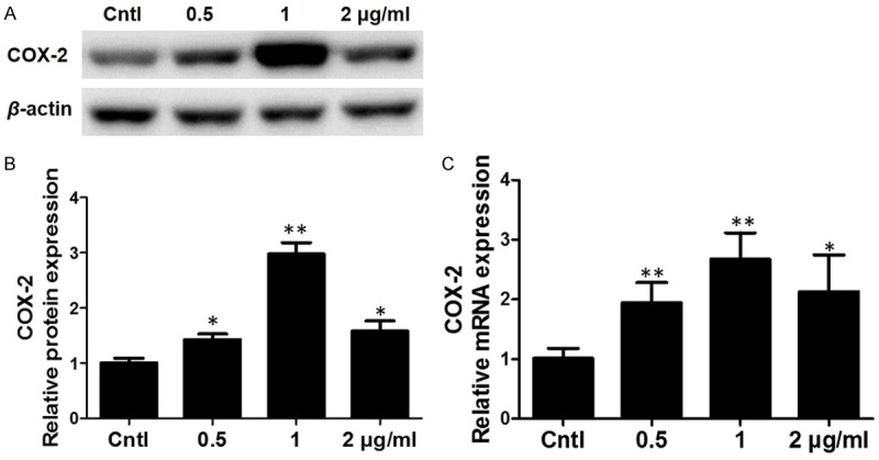
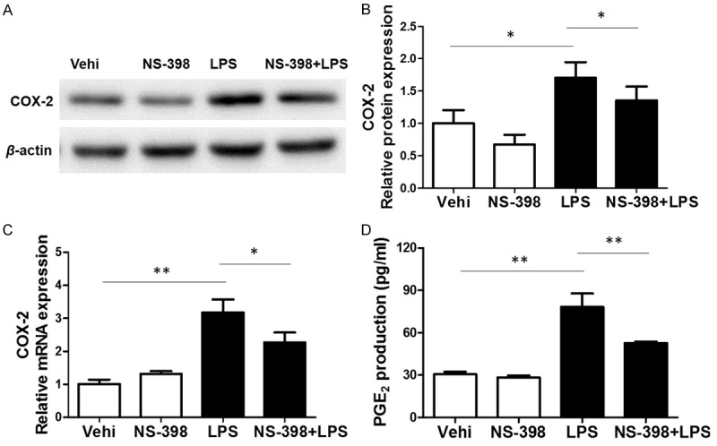
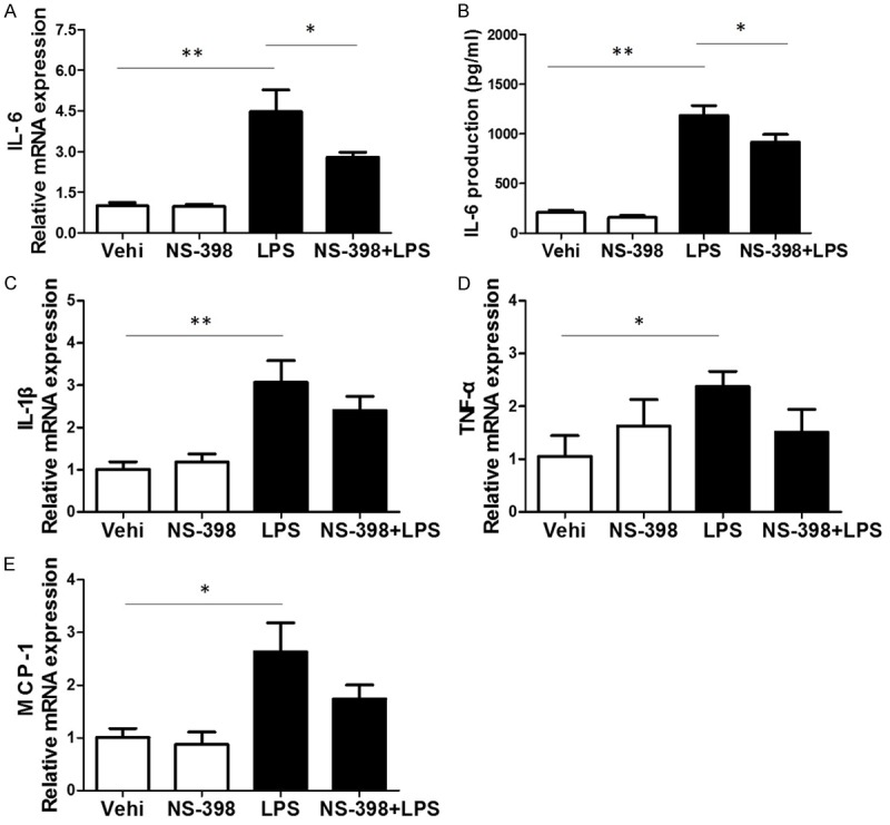
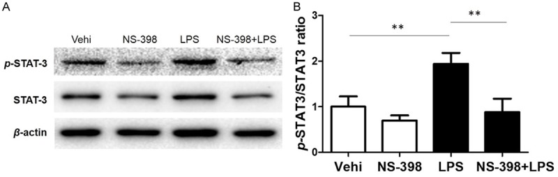
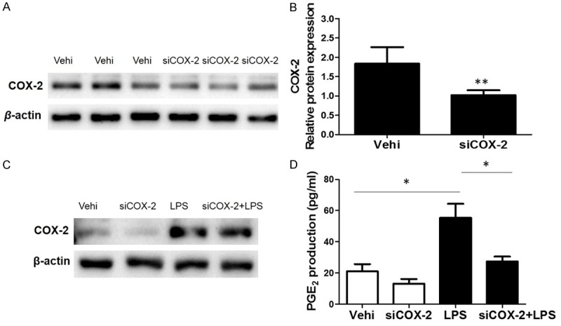
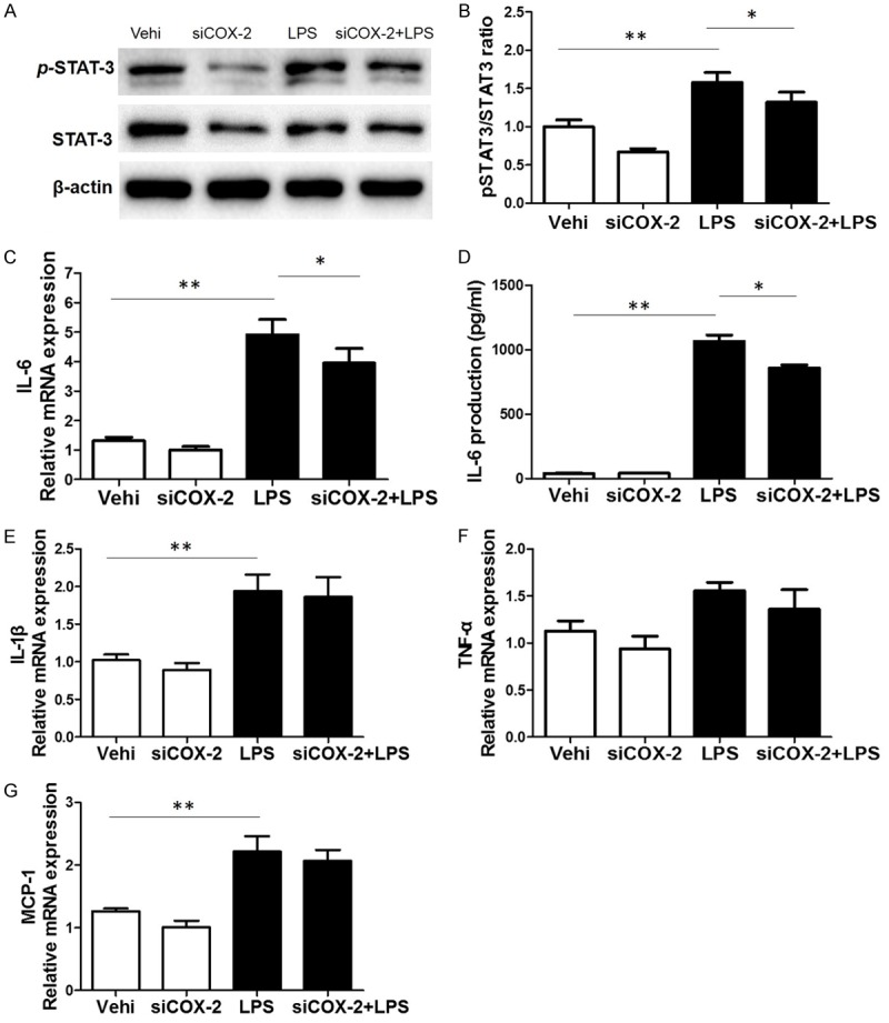
Many stimuli including lipopolysaccharide (LPS) could activate microglial cells to subsequently cause inflammatory nerve injury. However, the mechanism of LPS-induced neuroinflammation in microglial cells is still elusive. Thus, the present study was undertaken to examine the role of COX-2 in mediating the activation of Stat3 and the production of IL-6 in BV2 cells challenged with LPS. After 24 h treatment, LPS dose-dependently enhanced COX-2 expression at both mRNA and protein levels. Meanwhile, IL-6 with other inflammatory cytokines including IL-1β, TNF-α, and MCP-1 were similarly enhanced by LPS. Then a specific COX-2 inhibitor (NS-398) was administered to BV2 before LPS treatment. Significantly, COX-2 inhibition suppressed the upregulation of IL-6 at both mRNA and protein levels in line with the trend blockade on IL-1β, TNF-α, and MCP-1. Stat3 drives proinflammatory signaling pathways and contributes to IL-6 production via a transcriptional mechanism in many diseases. Here we found that inhibition of COX-2 entirely blocked LPS-induced Stat3 phosphorylation, which might contribute to the blockade of IL-6 production to some extent. Meanwhile, COX-2 siRNA approach largely reproduced the phenotypes shown by specific COX-2 inhibitor in LPS-treated BV2 cells. Together, these findings suggested that COX-2 might contribute to LPS-induced IL-6 production possibly through activating Stat3 signaling pathway in microglial cells.

Keywords: COX-2; IL-6; LPS; Microglial cells; Stat3.


Conflict of interest statement

None.

Figures

Figure 1
Figure 1
Effects of LPS on the expressions of inflammatory cytokines in BV2 cells. The mRNA levels of IL-6 (A), IL-1β (B), TNF-α (C) and MCP-1 (D) were upregulated by LPS treatment for 24 h at different doses. All values are means ± SD; n = 6 in each group. *P < 0.05 versus control, **P < 0.01 versus control.
Figure 2
Figure 2
Effects of LPS on the expression of COX-2 in BV2 cells. A. Western blotting analysis of COX-2 in BV2 cells. B. Quantitative analysis of COX-2 Western blots. C. qRT-PCR analysis of COX-2 in a dose-dependent experiment. All values are means ± SD; n = 6 in each group. *P < 0.05 versus control, **P < 0.01 versus control.
Figure 3
Figure 3
Effects of COX-2 inhibitor (NS-398) on COX-2 expression and PGE2 production in BV2 cells challenged with LPS. A. Western blotting analysis of COX-2 in BV2 cells. B. Quantitative analysis of COX-2 Western blots. C. COX-2 mRNA levels were elevated by LPS treatment. D. The levels of PGE2 in cell culture media were measured via ELISA assay. All values are means ± SD; n = 6 in each group. *P < 0.05 versus control or LPS group, **P < 0.01 versus control or LPS group.
Figure 4
Figure 4
Effects of COX-2 inhibitor on the mRNA expressions of inflammatory cytokines in BV2 cells challenged with LPS. A. qRT-PCR analysis of IL-6. B. ELISA assay of IL-6 in the medium. C. qRT-PCR analysis of IL-1β. D. qRT-PCR analysis of TNF-α. E. qRT-PCR analysis of MCP-1. All values are means ± SD; n = 6 in each group. *P < 0.05 versus control or LPS group, **P < 0.01 versus control or LPS group.
Figure 5
Figure 5
Effect of COX-2 inhibition on LPS-induced Stat3 phosphorylation. A. Western blotting analysis of p-Stat3 and Stat3 in BV2 cells. B. The ratio of p-Stat3 to Stat3. All values are means ± SD; n = 6 in each group. *P < 0.05 versus control or LPS group, **P < 0.01 versus control or LPS group.
Figure 6
Figure 6
Silencing COX-2 ameliorated LPS-induced PGE2 production. To examine the role of COX-2 in LPS-induced PGE2 production, COX-2 siRNA was applied to BV2 cells. A. Protein levels of COX-2 in BV2 cells treated with COX-2 siRNA or negative control treatment. B. mRNA levels of COX-2 in BV2 cells treated with COX-2 siRNA or negative control treatment. C. Representative images of Western blots of COX-2 with or without COX-2 silencing in response to LPS treatment. D. EIA assay of PGE2 in medium. All values are means ± SD; n = 6 in each group. *P < 0.05 versus control or LPS group, **P < 0.01 versus control or LPS group.
Figure 7
Figure 7
Silencing COX-2 blunted LPS-induced Stat3 phosphorylation and IL-6 upregulation. A. Western blotting analysis of p-Stat3 and Stat3 in BV2 cells. B. The ratio of p-Stat3 to Stat3. C. The mRNA level of IL-6 was measured by qRT-PCR. D. IL-6 release in cell culture medium were measured by using a ELISA kit. E. qRT-PCR analysis of IL-1β. F. qRT-PCR analysis of TNF-α. G. qRT-PCR analysis of MCP-1. All values are means ± SD; n = 6 in each group. *P < 0.05 versus control or LPS group, **P < 0.01 versus control or LPS group.

References

    1. Perry VH, Teeling J. Microglia and macrophages of the central nervous system: the contribution of microglia priming and systemic inflammation to chronic neurodegeneration. Semin Immunopathol. 2013;35:601–612. - PMC - PubMed
    1. Weng L, Zhang H, Li X, Zhan H, Chen F, Han L, Xu Y, Cao X. Ampelopsin attenuates lipopolysaccharide-induced inflammatory response through the inhibition of the NF-kappaB and JAK2/STAT3 signaling pathways in microglia. Int Immunopharmacol. 2017;44:1–8. - PubMed
    1. Xia CY, Zhang S, Gao Y, Wang ZZ, Chen NH. Selective modulation of microglia polarization to M2 phenotype for stroke treatment. Int Immunopharmacol. 2015;25:377–382. - PubMed
    1. Okorji UP, Velagapudi R, El-Bakoush A, Fiebich BL, Olajide OA. Antimalarial drug artemether inhibits neuroinflammation in BV2 microglia through Nrf2-dependent mechanisms. Mol Neurobiol. 2016;53:6426–6443. - PubMed
    1. Moidunny S, Matos M, Wesseling E, Banerjee S, Volsky DJ, Cunha RA, Agostinho P, Boddeke HW, Roy S. Oncostatin M promotes excitotoxicity by inhibiting glutamate uptake in astrocytes: implications in HIV-associated neurotoxicity. J Neuroinflammation. 2016;13:144. - PMC - PubMed

LinkOut - more resources