

Paeoniflorin exerts protective effect on radiation-induced hepatic fibrosis in rats via TGF-β1/Smads signaling pathway
- PMID: 29636890
- PMCID: PMC5883141
Paeoniflorin exerts protective effect on radiation-induced hepatic fibrosis in rats via TGF-β1/Smads signaling pathway
Abstract
Aim: This study aimed to investigate the protective effects of paeoniflorin (PAE) on radiation-induced hepatic fibrosis in a rat model.
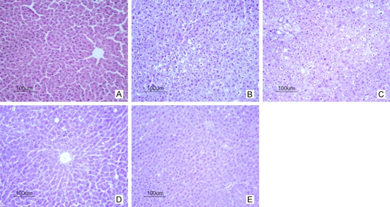
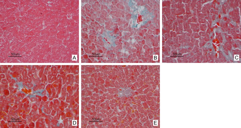
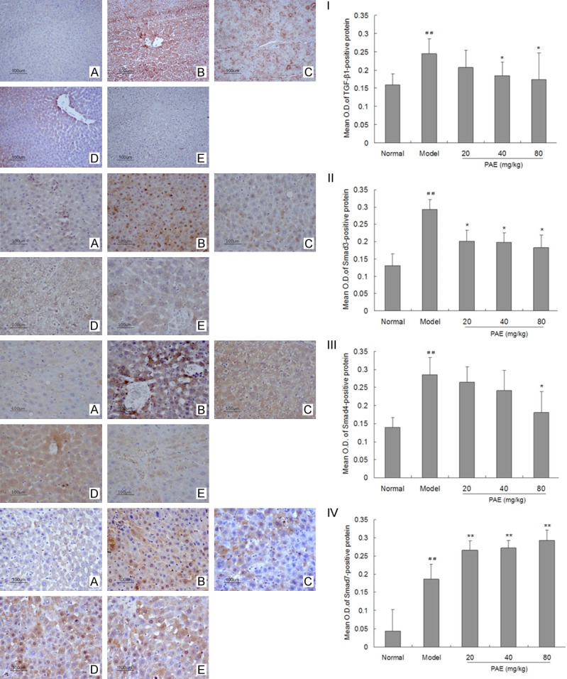
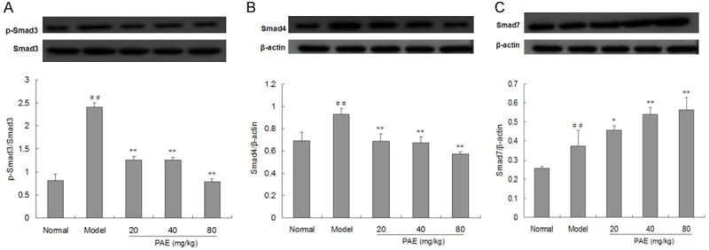
Methods: Fifty healthy male Sprague-Dawley rats were randomly assigned to normal control group, hepatic fibrosis group, and PAE treatment groups. X-ray exposure was employed to establish radiation-induced hepatic fibrosis model. PAE was administered once daily, and rats were sacrificed at week 26 after irradiation. The liver histopathology was evaluated under a light microscope after HE staining and Masson staining. Meanwhile, the protein expression of transforming growth factor-beta 1 (TGF-β1), Smad3/4 and Smad7 was detected by immunohistochemistry.
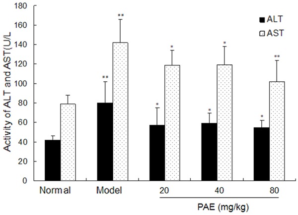
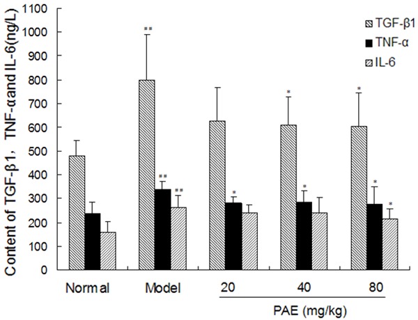
Results: Radiation-induced liver damage and collagen deposition were observed in the model group as compared to normal control group, but PAE treatment significantly attenuated the liver injury and reduce collagen deposition (P<0.05 or 0.01). The hepatic hydroxyproline content and serum levels of TGF-β1, hyaluronic acid, ro-collagen type III and laminin markedly increased in model group as compared to control group (P<0.01), but they decreased dramatically after PAE treatment. The expression of TGF-β1, Smad3/4 and Smad7 in the liver increased significantly in model group as compared to control group (P<0.01), and PAE could down-regulate the expression of Smad3/4 and up-regulate Smad7 expression (P<0.05 or 0.01). The activities of serum amino-transferase and aspartate aminotransferase were significantly higher in hepatic fibrosis group than in normal control group, but PAE treatment markedly reduced them (P<0.05).
Conclusion: PAE can inhibit the radiation induced hepatic fibrosis via regulating TGF-β1/Smads signaling pathway.
Keywords: Paeoniflorin; Smads; radiation-induced hepatic fibrosis; transforming growth factor-beta 1.
Conflict of interest statement
None.
Figures
References
-
- Huang HZ, Liang SX, Li YF. Progress in study on the dose of radiation tolerance of the liver. Guangxi Med J. 2009;31:1018–1020.
-
- Fidler IJ. Blockade of the TGF-beta superfamily by Smad7: breaking a link in the metastatic chain. J Natl Cancer Inst. 2005;97:1714–1715. - PubMed
LinkOut - more resources
Full Text Sources
Other Literature Sources
Research Materials