. 2018 Oct;374(1):149-163.
doi: 10.1007/s00441-018-2836-8. Epub 2018 Apr 10.

Effect of TGFβ1, TGFβ3 and keratinocyte conditioned media on functional characteristics of dermal fibroblasts derived from reparative (Balb/c) and regenerative (Foxn1 deficient; nude) mouse models

Affiliations

Effect of TGFβ1, TGFβ3 and keratinocyte conditioned media on functional characteristics of dermal fibroblasts derived from reparative (Balb/c) and regenerative (Foxn1 deficient; nude) mouse models

Joanna Bukowska et al. Cell Tissue Res. 2018 Oct.

Abstract

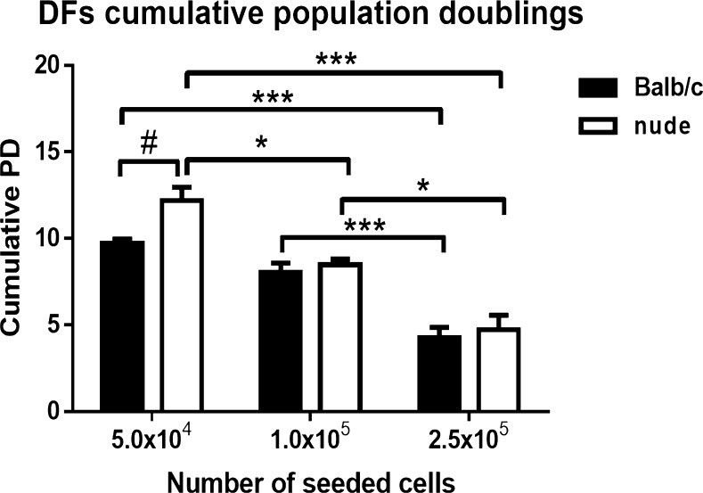
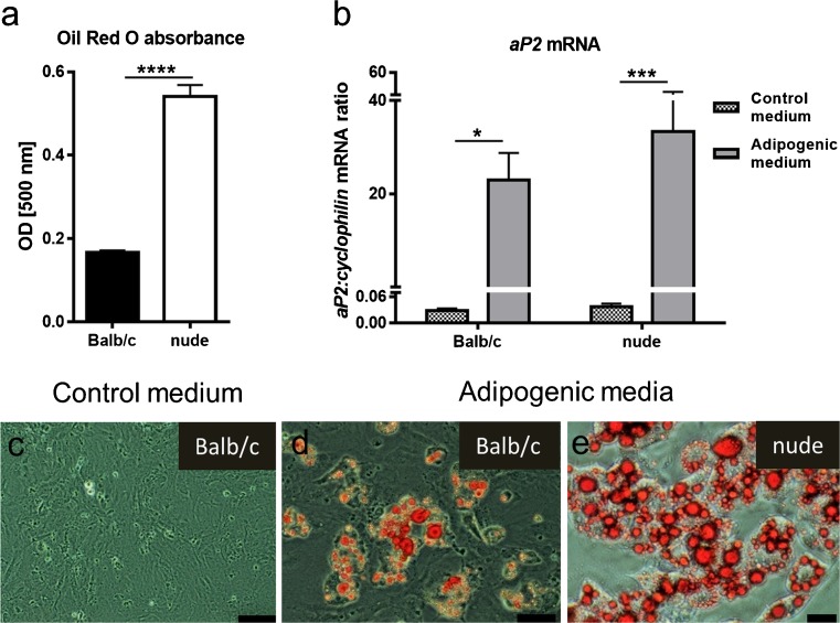
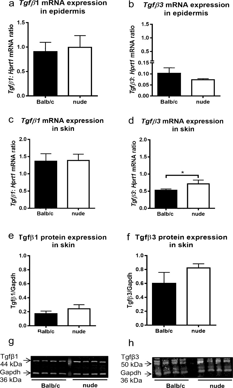
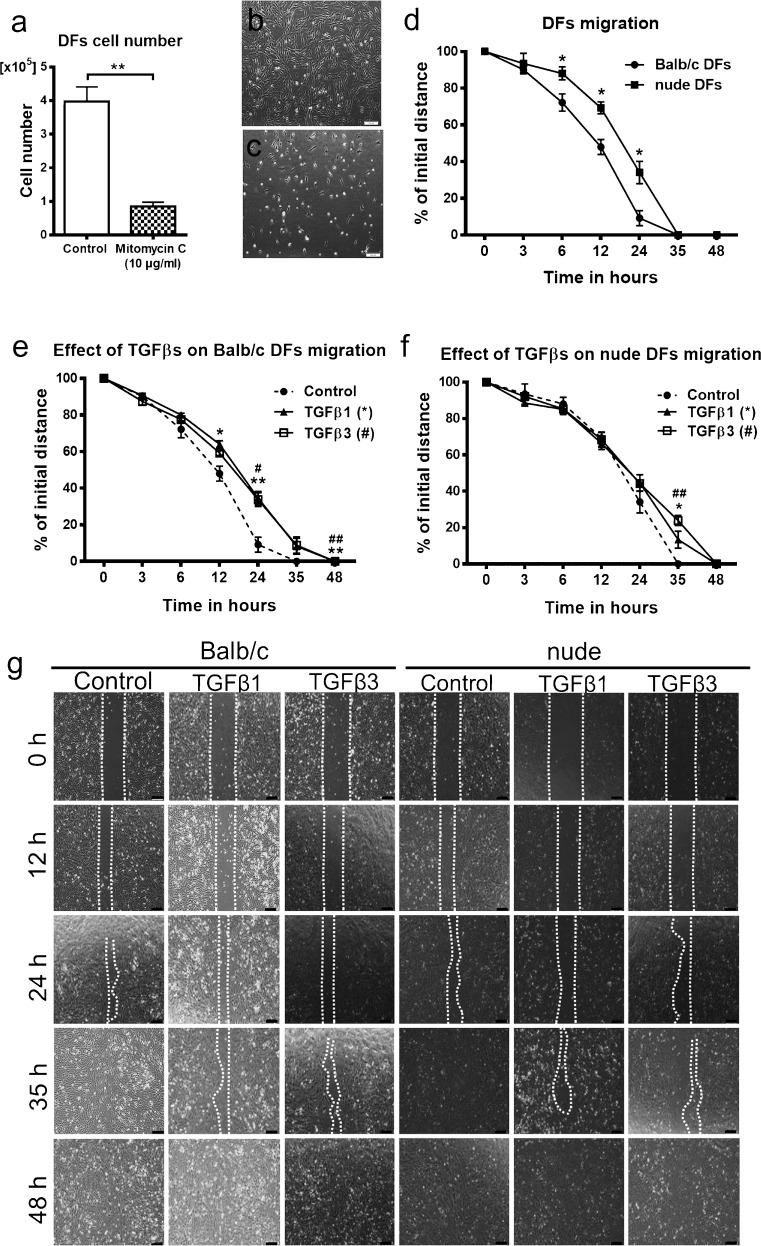
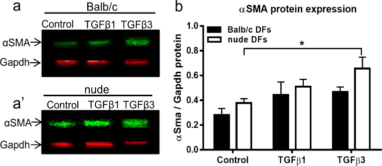
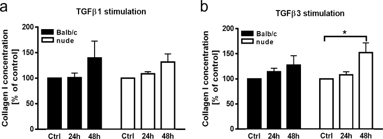
Skin injuries in mammals are healed through repair or regeneration. Our previous studies demonstrated that deficient expression of the transcription factor Foxn1 in epidermis of nude mice accounts for their skin's pronounced regenerative properties. Since homeostasis within the skin depends on complex interactions between the epidermal and underlying dermal layers, the present study characterizes and compares isolated dermal fibroblasts (DFs) between regenerative nude (Foxn1 deficient) mice and their wild-type Balb/c counterparts. Nude DFs exhibited a higher cumulative number of population doublings (cumulative PD) at low seeding density and increased adipogenic differentiation capacity relative to their Balb/c DF counterparts. Nude DFs displayed reduced migration and gel contraction, functional features associated with wound healing. The comparison of transforming growth factor β family (TGFβ) expression showed significantly higher levels of Tgfβ3 transcript between nude and Balb/c mice but no differences were detected for Tgfβ1. Nude DFs were specifically sensitive to the presence of the pro-regenerative TGFβ3 isoform, showing increased collagen I deposition and alpha smooth muscle actin expression. Viability of Balb/c DFs was stimulated by keratinocyte conditioned media (KCM) from Balb/c (Foxn1 active) but inhibited by nude (Foxn1 deficient) KCM. In contrast, nude DFs did not respond to either KCMs with respect to their metabolic activity. Collectively, the enhanced plasticity and greater sensitivity of nude DFs to TGFβ3 stimulation are indicative of and consistent with their pro-regenerative characteristics. These data support the hypothesis that epidermal Foxn1 plays a critical role in determining the DFs regenerative phenotype.

Keywords: Dermal fibroblasts; Foxn1; Skin; TGFβ; Wound healing.


Conflict of interest statement

Conflict of interest

The authors declare that they have no conflict of interest.

Figures

Fig. 1
Fig. 1
DFs derived from nude mice demonstrate the highest increase in cumulative PD at the lowest cell plating density. Comparison of cumulative population doublings (PD) of DFs derived from the skin of Balb/c and nude mice seeded in 24-well plates at densities 5.0 × 104, 1.0 × 105 and 2.5 × 105 per well. The results are shown as the mean ± SEM. Duplicate wells were used and the experiment was repeated 5 times (n = 15 animals; 3 animals per repeat). Asterisks indicate statistically significant differences among DFs seeded at different densities (*p < 0.05; ***p < 0.001). Hashes indicate significant differences between Balb/c and nude DFs seeded at the same density (#p < 0.05)
Fig. 2
Fig. 2
Nude DFs show an increase in adipogenic differentiation potential. Adipogenic capacity of DFs derived from the skin of Balb/c and nude mice seeded in 24-well plates at a density of 5.0 × 104 cells/well was measured by spectrophotometric analysis of Oil Red O staining (a) and on mRNA expression levels of adipose tissue marker aP2 (b). Morphology of Oil Red O stained cells cultured with control (c) or adipogenic differentiation media (d, e) within 6 days. The results are shown as the mean ± SEM. Triplicate wells were used, and the experiment was repeated 5 times (n = 15 animals; 3 animals per repeat). The asterisks indicate significant differences between groups (*p < 0.05; ***p < 0.001; ****p < 0.0001). Scale bars 100 μm (c), 50 μm (d) and 20 μm (e)
Fig. 3
Fig. 3
TGFβ1 and TGFβ3 expressions in the epidermis and in the skin tissues of newborn nude and Balb/c mice. Quantitative analysis of Tgfβ1 (a, c) and Tgfβ3 (b, d) mRNA expression in the epidermis (a, b) and in the skin (c, d). Western blots densitometry analysis (e, f) and representative blots (g, h) of TGFβ1 (e, g) and TGFβ3 (f, h) protein expression in the skin. The results are shown as the mean ± SEM of six animals per group (n = 6), each performed in duplicate. The asterisks indicate significant differences between groups (*p < 0.05)
Fig. 4
Fig. 4
The effect of TGFβ1 (a), TGFβ3 (b) and KCMs collected from Foxn1 active Balb/c (c) and Foxn1 deficient nude (d) keratinocytes on viability of Balb/c and nude DFs seeded in 96-well plates at a density of 2.0 × 104 per well. Both TGFβ isoforms (a, b) increase metabolic activity of Balb/c (black bars) DFs with very limited effect on nude DFs (white bars) viability. Stimulation with KCM collected from Balb/c mice promotes viability of Balb/c DFs (c), whereas KCM collected from nude keratinocytes shows an inhibitory impact on these cells (d). Both KCMs have no effect of nude DF metabolic activity (c, d). All values are expressed as percent of control. The results are shown as the mean ± SEM. Duplicate wells were used and the experiment was repeated 4 times (n = 12 animals; 3 animals per repeat). The asterisks indicate significant differences relative to the control, non-stimulated cultures (*p < 0.05; **p < 0.01; ***p < 0.001; ****p < 0.0001). Hashes show significant differences between Balb/c and nude DFs (#p < 0.05)
Fig. 5
Fig. 5
Migratory abilities of Balb/c and nude DFs. Inhibitory effect of mitomycin C (10 μg/ml) on DFs manifested by the decrease in the cell number (a) and the cells distribution in the culture plate (b, c). Wound healing (scratch) in vitro assay (d, e, f, g) shows faster migration of Balb/c than nude DFs (d). Both TGFβ1 and TGFβ3 isoforms suppress the motility of Balb/c and nude DFs plated in 12-well plates at a density of 2.5 × 105 per well (e, f, g). The results are shown as the mean ± SEM. Duplicate wells were used and the experiment was repeated 4 times (n = 12 animals; 3 animals per repeat). The asterisks indicate significant differences between mitomycin-treated and control, untreated DFs (a); Balb/c and nude DFs (d); TGFβ1-treated and control Balb/c DFs (e); TGFβ1-treated and control nude DFs (f) (*p < 0.05; **p < 0.01). Hashes show significant differences in TGFβ3-treated Balb/c (e) or nude (f) DFs relative to the control (#p < 0.05; ##p < 0.01). Representative images depicting the inhibitory effects of TGFβ1 and TGFβ3 on the migration of Balb/c and nude DFs at different time points (0, 6, 12, 24, 35, 48 h) within 48 h of stimulation (g). Scale bars 200 μm (b, c) and 100 μm (g)
Fig. 6
Fig. 6
Comparison of contractile abilities of Balb/c and nude DFs seeded at a density of 6.0 × 104 cells per 400 μl of collagen lattices in a 24-well plate. Representative images show the contractile abilities of Balb/c and nude DFs cultured with control medium, KCMs, or with control media supplemented with TGFβ (a). DFs were cultured in control (unstimulated) medium, (b), KCMs collected from Foxn1 active Balb/c and Foxn1deficient nude keratinocytes (c, d). DFs from Balb/c (e) and nude (f) were cultured in medium supplemented with TGFβ1 or TGFβ3. KCMs derived from Balb/c and nude keratinocytes enhance contraction of collagen lattices populated by Balb/c (c) and nude (d) DFs. Neither TGFβ1 nor TGFβ3 treatment affects matrix size populated by Balb/c (e) or nude (f) DFs. All values are expressed as percentage of control. The results are shown as the mean ± SEM. Duplicate wells were used and the experiment was repeated three times (n = nine animals; three animals per repeat). The asterisks indicate significant differences between Balb/c and nude DFs (b); Balb/c (c) and nude (d) DFs treated with Balb/c derived KCMs relative to the control (*p < 0.05; **p < 0.01; ***p < 0.001). Hashes show significant differences in Balb/c (c) and nude (d) DFs treated with nude-derived KCM relative to the control (#p < 0.05; ##p < 0.01). The asterisks (****p < 0.001) above the graphs –e) (c-f) show differences between day 0 relative to day 9 (day 0 vs day 9)
Fig. 7
Fig. 7
Representative Western blots (a, a’) and densitometry analysis (b) for total αSMA protein content detected in Balb/c and nude DFs. Cells were plated at a density of 2.5 × 105 on 35-mm Petri dishes and stimulated with TGFβ1 or TGFβ3 (10 ng/ml each). Treatment with TGFβ3 increases αSMA expression exclusively in nude DFs (white bars). The results are shown as the mean ± SEM of three animals per group (n = 3). The asterisk indicates significant differences between groups relative to the control (*p < 0.05)
Fig. 8
Fig. 8
The effect of TGFβ1 (a) and TGFβ3 (b) (10 ng/ml each) on collagen I protein concentration in the culture media collected from Balb/c (black bars) and nude (white bars) DFs plated in 24-well plates at a density of 4.5 × 105 per well. Exposition to TGFβ3 stimulates nude DFs to collagen I secretion after 48 h of treatment (b). All values are expressed as percent of the control. The results are shown as the mean ± SEM. Duplicate wells were used and the experiment was repeated 4 times (n = 12 animals; 3 animals per repeat). The asterisk indicates significant differences between groups relative to the control (*p < 0.05)

Similar articles

Cited by

References

    1. Ali N, Zirak B, Rodriguez RS, Pauli ML, Truong HA, Lai K, Ahn R, Corbin K, Lowe MM, Scharschmidt TC, Taravati K, Tan MR, Ricardo-Gonzalez RR, Nosbaum A, Bertolini M, Liao W, Nestle FO, Paus R, Cotsarelis G, Abbas AK, Rosenblum MD. Regulatory T cells in skin facilitate epithelial stem cell differentiation. Cell. 2017;169:1119–1129. doi: 10.1016/j.cell.2017.05.002. - DOI - PMC - PubMed
    1. Ansel JC, Tiesman JP, Olerud JE, Krueger JG, Krane JF, Tara DC, Shipley GD, Gilbertson D, Usui ML, Hart CE. Human keratinocytes are a major source of cutaneous platelet-derived growth factor. J Clin Invest. 1993;92:671–678. doi: 10.1172/JCI116636. - DOI - PMC - PubMed
    1. Chen X, Thibeault SL. Response of fibroblasts to transforming growth factor-beta1 on two-dimensional and in three-dimensional hyaluronan hydrogels. Tissue Eng A. 2012;18:2528–2538. doi: 10.1089/ten.tea.2012.0094. - DOI - PMC - PubMed
    1. Chen W, Fu X, Ge S, Sun T, Zhou G, Jiang D, Sheng Z. Ontogeny of expression of transforming growth factor-beta and its receptors and their possible relationship with scarless healing in human fetal skin. Wound Repair Regen. 2005;13:68–75. doi: 10.1111/j.1067-1927.2005.130109.x. - DOI - PubMed
    1. Clark RA, McCoy GA, Folkvord JM, McPherson JM. TGF-beta 1 stimulates cultured human fibroblasts to proliferate and produce tissue-like fibroplasia: a fibronectin matrix-dependent event. J Cell Physiol. 1997;170:69–80. doi: 10.1002/(SICI)1097-4652(199701)170:1<69::AID-JCP8>3.0.CO;2-J. - DOI - PubMed

MeSH terms

LinkOut - more resources