

S-Layer Protein-Based Biosensors
- PMID: 29641511
- PMCID: PMC6023001
- DOI: 10.3390/bios8020040
S-Layer Protein-Based Biosensors
Abstract
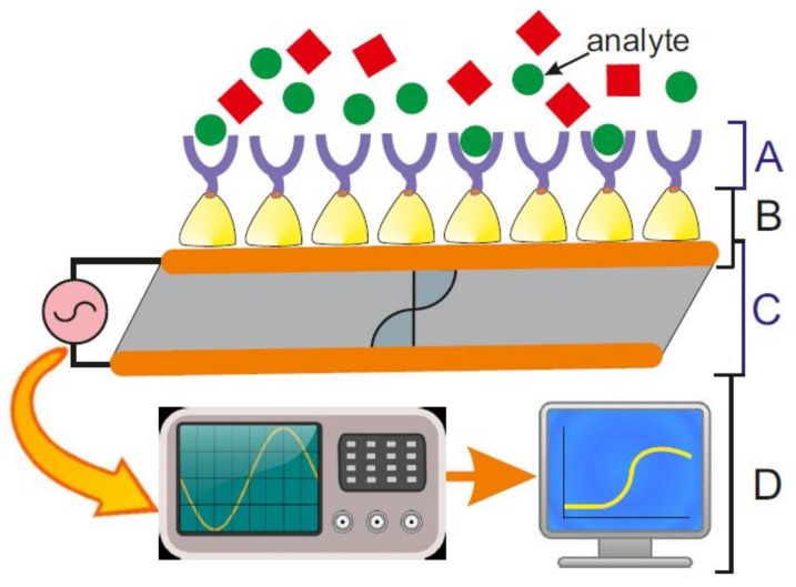
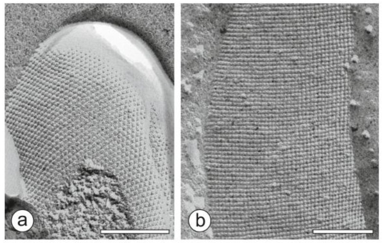
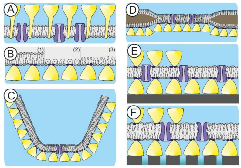
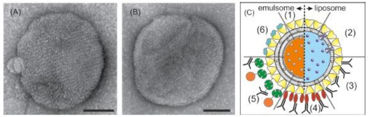
The present paper highlights the application of bacterial surface (S-) layer proteins as versatile components for the fabrication of biosensors. One technologically relevant feature of S-layer proteins is their ability to self-assemble on many surfaces and interfaces to form a crystalline two-dimensional (2D) protein lattice. The S-layer lattice on the surface of a biosensor becomes part of the interface architecture linking the bioreceptor to the transducer interface, which may cause signal amplification. The S-layer lattice as ultrathin, highly porous structure with functional groups in a well-defined special distribution and orientation and an overall anti-fouling characteristics can significantly raise the limit in terms of variety and the ease of bioreceptor immobilization, compactness of bioreceptor molecule arrangement, sensitivity, specificity, and detection limit for many types of biosensors. The present paper discusses and summarizes examples for the successful implementation of S-layer lattices on biosensor surfaces in order to give a comprehensive overview on the application potential of these bioinspired S-layer protein-based biosensors.
Keywords: S-layer protein; biomimetics; bioreceptor; biosensor; crystalline 2D protein lattice; linking matrix; lipid membrane platform.
Conflict of interest statement
The author declares no conflict of interest.
Figures
Similar articles
-
Electrochemical Biosensors Based on S-Layer Proteins.Sensors (Basel). 2020 Mar 19;20(6):1721. doi: 10.3390/s20061721. Sensors (Basel). 2020. PMID: 32204503 Free PMC article. Review.
-
2D crystalline protein layers as immobilization matrices for the development of DNA microarrays.Biosens Bioelectron. 2013 Feb 15;40(1):32-7. doi: 10.1016/j.bios.2012.05.037. Epub 2012 Jun 7. Biosens Bioelectron. 2013. PMID: 22727519
-
S-layer proteins as key components of a versatile molecular construction kit for biomedical nanotechnology.Mini Rev Med Chem. 2006 Aug;6(8):909-20. doi: 10.2174/138955706777935026. Mini Rev Med Chem. 2006. PMID: 16918497 Review.
-
Biosensing for the environment and defence: aqueous uranyl detection using bacterial surface layer proteins.Sensors (Basel). 2010;10(5):4739-55. doi: 10.3390/s100504739. Epub 2010 May 10. Sensors (Basel). 2010. PMID: 22399904 Free PMC article.
-
Characterization of a bacterial self-assembly surface layer protein and its application as an electrical nanobiosensor.J Nanosci Nanotechnol. 2011 Jan;11(1):402-7. doi: 10.1166/jnn.2011.3264. J Nanosci Nanotechnol. 2011. PMID: 21446464
Cited by
-
S-Layer Ultrafiltration Membranes.Membranes (Basel). 2021 Apr 8;11(4):275. doi: 10.3390/membranes11040275. Membranes (Basel). 2021. PMID: 33918014 Free PMC article. Review.
-
Polymer-Lipid Hybrid Materials.Chem Rev. 2021 Nov 24;121(22):13996-14030. doi: 10.1021/acs.chemrev.1c00755. Epub 2021 Nov 9. Chem Rev. 2021. PMID: 34752064 Free PMC article. Review.
-
Bio-fabrication of thermozyme-based nano-biosensors: their components and present scenario.J Mater Sci Mater Electron. 2022;33(8):5523-5533. doi: 10.1007/s10854-022-07741-9. Epub 2022 Jan 29. J Mater Sci Mater Electron. 2022. PMID: 38624939 Free PMC article.
-
Microscale and Nanoscale Biosensors.Biosensors (Basel). 2018 Jul 6;8(3):66. doi: 10.3390/bios8030066. Biosensors (Basel). 2018. PMID: 29986425 Free PMC article.
-
Molecular assemblies built with the artificial protein Pizza.J Struct Biol X. 2020 May 28;4:100027. doi: 10.1016/j.yjsbx.2020.100027. eCollection 2020. J Struct Biol X. 2020. PMID: 32647829 Free PMC article.
References
-
- Dugas V., Elaissari A., Chevalier Y. Recognition Receptors in Biosensors. Springer; New York, NY, USA: 2010. Surface sensitization techniques and recognition receptors immobilization on biosensors and microarrays; pp. 47–134.
-
- Schmidt J.J., Montemagno C.D. Bionanomechanical systems. Annu. Rev. Mater. Res. 2004;34:315–337. doi: 10.1146/annurev.matsci.34.040203.115827. - DOI
MeSH terms
Substances
Grants and funding
LinkOut - more resources
Full Text Sources
Other Literature Sources
Molecular Biology Databases
