Melatonin enhances antioxidant molecules in the placenta, reduces secretion of soluble fms-like tyrosine kinase 1 (sFLT) from primary trophoblast but does not rescue endothelial dysfunction: An evaluation of its potential to treat preeclampsia
- PMID: 29641523
- PMCID: PMC5894956
- DOI: 10.1371/journal.pone.0187082
Melatonin enhances antioxidant molecules in the placenta, reduces secretion of soluble fms-like tyrosine kinase 1 (sFLT) from primary trophoblast but does not rescue endothelial dysfunction: An evaluation of its potential to treat preeclampsia
Abstract
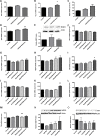
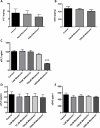
Preeclampsia is one of the most serious complications of pregnancy. Currently there are no medical treatments. Given placental oxidative stress may be an early trigger in the pathogenesis of preeclampsia, therapies that enhance antioxidant pathways have been proposed as treatments. Melatonin is a direct free-radical scavenger and indirect antioxidant. We performed in vitro assays to assess whether melatonin 1) enhances the antioxidant response element genes (heme-oxygenase 1, (HO-1), glutamate-cysteine ligase (GCLC), NAD(P)H:quinone acceptor oxidoreductase 1 (NQO1), thioredoxin (TXN)) or 2) alters secretion of the anti-angiogenic factors soluble fms-like tyrosine kinase-1 (sFLT) or soluble endoglin (sENG) from human primary trophoblasts, placental explants and human umbilical vein endothelial cells (HUVECs) and 3) can rescue TNF-α induced endothelial dysfunction. In primary trophoblast melatonin treatment increased expression of the antioxidant enzyme TXN. Expression of TXN, GCLC and NQO1 was upregulated in placental tissue with melatonin treatment. HUVECs treated with melatonin showed an increase in both TXN and GCLC. Melatonin did not increase HO-1 expression in any of the tissues examined. Melatonin reduced sFLT secretion from primary trophoblasts, but had no effect on sFLT or sENG secretion from placental explants or HUVECs. Melatonin did not rescue TNF-α induced VCAM-1 and ET-1 expression in endothelial cells. Our findings suggest that melatonin induces antioxidant pathways in placenta and endothelial cells. Furthermore, it may have effects in reducing sFLT secretion from trophoblast, but does not reduce endothelial dysfunction. Given it is likely to be safe in pregnancy, it may have potential as a therapeutic agent to treat or prevent preeclampsia.
Conflict of interest statement
Figures


References
-
- MacKay AP, Berg CJ, Atrash HK. Pregnancy-related mortality from preeclampsia and eclampsia. Obstet Gynecol. 2001;97(4):533–8. . - PubMed
-
- Mol BW, Roberts CT, Thangaratinam S, Magee LA, de Groot CJ, Hofmeyr GJ. Pre-eclampsia. Lancet. 2015. doi: 10.1016/S0140-6736(15)00070-7 . - DOI - PubMed
-
- Young BC, Levine RJ, Karumanchi SA. Pathogenesis of preeclampsia. Annu Rev Pathol. 2010;5:173–92. Epub 2010/01/19. doi: 10.1146/annurev-pathol-121808-102149 . - DOI - PubMed
-
- Powe CE, Levine RJ, Karumanchi SA. Preeclampsia, a disease of the maternal endothelium: the role of antiangiogenic factors and implications for later cardiovascular disease. Circulation. 2011;123(24):2856–69. Epub 2011/06/22. doi: 10.1161/CIRCULATIONAHA.109.853127 ; PubMed Central PMCID: PMC3148781. - DOI - PMC - PubMed
-
- Redman CW, Sargent IL. Latest advances in understanding preeclampsia. Science. 2005;308(5728):1592–4. doi: 10.1126/science.1111726 . - DOI - PubMed
Publication types
MeSH terms
Substances
LinkOut - more resources
Full Text Sources
Other Literature Sources
Medical
Miscellaneous

