

Multiple blood flow measurements before and after carotid artery stenting via phase-contrast magnetic resonance imaging: An observational study
- PMID: 29641548
- PMCID: PMC5895018
- DOI: 10.1371/journal.pone.0195099
Multiple blood flow measurements before and after carotid artery stenting via phase-contrast magnetic resonance imaging: An observational study
Abstract
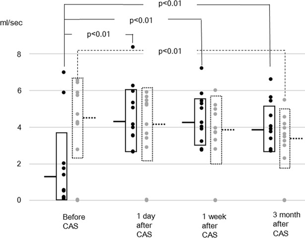
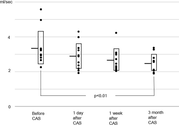
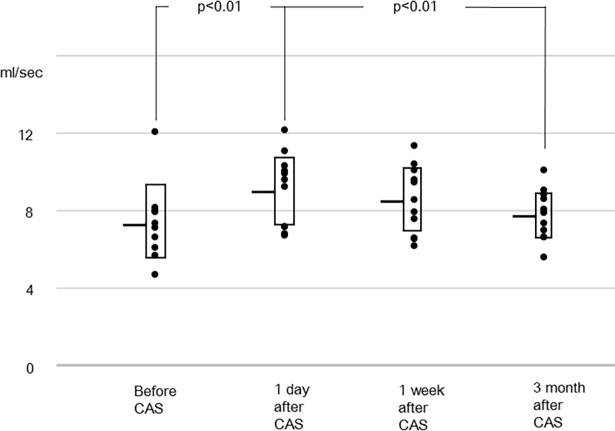
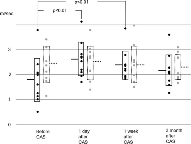
After carotid artery stenting, the procurement of information about blood flow redistribution among brain-feeding arteries and its time trend is essential to understanding a patient's physiological background and to determine their care regimen. Cerebral blood flow has been measured twice following carotid artery stenting in few previous studies, with some discrepancies in the results. The purpose of this study was to measure cerebral blood flow at multiple time points after carotid artery stenting, and to elucidate the time trend of cerebral blood flow and redistribution among arteries. Blood flow rates in 11 subjects were measured preoperatively, at one day, one week, and about three months, respectively after carotid artery stenting by using phase-contrast magnetic resonance imaging. The target vessels were the bilateral internal carotid arteries, the basilar artery, and the bilateral middle cerebral arteries. Lumen was semi-automatically defined using an algorithm utilizing pulsatility. The results showed that blood flow rates in the stented internal carotid artery and the ipsilateral middle cerebral artery increased following carotid artery stenting. Blood flow rates in the contralateral internal carotid artery and the basilar artery gradually declined, and they were lower than the preoperative values at three months after stenting. The sum of blood flow rates of the bilateral internal carotid arteries and the basilar artery increased after carotid artery stenting, and then decreased over the next three months. There was no significant change in the blood flow rate in the contralateral middle cerebral artery. From these results, it was concluded that redistribution among the bilateral internal carotid arteries and the basilar artery occurs after carotid artery stenting, and that it takes months thereafter to reach another equilibrium.
Conflict of interest statement
Figures
References
-
- Mas JL, Arquizan C, Calvet D, Viguier A, Albucher JF, Piquet P, et al. Long-term follow-up study of endarterectomy versus angioplasty in patients with symptomatic severe carotid stenosis trial. Stroke. 2014;45: 2750–6. doi: 10.1161/STROKEAHA.114.005671 - DOI - PubMed
-
- Bonati LH, Dobson J, Featherstone RL, Ederle J, van der Worp HB, de Borst GJ, et al. Long-term outcomes after stenting versus endarterectomy for treatment of symptomatic carotid stenosis: the International Carotid Stenting Study (ICSS) randomised trial. Lancet. 2015;385: 529–38. doi: 10.1016/S0140-6736(14)61184-3 - DOI - PMC - PubMed
-
- White CJ. Carotid artery stenting. J Am Coll Cardiol. 2014;64: 722–31. doi: 10.1016/j.jacc.2014.04.069 - DOI - PubMed
-
- Musiałek P, Grunwald IQ. How asymptomatic is "asymptomatic" carotid stenosis? Resolving fundamental confusion(s)—and confusions yet to be resolved. Pol Arch Intern Med. 2017;127:718–719. doi: 10.20452/pamw.4157 - DOI - PubMed
Publication types
MeSH terms
LinkOut - more resources
Full Text Sources
Other Literature Sources
Medical
