

Memory deficiency, cerebral amyloid angiopathy, and amyloid-β plaques in APP+PS1 double transgenic rat model of Alzheimer's disease
- PMID: 29641600
- PMCID: PMC5895023
- DOI: 10.1371/journal.pone.0195469
Memory deficiency, cerebral amyloid angiopathy, and amyloid-β plaques in APP+PS1 double transgenic rat model of Alzheimer's disease
Abstract
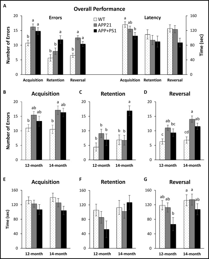
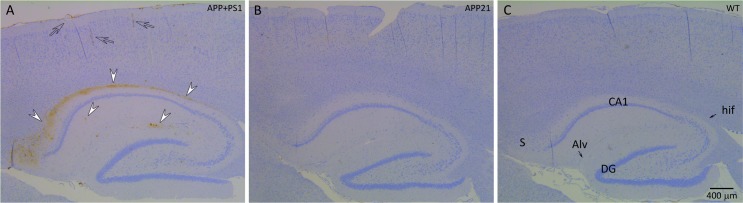
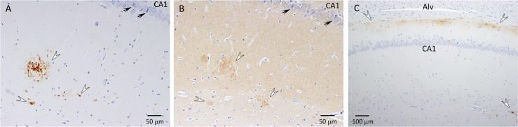
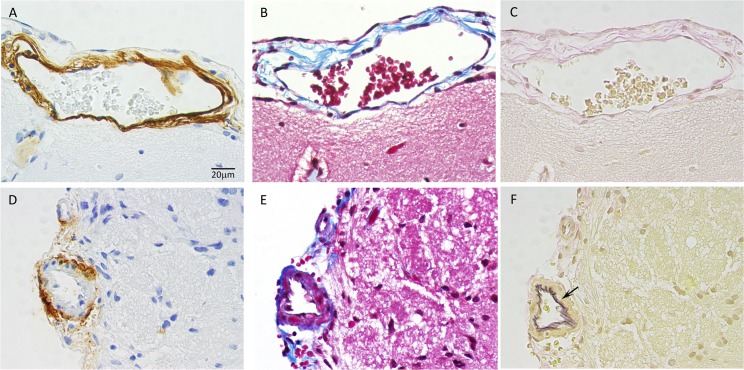
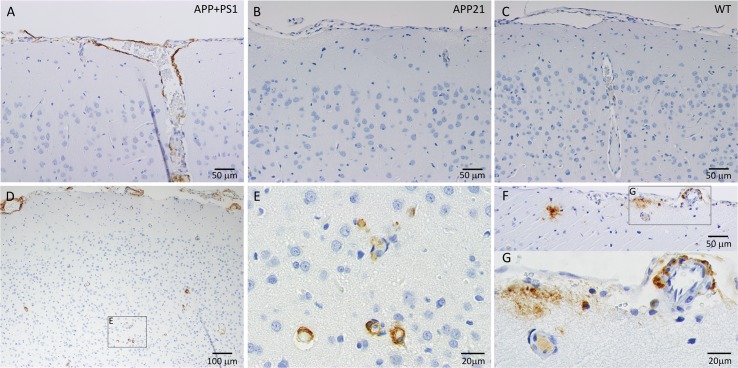
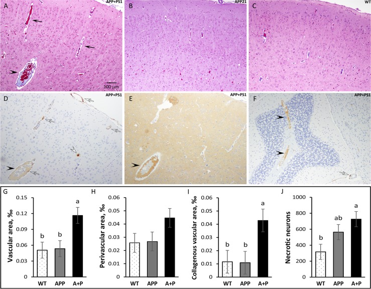
Transgenic rat models of Alzheimer's disease were used to examine differences in memory and brain histology. Double transgenic female rats (APP+PS1) over-expressing human amyloid precursor protein (APP) and presenilin 1 (PS1) and single transgenic rats (APP21) over-expressing human APP were compared with wild type Fischer rats (WT). The Barnes maze assessed learning and memory and showed that both APP21 and APP+PS1 rats made significantly more errors than the WT rats during the acquisition phase, signifying slower learning. Additionally, the APP+PS1 rats made significantly more errors following a retention interval, indicating impaired memory compared to both the APP21 and WT rats. Immunohistochemistry using an antibody against amyloid-β (Aβ) showed extensive and mostly diffuse Aβ plaques in the hippocampus and dense plaques that contained tau in the cortex of the brains of the APP+PS1 rats. Furthermore, the APP+PS1 rats also showed vascular changes, including cerebral amyloid angiopathy with extensive Aβ deposits in cortical and leptomeningeal blood vessel walls and venous collagenosis. In addition to the Aβ accumulation observed in arterial, venous, and capillary walls, APP+PS1 rats also displayed enlarged blood vessels and perivascular space. Overall, the brain histopathology and behavioral assessment showed that the APP+PS1 rats demonstrated behavioral characteristics and vascular changes similar to those commonly observed in patients with Alzheimer's disease.
Conflict of interest statement
Figures
Similar articles
-
Presenilin 1 transgene addition to amyloid precursor protein overexpressing transgenic rats increases amyloid beta 42 levels and results in loss of memory retention.BMC Neurosci. 2016 Jul 7;17(1):46. doi: 10.1186/s12868-016-0281-8. BMC Neurosci. 2016. PMID: 27388605 Free PMC article.
-
Lack of P-glycoprotein Results in Impairment of Removal of Beta-Amyloid and Increased Intraparenchymal Cerebral Amyloid Angiopathy after Active Immunization in a Transgenic Mouse Model of Alzheimer's Disease.Curr Alzheimer Res. 2017;14(6):656-667. doi: 10.2174/1567205013666161201201227. Curr Alzheimer Res. 2017. PMID: 27915995
-
The effect of focal brain injury on beta-amyloid plaque deposition, inflammation and synapses in the APP/PS1 mouse model of Alzheimer's disease.Exp Neurol. 2015 May;267:219-29. doi: 10.1016/j.expneurol.2015.02.034. Epub 2015 Mar 4. Exp Neurol. 2015. PMID: 25747037
-
Modeling Alzheimer's disease in transgenic mice: effect of age and of presenilin1 on amyloid biochemistry and pathology in APP/London mice.Exp Gerontol. 2000 Sep;35(6-7):831-41. doi: 10.1016/s0531-5565(00)00149-2. Exp Gerontol. 2000. PMID: 11053674 Review.
-
Metabolism of presenilin 1: influence of presenilin 1 on amyloid precursor protein processing.Neurobiol Aging. 1998 Jan-Feb;19(1 Suppl):S15-8. doi: 10.1016/s0197-4580(98)00026-8. Neurobiol Aging. 1998. PMID: 9562461 Review.
Cited by
-
Brain endothelial cell activation and dysfunction associate with and contribute to the development of enlarged perivascular spaces and cerebral small vessel disease.Histol Histopathol. 2024 Dec;39(12):1565-1586. doi: 10.14670/HH-18-792. Epub 2024 Jul 10. Histol Histopathol. 2024. PMID: 39051093 Review.
-
White matter inflammation and cognitive function in a co-morbid metabolic syndrome and prodromal Alzheimer's disease rat model.J Neuroinflammation. 2020 Jan 21;17(1):29. doi: 10.1186/s12974-020-1698-7. J Neuroinflammation. 2020. PMID: 31964387 Free PMC article.
-
Structural changes in cerebral microvasculature induced by ferroptosis contribute to blood-brain barrier disruption in Alzheimer's disease: an autopsy study.Alzheimers Dement. 2025 Apr;21(4):e70103. doi: 10.1002/alz.70103. Alzheimers Dement. 2025. PMID: 40189804 Free PMC article.
-
Blood-brain barrier leakage in Alzheimer's disease: From discovery to clinical relevance.Pharmacol Ther. 2022 Jun;234:108119. doi: 10.1016/j.pharmthera.2022.108119. Epub 2022 Jan 30. Pharmacol Ther. 2022. PMID: 35108575 Free PMC article. Review.
-
Zebrafish as a Potential Model for Neurodegenerative Diseases: A Focus on Toxic Metals Implications.Int J Mol Sci. 2023 Feb 8;24(4):3428. doi: 10.3390/ijms24043428. Int J Mol Sci. 2023. PMID: 36834835 Free PMC article. Review.
References
-
- Alzheimer’s Association. 2017 Alzheimer’s Disease Facts and Figures. Alzheimers Dement 2017;13:325–73.
-
- Vassar R. BACE1: the beta-secretase enzyme in Alzheimer's disease. J Mol Neurosci. 2004;23:105–14. doi: 10.1385/JMN:23:1-2:105 - DOI - PubMed
-
- Attems J. Sporadic cerebral amyloid angiopathy: pathology, clinical implications, and possible pathomechanisms. Acta Neuropathol. 2005;110:345–59. doi: 10.1007/s00401-005-1074-9 - DOI - PubMed
-
- Weller RO, Boche D, Nicoll JA. Microvasculature changes and cerebral amyloid angiopathy in Alzheimer's disease and their potential impact on therapy. Acta Neuropathol. 2009;118:87–102. doi: 10.1007/s00401-009-0498-z - DOI - PubMed
-
- Fanselow MS, Dong HW. Are the dorsal and ventral hippocampus functionally distinct structures? Neuron. 2010;65(1):7–19. doi: 10.1016/j.neuron.2009.11.031 - DOI - PMC - PubMed
Publication types
MeSH terms
Substances
Grants and funding
LinkOut - more resources
Full Text Sources
Other Literature Sources
Medical
