. 2018 Apr 11;14(4):e1007310.
doi: 10.1371/journal.pgen.1007310. eCollection 2018 Apr.

Effector gene birth in plant parasitic nematodes: Neofunctionalization of a housekeeping glutathione synthetase gene

Affiliations

Effector gene birth in plant parasitic nematodes: Neofunctionalization of a housekeeping glutathione synthetase gene

Catherine J Lilley et al. PLoS Genet. .

Abstract

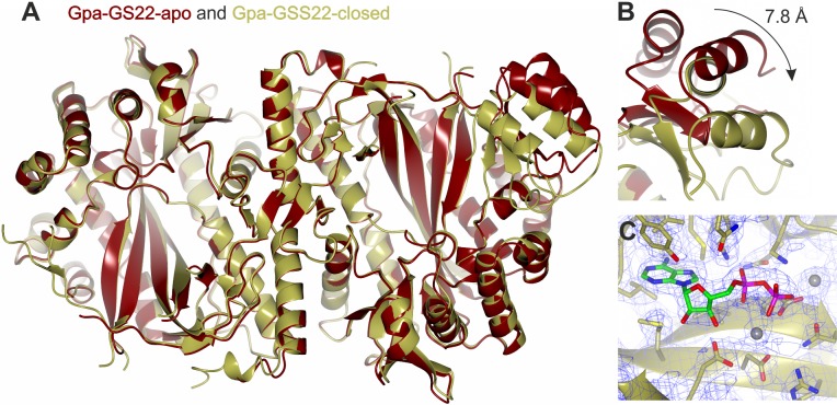
Plant pathogens and parasites are a major threat to global food security. Plant parasitism has arisen four times independently within the phylum Nematoda, resulting in at least one parasite of every major food crop in the world. Some species within the most economically important order (Tylenchida) secrete proteins termed effectors into their host during infection to re-programme host development and immunity. The precise detail of how nematodes evolve new effectors is not clear. Here we reconstruct the evolutionary history of a novel effector gene family. We show that during the evolution of plant parasitism in the Tylenchida, the housekeeping glutathione synthetase (GS) gene was extensively replicated. New GS paralogues acquired multiple dorsal gland promoter elements, altered spatial expression to the secretory dorsal gland, altered temporal expression to primarily parasitic stages, and gained a signal peptide for secretion. The gene products are delivered into the host plant cell during infection, giving rise to "GS-like effectors". Remarkably, by solving the structure of GS-like effectors we show that during this process they have also diversified in biochemical activity, and likely represent the founding members of a novel class of GS-like enzyme. Our results demonstrate the re-purposing of an endogenous housekeeping gene to form a family of effectors with modified functions. We anticipate that our discovery will be a blueprint to understand the evolution of other plant-parasitic nematode effectors, and the foundation to uncover a novel enzymatic function.


Conflict of interest statement

The authors have declared that no competing interests exist.

Figures

Fig 1
Fig 1. Two successive glutathione synthetase gene family expansions have occurred during the evolution of cyst and reniform nematode obligate biotrophic interactions.
A) A representation of a Bayesian phylogenetic tree based on a protein alignment of 180 glutathione synthetase (GS) like genes from eleven nematode species. The phylogeny is divided into three well supported Clades (node labels indicate support values for 2.5 million iterations, branch line width is scaled by support). Clade 1 (green) contains at least one sequence from each nematode species analysed. Clade 2 (blue) contains the first expansion of GS genes and exclusively comprises sequences from nematode species which spend the majority of their life within their plant host (and includes both migratory and sedentary endo-parasites). Clade 3 (red) contains the second, more recent and more diverse expansion and comprises cyst and reniform nematode genes only. Expression data for G. pallida, G. rostochiensis, R. reniformis, N. aberrans, H. avenae, and H. schachtii is shown adjacent to each sequence, as the fold-change between pre/non-parasitic and parasitic stages. See S2 Fig for a more detailed analysis of G. pallida GS-like gene expression across the life cycle. Black bars indicate those sequences that encode a canonical signal peptide for secretion. B) Schematic phylogeny of the phylum Nematoda [26, 72] with a key depicting the different modes of parasitism, and the species analysed: Lel–Longidorus elongatus; CelCaenorhabditis elegans; Bxy–Bursaphelenchus xylophilus; MinMeloidogyne incognita; PpePratylenchus penetrans; NabNacobbus aberrans; RreRotylenchulus reniformis; GpaGlobodera pallida; GroGlobodera rostochiensis; HavHeterodera avenae; HscHeterodera schachtii. Coloured bars indicate the GS Clades that those species contribute sequences to.
Fig 2
Fig 2. New GS-like paralogues are redeployed during parasitism as effectors.
A) Examples of spatial expression patterns (in situ hybridisation) of GS-like gene members of Clades 1, 2, and 3 (See S3 Fig for additional examples and negative controls). Globodera pallida Clade 1 and 2 GS genes are expressed in the intestine (white arrowheads). Clade 3 GS-like genes are specifically expressed in the dorsal gland cell (Dg) of G. pallida, G. rostochiensis and R. reniformis (left to right). Scale bars = 100 μm. B) An affinity-purified antibody raised against G. pallida Clade 3 GS-like effector Gpa-GSS17 recombinant protein was used for immunolocalisation (green fluorescence). Gpa-GSS17 is localised in the dorsal gland cell (Dg) of parasitic nematodes, the cytoplasmic gland extension and the ampulla at the base of the stylet where secretions accumulate prior to their release (top) Scale bars = 50 μm. The same native protein was localised within the syncytial feeding site (asterisks) induced by the nematode in a potato root (green fluorescence, bottom left). No such localisation was seen with the 2o antibody control (bottom right). Cell walls are stained blue. C) Western blot to determine antibody specificity. The anti-Gpa-GSS17 antibody specifically recognises Gpa-GSS17, and does not recognise other Clade 3 GS-like proteins tested, other plant-parasitic nematode GS proteins tested (Clades 1 and 2), nor the endogenous potato GS (St-GSS1).
Fig 3
Fig 3. Cyst nematode GS-like genes are re-purposed to carry out a novel function.
A) Purified protein for St-GSS1, Gpa-GSS1, 5, 12, 17, 22, 24 and 30 were tested for glutathione synthetase activity by measuring phosphate release from ATP in the presence of canonical substrates (γ-EC, glycine and ATP). To determine specific activity, rates are presented with subtraction of buffer controls in S2 Table. B) and C) Comparison of residues in the di-peptide binding pocket of Gpa-GSS22-closed and St-GSS-closed, with inset the variation at these positions in all other G. pallida Clade 3 GS-like effectors. B) In St-GSS1, the cysteine of the di-peptide substrate (γ-EC) is coordinated by the side chain of an arginine (top left), and the backbone of two serines (bottom left and bottom right). The arginine is conserved in Gpa-GSS22 and all GS-like effectors (inset). The two serines are not conserved in sequence in either Gpa-GSS22 or the remainder of Clade 3 (inset), but the equivalent residues are preferentially small and uncharged amino acids that do not vary greatly in the remainder of Clade 3 (inset). C) In St-GSS1, the glutamic acid of γ-EC is coordinated exclusively by side chain interactions with a number of charged residues. All of these residues are different in Gpa-GSS22, and these positions are highly variable across Clade 3 (inset).
Fig 4
Fig 4. The crystal structure of Globodera pallida GS-like effector Gpa-GSS22.
A) The crystal structure of Gpa-GSS22 is composed of a homodimeric molecule in both its open (apo (red)) and ADP-bound closed (gold) conformations. B) The two helix bundle that constitutes the ATP grasp fold undergoes a 7.8 Å conformational change on binding ADP to close over the active site. C) The presence of electron density (blue mesh) in the active site is consistent with a single ADP molecule and two magnesium ions per subunit.
Fig 5
Fig 5. Comparison of residues in the glutamate binding pocket of canonical GS with the same positions in nematode GS-like effectors.
The canonical arrangement in St-GSS1 surrounding the glutamic acid of γ-EC has been conserved for ~1 billion years of evolution in three kingdoms: Plantae (Solanum tuberosum St-GSS1-closed, PDB 5OES), Fungi (Saccharomyces cerevisiae, PDB 1M0T), and Animalia (Homo sapiens GSS1, PDB 2HGS). Both solved structures of GS-like effectors (Gpa-GSS22 and Gpa-GSS30) show a non-canonical arrangement that is highly unusual among Eukaryotes. Together with the conservation in the remainder of the di-peptide substrate binding pocket, and the conservation in the entire ATP binding pocket, this diversification likely indicates novel substrate usage.
Fig 6
Fig 6. The detection of novel thiols in syncytial feeding sites induced by cyst nematodes.
A) Free thiols, stained with ThiolTracker Violet, accumulate in the cytoplasm of syncytia induced by the cyst nematode Heterodera schachtii in Arabidopsis roots throughout the infection process (7, 14 and 21 days post infection). N indicates nematode. Scale bars = 100 μm. B) Optical cross section through the feeding site using the same stain. Arrows indicate partially dissolved cell wall. C) Analysis of low molecular weight thiols extracted from syncytia formed in potato by Globodera pallida (blue), control uninfected potato root (red), female nematodes (orange), and glutathione standard (black), by hydrophilic interaction liquid chromatography. Example plant-specific peaks are indicated with red arrows, syncytia-specific peaks with blue arrows, and a plant-specific peak increased in abundance in syncytia with a black arrow. D) The same sample used in panel C was separated at higher resolution using a shallower elution gradient. The peak at approximately 1.8 minutes in panel C (asterisk) corresponds to approximately 4 minutes in panel D (asterisk). This peak is absent in control roots, absent in nematode tissue, and highly abundant in syncytial feeding site material. No corresponding peak in the mass spectrum trace was identified (bottom).
Fig 7
Fig 7. Impairing plant glutathione synthetic capacity is independently detrimental to syncytium and cyst nematode development.
Both syncytia (A) and nematodes (B) are significantly smaller on pad2-1 mutant Arabidopsis roots than on wild-type (Student’s T-test p ≤ 0.001, n = 82 and 147 for syncytia and nematodes respectively). Error bars indicate standard error of the mean. (C) In both pad2-1 and wild-type roots, syncytium and nematode size significantly co-vary (Pearson’s correlation, p ≤ 0.05 and 0.001, n = 49 and 66 respectively), however the correlation is weak and most of the variation in nematode size (83–89%) is not explained by syncytium size. (D) Example images of unsuccessful nematode infection on pad2-1 roots (middle) and successful infection on both pad2-1 (bottom) and wild-type roots (top). Black and white arrows indicate syncytial and nematode boundaries respectively, blue arrows indicate an aborted syncytium and surrounding areas, scale bars indicate 100 μm. E) and F) In the presence of 1 mM BSO, syncytia are initiated but not properly maintained. Apoptosis, indicated by blue arrows and evidenced by propidium iodide staining (red), occurs in the local area of the syncytium (blue arrows) and spreads often non-distal to the site of infection; a similar, but more severe and frequent phenotype than observed in pad2-1 roots. Scale bars indicate 100 μm. (G) and (H) J2 stage nematodes incubated for the normal root invasion period (48 hours) on water agar plates containing 1 mM BSO are unaffected in (G) mortality (n = 68) or (H) motility (Mann-Whitney U Test, p = 0.408 n = 20).

References

    1. van Megen H, van den Elsen S, Holterman M, Karssen G, Mooyman P, Bongers T, et al. A phylogenetic tree of nematodes based on about 1200 full-length small subunit ribosomal DNA sequences. Nematology. 2009;11:927–50.
    1. Jones JT, Haegeman A, Danchin EGJ, Gaur HS, Helder J, Jones MGK, et al. Top 10 plant-parasitic nematodes in molecular plant pathology. Mol Plant Pathol. 2013;14(9):946–61. doi: 10.1111/mpp.12057 - DOI - PMC - PubMed
    1. Blaxter M, Koutsovoulos G. The evolution of parasitism in Nematoda. Parasitology. 2015;142(S1):S26–S39. - PMC - PubMed
    1. Holterman M, Karegar A, Mooijman P, van Megen H, van den Elsen S, Vervoort MT, et al. Disparate gain and loss of parasitic abilities among nematode lineages. PLoS ONE. 2017;12(9):e0185445 doi: 10.1371/journal.pone.0185445 - DOI - PMC - PubMed
    1. Holterman M, Karssen G, van den Elsen S, van Megen H, Bakker J, Helder J. Small Subunit rDNA-based phylogeny of the Tylenchida sheds light on relationships among some high-impact plant-parasitic nematodes and the evolution of plant feeding. Phytopathology. 2009;99(3):227–35. doi: 10.1094/PHYTO-99-3-0227 - DOI - PubMed

Publication types

Substances