Review
. 2018 Jun 4:20:403-429.
doi: 10.1146/annurev-bioeng-062117-121113. Epub 2018 Apr 11.

Physiology and Engineering of the Graded Interfaces of Musculoskeletal Junctions

Affiliations
Review

Physiology and Engineering of the Graded Interfaces of Musculoskeletal Junctions

Edward D Bonnevie et al. Annu Rev Biomed Eng. .

Abstract

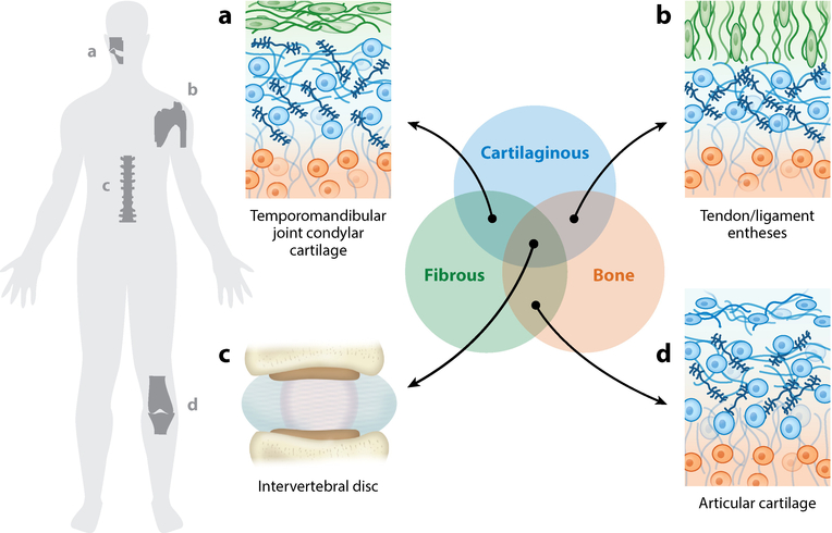
The connective tissues of the musculoskeletal system can be grouped into fibrous, cartilaginous, and calcified tissues. While each tissue type has a distinct composition and function, the intersections between these tissues result in the formation of complex, composite, and graded junctions. The complexity of these interfaces is a critical aspect of their healthy function, but poses a significant challenge for their repair. In this review, we describe the organization and structure of complex musculoskeletal interfaces, identify emerging technologies for engineering such structures, and outline the requirements for assessing the complex nature of these tissues in the context of recapitulating their function through tissue engineering.

Keywords: articular cartilage; bone; gradients; interfaces; intervertebral disc; temporomandibular joint; tendon.


Figures

Figure 1
Figure 1
Graded material connections are universal in the musculoskeletal system. In a simplified context, connective tissues can be grouped into cartilaginous (blue), fibrous (green), and calcified (i.e., bone) (orange) tissue types. The connections between these dissimilar materials are present throughout the body, and their mechanical importance and diversity pose obstacles for tissue engineering functional replacements. Examples include (a) the graded structure from the surface to the bone of the condylar cartilage in the temporomandibular joint; (b) the interface between tendon/ligament and bone; (c) the multiple interfaces among fibrous, cartilaginous, and bony tissue represented in the intervertebral disc of the spine; and (d) the interface between articular cartilage and bone (i.e., enthesis).
Figure 2
Figure 2
Articular cartilage lines the ends of long bones (highlighted in red). This tissue exhibits a depth-dependent (a) biochemical and (b) mechanical profile—a compliant superficial zone transitions to stiffer and proteoglycan-rich middle and deep zones (2, 3, 26, 32). (c) Cellular, collagen (blue), and proteoglycan (navy) content evolves over the depth from the articular surface to subchondral bone (orange); between the deep zone and subchondral bone, stiffness increases rapidly in a mineralization-dependent manner (5). (d) Fourier transform infrared spectroscopic mapping of healthy and arthritic cartilage reveals that this depth-dependent profile is disrupted in disease and degeneration (colors indicate increasing content and organization from blue to red). Abbreviations: DZ, deep zone; OA, osteoarthritis; SZ, superficial zone. Panel d adapted with permission from Reference .
Figure 3
Figure 3
Advances in generating and assessing graded cartilage structures. (a) Photocrosslinkable materials have emerged as a tool to generate material gradients. Steinmetz et al. (51) tuned polymerization times to demonstrate that a gradient between two materials can be controlled to produce (left) gradual to (right) abrupt material gradients. (b) Lu et al. (62) revealed that a composite material implant (dashed boxes) generated native-like tissue when coupled with cartilage- and bone-promoting growth factors (IGF-1 and BMP-2, respectively). (c) Griffin et al. (58) utilized elastography techniques to determine spatially varying mechanical properties of native and engineered tissue (26, 58). By analyzing the strain-mapped images of undeformed tissue before and after application of shear, the authors found that implanted cartilage (MACI®; blue) did not recapitulate native-like, graded properties (control; gray) even after 53 weeks of implantation. Spectroscopic techniques such as (d) Raman spectroscopy and (e) traditional histology enable quantitative analysis of the spatially varying biochemical content of native and engineered tissue. Abbreviations: BMP, bone morphogenetic protein; IGF, insulin-like growth factor. Panel a adapted from Reference with permission. Panel b adapted from Reference with permission. Panel c adapted from Reference with permission. Panels d and e adapted from Reference with permission; these are unofficial adaptations from an article that appeared in an ACS publication. ACS has not endorsed the content of these adaptations or the context of their use.
Figure 4
Figure 4
Tendon and ligament entheses attach fibrous tissue to bone, e.g., the Achilles tendon (highlighted in red). These structures represent a complex interface with gradation in (a) extracellular matrix components that functionally correspond to (b) location-dependent mechanical properties. Of note is the inclusion of a compliant region (gray) between the fibrous tissue (green) and bone (orange) (8, 74). (c) This compliant inclusion is fibrocartilaginous in nature and exhibits spatially varying collagen makeup that shifts from type I (green) to type II (blue) collagen before transitioning to bone (orange). (d) Fourier transform infrared spectroscopic mapping of the enthesis reveals this spatial evolution of proteoglycans, collagen, and mineral content in the transition from fibrous tissue to bone. Abbreviation: a.u., arbitrary units. Panel d adapted from Reference with permission.
Figure 5
Figure 5
(a) Electrospinning of synthetic polymeric materials, where a charged polymeric solution is collected onto a grounded substrate, is a promising technique to fabricate fibrous tissues with defined organization when deposited onto a spinning mandrel with surface velocity (V). (b) Aligned, fibrous materials can also be synthesized using natural materials such as collagen coupled with cell-based remodeling. Cell-mediated compaction of these gels has emerged as a powerful tool to instill organization into gels with predefined boundary conditions. (c,d) Mineralization of electrospun scaffolds can be used to mimic the graded material properties of the tendon enthesis. Li et al. (103) demonstrated that introducing a mineralization gradient within a scaffold (c) provides spatial gradation of mechanical properties, as revealed by strain mapping (d). Panel a adapted from Reference with permission. Panels c and d adapted from Reference with permission.
Figure 6
Figure 6
The cartilage of the temporomandibular condylar cartilage (TMJ; highlighted in red) is distinct from typical articular cartilage due to the inclusion of a prominent fibrous superficial zone. This tissue exhibits (a) a biochemical gradient and (b) a mechanical profile where (c) a fibrous (green) type I collagen–rich superficial zone is stiff in tension compared with the cartilaginous middle and deep zones (blue), which then transition to comparatively rigid bone (orange). (d) The superficial zone exhibits location-dependent orientation and maintains a defined anterior–posterior orientation of type I collagen fibers. (e) Model-based evidence suggests that the increased tensile properties of the superficial zone shield the deeper cartilaginous tissue from elevated matrix stresses. (f,g) Efforts to engineer the TMJ condylar cartilage include methods to produce defined biochemical gradients in engineered composites (111). (f) Dormer et al. (111) revealed that introducing a gradient of biochemical cues within a tissue-engineered construct promoted both cartilage (Safranin O) and bone (Alizarin Red) formation. (g) Hollister et al. (112) focused on the reconstruction of the mandible using three-dimensional (3D) printing techniques. Abbreviation: CT, computed tomography. Panel d adapted from Reference with permission. Panel e adapted from Reference with permission. Panels f and g adapted from References and , respectively, with permission.
Figure 7
Figure 7
The intervertebral disc maintains a composite graded structure with connections among cartilaginous, fibrous, and bone tissues. Variation in material connections exists along orthogonal axes. In the coronal plane schematic (c), the inner region of the disc, the nucleus pulposus (NP), has a gel-like morphology that is rich in proteoglycans. This region transitions to a dense fibrous tissue at its lateral periphery, the annulus fibrosus (AF). In the superior and inferior directions, the NP transitions first to a cartilaginous end plate, which then transitions to bone. The transition from AF to NP maintains (a) gradations in tissue compositions that relate to (d) location-dependent loading patterns. Due to the composite makeup and ordered structure of the disc, the NP experiences compressive loading whereas the AF experiences tensile loading. As the NP transitions to cartilage and then to bone, there also exist gradations in (b) biochemical makeup and (e) structure. Panel d adapted from Reference with permission. Panel e adapted from Reference with permission.
Figure 8
Figure 8
(a) (Top) Creating layered sheets of electrospun nanofibrous scaffolds (NFS) recapitulates the lamellar collagen structure of the native annulus fibrosus (AF), maintaining the angle-ply structure as revealed by polarized light microscopy. (Middle) By winding these NFS into multilayer lamellar structures, and incorporating this engineered AF tissue with a central hydrogel, Nerurkar et al. (137) developed disc-like composite structures. (Bottom) Following culture, microscopy techniques revealed that cells remain viable and deposit substantial extracellular matrix, leading to native disc-like structure and function. (b) (Top) Bowles et al. (152) demonstrated that discs formed through cell-mediated, self-assembled collagen compaction show collagen and cellular reorganization around the nucleus, reminiscent of the aligned fibrous structure of the AF. (Middle) Through the incorporation of both AF and nucleus pulposus (NP) cells in a tissue engineering approach, disc-like composites have been fabricated by allowing a collagen gel to contract around an inner alginate gel core. (Bottom) Moriguchi et al. (138) showed that this technique can recapitulate the structure of canine discs. Abbreviation: IVD, intervertebral disc. Panel a adapted from References and with permission. Panel b adapted from References and with permission.

Similar articles

Cited by

References

    1. Delale F 1984. Stress singularities in bonded anisotropic materials. Int. J. Solids Struct. 20:31–40
    1. Jeffery AK, Blunn GW, Archer CW, Bentley G. 1991. Three-dimensional collagen architecture in bovine articular cartilage. J. Bone Joint Surg. Br. 73:795–801 - PubMed
    1. Klein TJ, Chaudhry M, Bae WC, Sah RL. 2007. Depth-dependent biomechanical and biochemical properties of fetal, newborn, and tissue-engineered articular cartilage. J. Biomech. 40:182–90 - PubMed
    1. Buckley MR, Gleghorn JP, Bonassar LJ, Cohen I. 2008. Mapping the depth dependence of shear properties in articular cartilage. J. Biomech. 41:2430–37 - PubMed
    1. Ferguson VL, Bushby AJ, Boyde A. 2003. Nanomechanical properties and mineral concentration in articular calcified cartilage and subchondral bone. J. Anat. 203:191–202 - PMC - PubMed

Publication types

LinkOut - more resources