. 2018 Apr 10;23(2):485-498.
doi: 10.1016/j.celrep.2018.03.034.

ADAMTS9-Regulated Pericellular Matrix Dynamics Governs Focal Adhesion-Dependent Smooth Muscle Differentiation

Affiliations

ADAMTS9-Regulated Pericellular Matrix Dynamics Governs Focal Adhesion-Dependent Smooth Muscle Differentiation

Timothy J Mead et al. Cell Rep. .

Abstract

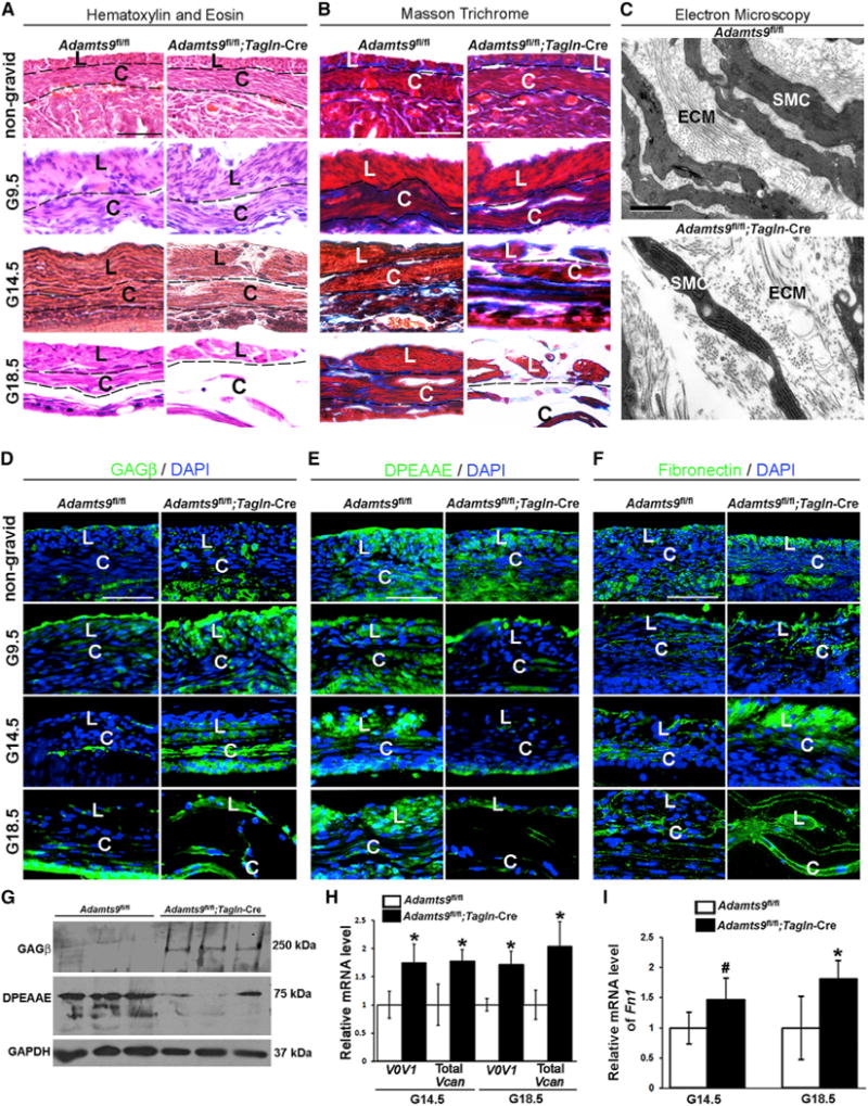
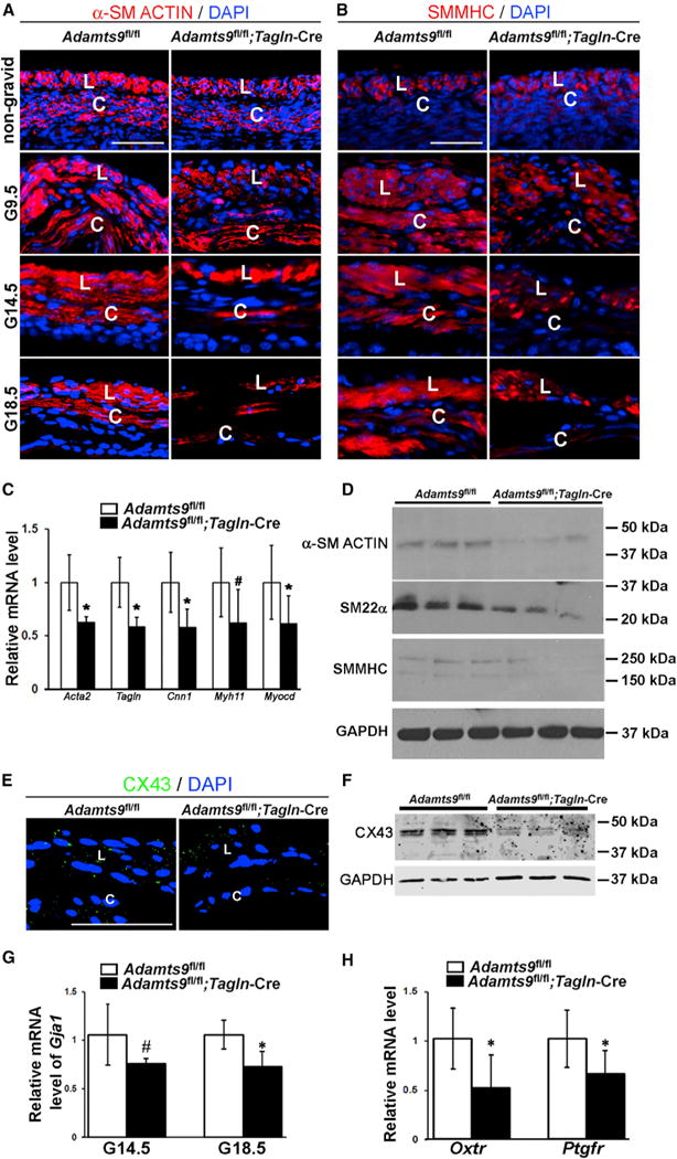
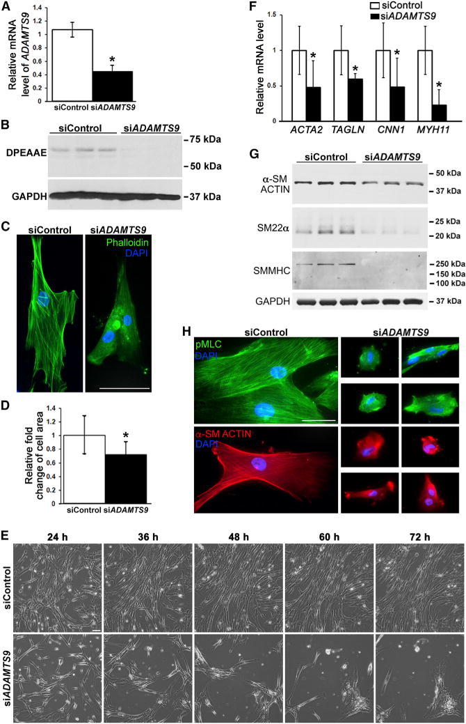
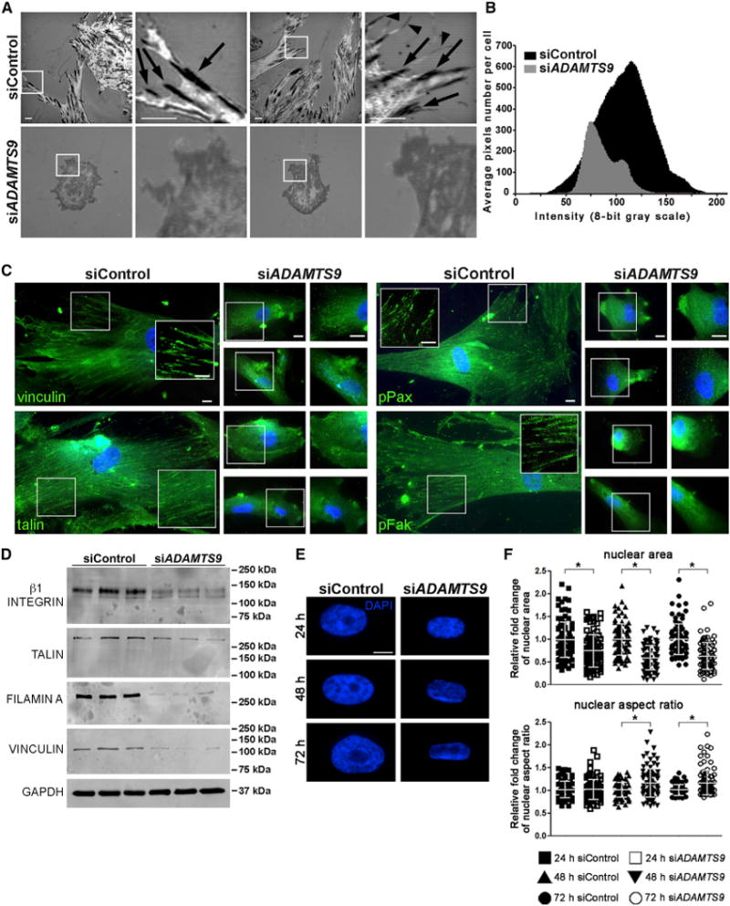
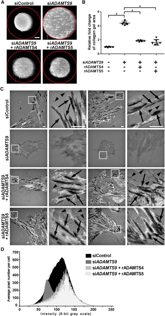
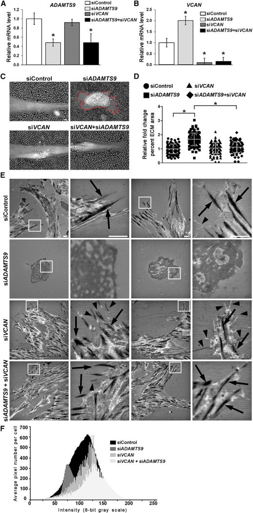
Focal adhesions anchor cells to extracellular matrix (ECM) and direct assembly of a pre-stressed actin cytoskeleton. They act as a cellular sensor and regulator, linking ECM to the nucleus. Here, we identify proteolytic turnover of the anti-adhesive proteoglycan versican as a requirement for maintenance of smooth muscle cell (SMC) focal adhesions. Using conditional deletion in mice, we show that ADAMTS9, a secreted metalloprotease, is required for myometrial activation during late gestation and for parturition. Through knockdown of ADAMTS9 in uterine SMC, and manipulation of pericellular versican via knockdown or proteolysis, we demonstrate that regulated pericellular matrix dynamics is essential for focal adhesion maintenance. By influencing focal adhesion formation, pericellular versican acts upstream of cytoskeletal assembly and SMC differentiation. Thus, pericellular versican proteolysis by ADAMTS9 balances pro- and anti-adhesive forces to maintain an SMC phenotype, providing a concrete example of the dynamic reciprocity of cells and their ECM.

Keywords: extracellular matrix; focal adhesion; interference reflection microscopy; metalloprotease; myometrium; parturition; proteoglycan; proteolysis; smooth muscle; uterus.


Conflict of interest statement

DECLARATION OF INTERESTS

The authors declare no competing interests.

Figures

Figure 1
Figure 1. Altered Myometrial Structure and Versican and Fibronectin Dynamics in Adamts9-Deficient Myometrium
(A) H&E stain shows myometrial disruption, with increased intercellular space between uterine SMCs in gestational age (G) 14.5 days and G18.5 Adamts9fl/fl;Tagln-Cre uterus. (B) Masson trichrome stain shows reduced collagen staining (blue) in G18.5 Adamts9fl/fl;Tagln-Cre myometrium. (C) TEM at G18.5 shows increased intercellular distance between SMCs, sparse collagen fiber bundles, and abundant amorphous non-collagenous matrix in Adamts9fl/fl;Tagln-Cre myometrium. (D and E) Immunofluorescence showing versican accumulation (anti-GAGβ, green; nuclei stained blue by DAPI) from G9.5 to G18.5 (D) and reduced ADAMTS-cleaved versican (anti-DPEAAE, green) (E) in 3-week-old and gravid Adamts9fl/fl;Tagln-Cre myometrium. (F) Increased fibronectin staining (green; nuclei stained blue by DAPI) is seen in non-gravid and gravid Adamts9fl/fl;Tagln-Cre myometrium compared with control. (G) Representative western blots show increased versican (anti-GAGβ) and reduced cleaved versican (anti-DPEAAE) in three Adamts9fl/fl;Tagln-Cre uteri relative to their control littermates. (H) qRT-PCR shows increased Vcan mRNA in Adamts9fl/fl;Tagln-Cre uterus. n = 3. *p ≤ 0.01. V0V1 denotes the two major extra-neural splice variants V0 and V1. (I) qRT-PCR shows Fn1 mRNA is increased in the mutant uterus. n = 4. *p ≤ 0.01, #p ≤ 0.05. L, longitudinal myometrial layer; C, circular myometrial layer; ECM, extracellular matrix; SMC, smooth muscle cell. Scale bars, 50 μm in (A), (B), and (D)–(F) and 1 μm in (C). Error bars indicate SEM. See also Figures S1–S4.
Figure 2
Figure 2. Adamts9 Deletion Reduces Contractile Protein Content of Myometrial SMCs and Interferes with Gap Junction Formation and Expression of Parturition Hormone Receptors
(A) Reduced staining intensity of smooth muscle a-actin (SMA, red; nuclei, blue) in non-gravid and gravid Adamts9fl/fl;Tagln-Cre myometrium. (B) Reduced smooth muscle myosin heavy chain staining (SMMHC, red; nuclei, blue) in gravid Adamts9fl/fl;Tagln-Cre myometrium. (C) qRT-PCR shows reduction of Acta2, Tagln, Cnn1, Myh11, and Myocd mRNAs in G18.5 Adamts9fl/fl;Tagln-Cre myometrium, compared with control. n = 4. *p ≤ 0.01, #p ≤ 0.05. (D) Representative western blots for SMA, SM22a, and SMMHC show their reduction in three independent G18.5 gravid Adamts9fl/fl;Tagln-Cre uteri relative to littermate controls. GAPDH was used as a control. (E) Fewer anti-Cx43 stained speckles (green) indicate fewer gap junctions in Adamts9fl/fl;Tagln-Cre myometrium at G18.5. (F) Representative western blot shows reduced Cx43 in three pairs of G18.5 Adamts9fl/fl;Tagln-Cre and Adamts9fl/fl uterus controls. GAPDH was used as a control. (G) Gja1 mRNA levels are reduced in G14.5 and G18.5 Adamts9fl/fl;Tagln-Cre uterus. n = 4. *p ≤ 0.01, #p ≤ 0.05. (H) Reduced Oxtr and Ptgfr mRNA in G18.5 Adamts9fl/fl;Tagln-Cre uterus. n = 4. *p ≤ 0.01. Scale bars, 50 μm. Error bars indicate SEM. See also Figures S2–S4.
Figure 3
Figure 3. ADAMTS9 Knockdown Alters HUSMC Shape, Reduces Versican Proteolysis, and Impairs Contractile Protein Expression
(A) qRT-PCR shows reduced ADAMTS9 mRNA in siADAMTS9-transfected HUSMCs. n = 3. *p ≤ 0.01. (B) Western blot using anti-DPEAAE antibody shows reduced ADAMTS-cleaved versican in the medium of siADAMTS9-transfected cells. GAPDH was used as a loading control. (C and D) ADAMTS9 knockdown reduces stress fiber formation (phalloidin-FITC, green) and impairs cell spreading on plastic (C), quantitatively represented as a reduction of surface area in (D). n = 20. *p ≤ 0.01. (E) Still images from phase contrast microscopy time-lapse videos (see Videos S1 and S2) of HUSMCs on tissue-culture plastic. In contrast to well-spread control siRNA transfected cells, the siADAMTS9-transfected cells rounded and clumped together 24 hr after transfection until most had detached from the substratum. Contrast with Figure S6B, in which the siADAMTS9-transfected cells are plated on fibronectin. (F–H) qRT-PCR (F), western blot (G), and immunofluorescence (H) show reduction of smooth muscle contractile gene mRNAs, protein, and staining in HUSMC after Adamts9 knockdown. n = 3. GAPDH was used as a loading control in (G).*p ≤ 0.01. Scale bars, 50 μm. Error bars indicate SEM. See also Figures S5 and S6 and Videos S1 and S2.
Figure 4
Figure 4. ADAMTS9 Knockdown Eliminates Focal Adhesions in HUSMC and Alters Nuclear Dimensions
(A) IRM identifies focal adhesions as dark linear regions (arrows) as well as filopodia (arrowheads) at the cell-substratum interface in control siRNA-transfected HUSMC cultures, whereas focal adhesions are poorly formed or absent in siADAMTS9-transfected HUSMC (shown in gray, indicating non-focal close contacts). IRM imaging was done after plating on glass. (B) IRM image quantification of siControl cells showed a bell-shaped spread of very dark (0) to very light (255) signal, while siADAMTS9-transfected cells have overall dominance of gray signal intensity with fewer pixels per cell, indicating reduced cell area and an absence of very dark and very bright signal. n = 12 cells. (C) Reduced staining intensity of focal adhesion components vinculin, talin, phospho (p)-paxillin, and p-FAK (green, nuclei, blue) in siADAMTS9-transfected HUSMCs plated on plastic. The boxed regions with white borders are shown as higher magnification insets or alongside. (D) Western blots of siADAMTS9-transfected cells show decreased β1 integrin, talin, filamin A, and vinculin, compared with control siRNA-transfected cells. GAPDH is shown as a loading control. (E and F) DAPI-stained nuclei (E) from control or ADAMTS9 siRNA-transfected HUSMC plated on plastic were imaged to quantify the nuclear area and aspect ratio, which are significantly different in the two groups (F). Scale bars, 10 μm. Error bars indicate SEM. See also Videos S3 and S4.
Figure 5
Figure 5. siADAMTS9 Impairs Cellular Contractility and Exogenous ADAMTS4 and ADAMTS5 Restore siADAMTS9-Induced Changes in Cell Shape, Focal Adhesions, and Contractility
(A) Representative collagen gel contraction assays demonstrate reduced contractility of siADAMTS9-transfected HUSMCs and restoration of contractility after treatment with recombinant ADAMTS4 or ADAMTS5. The red circle indicates the initial size of the collagen gel. (B) Quantification of gel area demonstrates reduced gel contraction by siADAMTS9-transfected cells and its reversal by addition of exogenous ADAMTS4 or ADAMTS5. Each data point in the plot represents a single gel. n = 6. *p ≤ 0.01. (C) IRM shows that ADAMTS9 siRNA severely impairs focal adhesions, seen as dark, linear regions (arrows) in control siRNA-transfected cells and filopodia (arrowheads). These are restored by exogenous ADAMTS4 and ADAMTS5. HUSMCs were plated on glass for IRM. (D) Quantification of grayscale density in IRM-generated images from n = 10 ADAMTS4 and ADAMTS5-treated cells shows shifts to the left and right that are indicative of normal adhesive interactions and increased pixel numbers indicative of cell spreading. The data for control and siADAMTS9-treated cells are identical to that shown in Figure 4B. Scale bars, 1 mm in (A) and 10 μm in (C). Error bars indicate SEM. See also Figure S6.
Figure 6
Figure 6. Concurrent Inactivation of VCAN in siADAMTS9-Transfected Cells Restores Normal Pericellular Matrix Dimensions and Formation of Focal Adhesions
For a Figure360 author presentation of Figure 6, see https://doi.org/10.1016/j.celrep.2018.03.034). (A and B) qRT-PCR illustrates effective knockdown of ADAMTS9 (A) and VCAN (B). Note the statistically significant increase of VCAN RNA upon ADAMTS9 knockdown. *p ≤ 0.01. (C) Erythrocyte exclusion assay demonstrates increased pericellular matrix around siADAMTS9-transfected HUSMCs (red line). This is restored to normal by siVCAN. (D) Quantification of pericellular matrix area demonstrates a significant increase in siADAMTS9-transfected cells, which is reversed by siVCAN. n = 100. *p ≤ 0.01. (E) IRM shows that co-silencing of VCAN and ADAMTS9 restores focal adhesions (arrows) and filopodia (arrowheads) reduced in siADAMTS9-transfected cells. HUSMCs were plated on glass for IRM. (F) Quantification of grayscale density in IRM-generated images from n = 10 cells shows shifts to the left and right that are indicative of normal adhesive interactions and increased pixel numbers indicative of increased cell area. The data for control and siADAMTS9-transfected cells are identical to that shown in Figure 4B. Scale bars, 10 μm. Error bars indicate SEM. See also Figure S6.

Similar articles

Cited by

References

    1. Abercrombie M, Dunn GA. Adhesions of fibroblasts to substratum during contact inhibition observed by interference reflection microscopy. Exp Cell Res. 1975;92:57–62. - PubMed
    1. Benz BA, Nandadasa S, Takeuchi M, Grady RC, Takeuchi H, LoPilato RK, Kakuda S, Somerville RPT, Apte SS, Haltiwanger RS, Holdener BC. Genetic and biochemical evidence that gastrulation defects in Pofut2 mutants result from defects in ADAMTS9 secretion. Dev Biol. 2016;416:111–122. - PMC - PubMed
    1. Bidwell MC, Eitzman BA, Walmer DK, McLachlan JA, Gray KD. Analysis of messenger ribonucleic acid and protein for the ligands and receptors of the platelet-derived growth factor signaling pathway in the placenta, extraembryonic membranes, and uterus during the latter half of murine gestation. Endocrinology. 1995;136:5189–5201. - PubMed
    1. Bissell MJ, Aggeler J. Dynamic reciprocity: how do extracellular matrix and hormones direct gene expression? Prog Clin Biol Res. 1987;249:251–262. - PubMed
    1. Bonnans C, Chou J, Werb Z. Remodelling the extracellular matrix in development and disease. Nat Rev Mol Cell Biol. 2014;15:786–801. - PMC - PubMed

MeSH terms