. 2018 Apr 10;23(2):555-567.
doi: 10.1016/j.celrep.2018.03.062.

Environmental Enrichment and Social Isolation Mediate Neuroplasticity of Medium Spiny Neurons through the GSK3 Pathway

Affiliations

Environmental Enrichment and Social Isolation Mediate Neuroplasticity of Medium Spiny Neurons through the GSK3 Pathway

Federico Scala et al. Cell Rep. .

Abstract

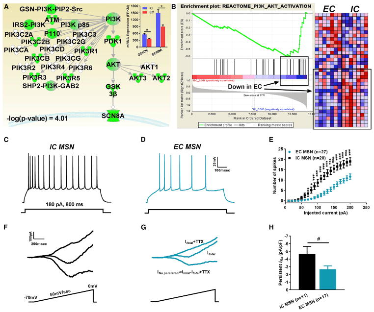
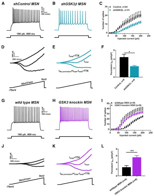
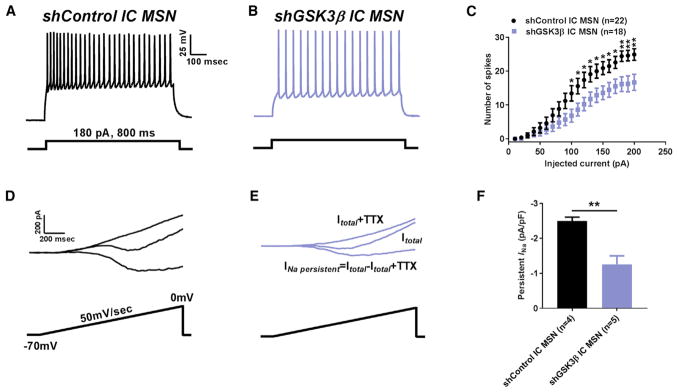
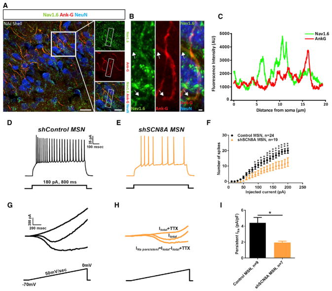
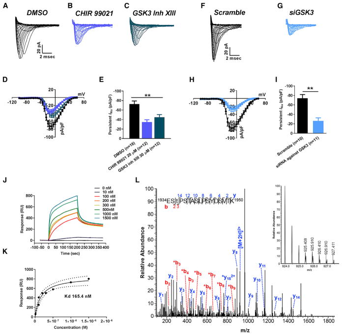
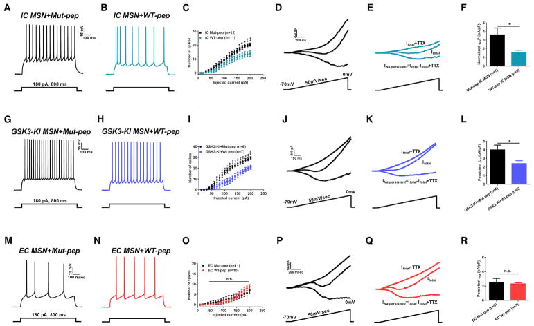
Resilience and vulnerability to neuropsychiatric disorders are linked to molecular changes underlying excitability that are still poorly understood. Here, we identify glycogen-synthase kinase 3β (GSK3β) and voltage-gated Na+ channel Nav1.6 as regulators of neuroplasticity induced by environmentally enriched (EC) or isolated (IC) conditions-models for resilience and vulnerability. Transcriptomic studies in the nucleus accumbens from EC and IC rats predicted low levels of GSK3β and SCN8A mRNA as a protective phenotype associated with reduced excitability in medium spiny neurons (MSNs). In vivo genetic manipulations demonstrate that GSK3β and Nav1.6 are molecular determinants of MSN excitability and that silencing of GSK3β prevents maladaptive plasticity of IC MSNs. In vitro studies reveal direct interaction of GSK3β with Nav1.6 and phosphorylation at Nav1.6T1936 by GSK3β. A GSK3β-Nav1.6T1936 competing peptide reduces MSNs excitability in IC, but not EC rats. These results identify GSK3β regulation of Nav1.6 as a biosignature of MSNs maladaptive plasticity.

Keywords: GSK3β; Nav1.6; enriched environment; isolated condition; medium spiny neurons; neuronal firing; persistent sodium current; plasticity; reward pathway.


Conflict of interest statement

DECLARATION OF INTERESTS

The authors declare no competing interests.

Figures

Figure 1
Figure 1. Unbiased Transcriptomic Screening Identifies GSK3β and SCN8A (Nav1.6) as EC-Sensitive Protecting Genes in the NAc
(A) Custom IPA pathway depicting EC/IC-regulated transcripts; RNA-seq analysis (top right inset) of GSK3β and SCN8A mRNA in EC versus IC conditions (Zhang et al., 2016b). (B) GSEA enrichment plot of PI3K/Akt/GSK3 Reactome pathway and corresponding heatmap in EC versus IC conditions (Zhang et al., 2016b). (C–E) Representative traces of APs in MSNs from IC rats (C), EC rats (D), and input-output curves (E). (F–H) Representative traces of MSN INaP from IC rats (F), EC rats (G), and bar graph (H). Data are represented as mean ± SEM. *p < 0.05, **p < 0.01, ***p < 0.005 with Student’s t test. #p < 0.05 with Mann-Whitney test.
Figure 2
Figure 2. Intrinsic Firing and Na+-Persistent Current of MSNs Are Bi-directionally Controlled by the GSK3 Pathway
(A–C) Representative traces of APs in NAc MSNs from shControl rats (A), shGSK3β rats (B), and input-output curves (C). (D–F) Representative traces of MSN INaP from shControl rats (D), shGSK3β rats (E), and bar graph (F). (G–I) Representative traces of APs in NAc MSNs from wild-type mice (G), GSK3 knockin mice (H), and input-output curves (I). (J–L) Representative traces of MSN INaP from wild-type mice (J), GSK3 KI mice (K), and bar graph (L). Data are represented as mean ± SEM. *p < 0.05, **p < 0.01, ***p < 0.005 with Student’s t test.
Figure 3
Figure 3. In Vivo Genetic Silencing of GSK3β Prevents Maladaptive Plasticity of MSNs
(A–C) Representative traces of APs MSNs from shControl rats (A), shGSK3β-expressing IC rats (B), and corresponding input-output curves (C). (D–F) Representative traces of MSNs INaP from shControl rats (D), shGSK3β IC rats (E), and bar graph (F). Data are represented as mean ± SEM. *p < 0.05, **p < 0.01 with Student’s t test.
Figure 4
Figure 4. Nav1.6 Is a Molecular Determinant of Intrinsic Firing in MSNs
(A) Confocal imaging of Nav1.6 (green), NeuN (blue), and ankyrin-G (red) at the axonal initial segment of neurons in the NAc. (B) Zoom inset of (A), arrows indicate starting and end points of axon initial segment (AIS). Scale bar, 1 μm. (C) Profile of Nav1.6 (green) channels and Ank (red) immunofluorescence intensity line scans along the AIS region in MSN. (D–F) Representative traces of APs in MSNs from shControl (D), shSCN8A (E), and corresponding input-output curves (F). (G–I) Representative traces of MSNs from shControl (G) and shSCN8A (F), respectively, and bar graphs (I). Data are represented as mean ± SEM. *p < 0.05, **p < 0.01, ***p < 0.005; Student’s t test.
Figure 5
Figure 5. In Vitro Studies of Functional Interaction between Nav1.6-Encoded Currents and GSK3
(A–C) Representative traces of transient INa recorded from Nav1.6-HEK293 cells treated with DMSO (A), CHIR 99021 (B), and GSK3 inhibitor XIII (C). (D and E) Nav1.6 current-voltage relationship of DMSO or CHIR 99021 or GSK3 inhibitor XIII treatment (D), and peak current density at −10 mV voltage step (E). (F and G) Representative traces of transient INa recorded from Nav1.6-HEK293 cells treated with siScramble (F) and siGSK3 (G). (H and I) Nav1.6 current-voltage relationship of siScramble versus siGSK3 (H), and peak current density at −10 mV voltage step (I). (J) Representative SPR sensorgram of GSK3β binding to Nav1.6 C-tail. (K) SPR fitting curve of GSK3β binding with Nav1.6 C-tail. (L) Higher energy collisional dissociation (HCD) fragmentation spectrum of the phosphopeptide EStPSTASLPSYDSVTK, encompassing residues 1934–1950 of the Nav1.6 C terminus. The presence of non-phosphorylated b2 (theoretical m/z of 217.08, observed m/z of 217.08) and y14 (theoretical m/z of 1,452.72, observed m/z of 1,452.72) ions along with phosphorylated b3 (theoretical m/z of 398.10, observed m/z of 398.10) confirms T1936 as the site of phosphorylation. The parent ion was fragmented with a co-eluting peptide within the ±2 Da window (inset) resulting in the mixed spectrum shown here. t, phosphothreonine; *, fragment with loss of phosphoric acid). Data are represented as mean ± SEM. *p < 0.05, **p < 0.01; Student’s t test.
Figure 6
Figure 6. Nav1.6-Based Peptide Restores Maladaptive Plasticity in IC Rats and GSK3 Knockin Mice
(A–C) Representative traces of APs in NAc MSN from IC rats treated with Mut-pep (A), WT-pep (B), and input-output curves (C). (D–F) Representative traces of INaP in NAc MSNs from IC rats treated with Mut-pep (D), WT-pep (E), and bar graph (F). (G–I) Representative traces of APs in NAc MSN from GSK3-KI with Mut-pep (G), WT-pep (H), and input-output curves (I). (J–L) Representative traces of INaP in NAc MSN from GSK3-KI treated with Mut-pep (J) and WT-pep (K), respectively, and bar graph (L). (M–O) Representative traces of APs in NAc MSN from EC rats treated with Mut-pep (M) and WT-pep (N), and input-output curves (O). (P–R) Representative traces of INaP in NAc MSN from EC rats treated with Mut-pep (P), WT-pep (Q), and bar graph (R). Data are represented as mean ± SEM. *p < 0.05, **p < 0.01, ***p < 0.005 with Student’s t test or one-way ANOVA with Bonferroni or Dunnett’s post hoc test.

References

    1. Ali SR, Liu Z, Nenov MN, Folorunso O, Singh AK, Scala F, Chen H, James TF, Alshammari M, Panova-Elektronova NI, et al. Functional modulation of voltage-gated sodium channels by a FGF14-based peptidomimetic. ACS Chem Neurosci. 2018 doi: 10.1021/acschemneuro.7b00399. Published online February 6, 2018. - DOI - PMC - PubMed
    1. Ataman B, Ashley J, Gorczyca M, Ramachandran P, Fouquet W, Sigrist SJ, Budnik V. Rapid activity-dependent modifications in synaptic structure and function require bidirectional Wnt signaling. Neuron. 2008;57:705–718. - PMC - PubMed
    1. Baek JH, Cerda O, Trimmer JS. Massspectrometry-based phosphoproteomics reveals multisite phosphorylation on mammalian brain voltage-gated sodium and potassium channels. Semin Cell Dev Biol. 2011;22:153–159. - PMC - PubMed
    1. Beaulieu JM. A role for Akt and glycogen synthase kinase-3 as integrators of dopamine and serotonin neurotransmission in mental health. J Psychiatry Neurosci. 2012;37:7–16. - PMC - PubMed
    1. Beck H, Yaari Y. Plasticity of intrinsic neuronal properties in CNS disorders. Nat Rev Neurosci. 2008;9:357–369. - PubMed

MeSH terms

Substances