. 2018 Apr 10;23(2):584-595.
doi: 10.1016/j.celrep.2018.03.061.

A Universal Approach to Optimize the Folding and Stability of Prefusion-Closed HIV-1 Envelope Trimers

Affiliations

A Universal Approach to Optimize the Folding and Stability of Prefusion-Closed HIV-1 Envelope Trimers

Lucy Rutten et al. Cell Rep. .

Abstract

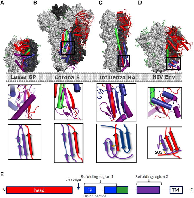
The heavily glycosylated native-like envelope (Env) trimer of HIV-1 is expected to have low immunogenicity, whereas misfolded forms are often highly immunogenic. High-quality correctly folded Envs may therefore be critical for developing a vaccine that induces broadly neutralizing antibodies. Moreover, the high variability of Env may require immunizations with multiple Envs. Here, we report a universal strategy that provides for correctly folded Env trimers of high quality and yield through a repair-and-stabilize approach. In the repair stage, we utilized a consensus strategy that substituted rare strain-specific residues with more prevalent ones. The stabilization stage involved structure-based design and experimental assessment confirmed by crystallographic feedback. Regions important for the refolding of Env were targeted for stabilization. Notably, the α9-helix and an intersubunit β sheet proved to be critical for trimer stability. Our approach provides a means to produce prefusion-closed Env trimers from diverse HIV-1 strains, a substantial advance for vaccine development.

Keywords: ConC_base; HIV; SOSIP; X-ray structure; chronic; envelope protein; hybrid sheet; stabilization; transmitted/founder; vaccine.


Conflict of interest statement

DECLARATION OF INTERESTS

These studies were funded by Janssen Vaccines and Prevention. L.R., D.T., D.v.M., S.B., N.M.S., A.K., I.J.M.B., H.S., and J.P.M.L. are employees at Janssen. L.R., D.T., N.M.S., A.K., and J.P.M.L. are inventors on an international patent application describing trimer stabilizing HIV envelope protein mutations. The remaining authors declare no competing interests.

Figures

Figure 1
Figure 1. HIV-1 Env Selection for Stabilization
(A and B) SEC profiles of GN lectin-purified HIV Env SOSIPs of (A) clade C consensus (ConC_SOSIP) and closest wild-type Env sequences indicated with GenBank numbers. The trimer peak is indicated with an asterisk, gp140 monomer peak is indicated with a degree symbol, gp120 monomer is indicated with a diamond, and aggregates are indicated with an at symbol. (B) clade C mosaic SOSIP 201C-433C (DS_sC4_SOSIP) and closest wild-type Env sequences indicated with the GenBank numbers. The trimer peak is indicated with an asterisk and gp140 monomer peak with a degree symbol. (C) Analysis of GN lectin-purified ConC_SOSIP on SDS-PAGE under reducing (+DTT) and non-reducing (−DTT) conditions compared with markers (M). (D) Negatively stained EM of ConC_SOSIP trimers with 2D-averaged classes (right).
Figure 2
Figure 2. Single Point Mutations that Enhance Yield and Stability of ConC SOSIP Trimers
(A) Schematic definition of regions of HIV-1 Env that move more than 5 Å between prefusion closed and CD4-bound forms are shown blue for gp120, green for gp41, and black if not defined in either conformation. Residues 31–511 form the mature gp120. Refolding region 1 encompasses residues 512–570. The base helix is residues 571–595. Refolding region 2 encompasses residues 596–664. Previously defined HR1 (residues 540–590) and HR2 (residues 624–664) are also indicated. (B) Location of structure-based mutation in the prefusion closed Env trimer. Two protomers are shown in surface representation (pink and peach) and the third protomer is shown in ribbon representation, with residues that move more than 5 Å between prefusion-closed and CD4-bound conformations in blue (for gp120) or in green (for gp41) and otherwise gray or black (if not defined in either conformation). (C) Comparison of expression level plotted against PGT145 binding for single point mutation variants of ConC_SOSIP Env using AlphaLISA in cell culture supernatant 3 days after transfection. Substitutions with highest increase in trimer yield and trimer fraction are labeled. Substitutions at the same positions have identical color and largest dot was selected for further evaluation. (D) SEC profiles using crude cell culture supernatants 3 days after transfection with the best single point mutation variants compared with backbone ConC_SOSIP (dotted gray line) (colored as in C). The trimer peak is indicated with an asterisk and gp140 monomer peak is indicated with a degree symbol. See also Table S1.
Figure 3
Figure 3. Combined Effect of Cumulative Substitutions
(A) SEC-MALS analysis (left) and temperature stability as measured by DSC (right) of Env variants with cumulative combination of amino acid substitutions in ConC_SOSIP. The trimer peak is indicated with an asterisk. For constructs with two melting event, a Tm1 (minor) and Tm2 (main) is indicated. Construct number 10 (ConC_base0) has a Glu instead of Gln at position 588. (B) DSC pattern of ConC_base0. (C) 17b binding in the presence (solid lines) and absence of CD4 (dotted lines). (D) Antibody binding levels measured with Octet after 0 and 4 weeks incubation at 37°C of ConC_base0. (E) SEC-MALS of purified ConC_base0 with SEC (purple) and molar mass trace. See also Figure S1B.
Figure 4
Figure 4. Crystal Structure of ConC_base0
(A) Mutations in ConC_base0 are shown in red ball-and-stick with molecular surface. gp120 and gp41 are shown in ribbon for one protomer with the color scheme shown in Figure 2B. The other two protomers are shown in surface representation, colored peach and pink. Residues on gp120 and gp41 potentially interacting with the stabilizing mutations are shown in ball-and-stick without molecular surface. (B) Detail of trimer base showing the functionally critical α-9 helix in dark green. Interaction of stabilizing residues 651F and 655I at the trimer interface (red). Residue E647 and K658 that when substituted to hydrophobic residues are also stabilizing (Figure 2) are indicated in orange. (C) Both protomers shown as ribbons showing the functionally critical α-9 helix in dark green of one protomer that interacts with α6 and intersubunit β sheet of the other protomer (colored as in A) Clustering of stabilizing residues indicated on both sides of the intersubunit β sheet. See also Figures S2–S4 and Table S2.
Figure 5
Figure 5. Prefusion Closed HIV Env_SOSIP Trimers Through Sequence Repair and Mutational Stabilization
(A) Universal concept for repairing HIV-1 Env sequence illustrated for strain C97ZA. Occurrence of highest consensus residue in the total HIV-1 database (top bars) and strain C97ZA residue (bottom bars) sorted from low to high occurrence percentage of C97ZA residue position. C97ZA sequence positions to be substituted to consensus were selected based on the following criteria: positions with a C97ZA residue that occurs <2% in consensus Env sequence (blue bars); positions with a C97ZA residue that occur between 2% and 7.5% in consensus Env and are buried or partly buried (magenta bars); positions that are exposed and hydrophobic in C97ZA and hydrophilic in consensus (yellow bars); and a position that is a PNGS in the consensus (green bar). (B) AlphaLISA signals in cell culture supernatant for all SOSIP variants normalized to the ConC_SOSIP for broadly neutralizing (left) and non-neutralizing or strain-specific (right) mAbs. For the non-bNAbs the intensities are further normalized for expression levels. (C) Analytical SEC profile of control Env_SOSIPs (red line) repaired according to the concept described in a (blue line) and additional stabilizing substitutions according to Table S3 (green line). Analysis was performed on crude cell culture supernatant instead of purified protein. The trimer peaks are indicated with asterisks. (D) Repaired and stabilized (REP & STAB) C97ZA_SOSIP compared with CZA97_SOSIP.4.2-M6.IT-2 (Ringe et al., 2017) according to the approach previously described. The upper panel shows the analytical SEC pattern on crude supernatant and the lower panel shows the AlphaLISA signals normalized to those of ConC_SOSIP. See also Figures S5 and S6 and Table S3–S5.
Figure 6
Figure 6. Intersubunit Sheets of Head Domain and Fusion Domain in Type I Fusion Proteins
(A–D) Prefusion trimer structures are shown with 2 protomers as light and dark gray surface representations and one protomer in ribbon with helices represented as cylinders for Lassa GP (A) (Hastie et al., 2017), MERS S (B) (Pallesen et al., 2017), Influenza HA (C), and HIV Env (6CK9) (D). Color coding of structural elements in the fusion protein is based on refolding region 1 (blue), base helix (green), refolding region 2 (purple), and head domain (red) according to (E), with inserts depicting the intersubunit sheet composed of part of the head region (red) and refolding region 2 or both refolding regions. Detachment or splaying of the head is expected to destabilize the hybrid sheet and to effect refolding. (E) Color scheme.

Similar articles

Cited by

References

    1. Binley JM, Sanders RW, Clas B, Schuelke N, Master A, Guo Y, Kajumo F, Anselma DJ, Maddon PJ, Olson WC, Moore JP. A recombinant human immunodeficiency virus type 1 envelope glycoprotein complex stabilized by an intermolecular disulfide bond between the gp120 and gp41 subunits is an antigenic mimic of the trimeric virion-associated structure. J Virol. 2000;74:627–643. - PMC - PubMed
    1. Carlson JM, Schaefer M, Monaco DC, Batorsky R, Claiborne DT, Prince J, Deymier MJ, Ende ZS, Klatt NR, DeZiel CE, et al. HIV transmission. Selection bias at the heterosexual HIV-1 transmission bottleneck. Science. 2014;345:1254031. - PMC - PubMed
    1. Chuang GY, Geng H, Pancera M, Xu K, Cheng C, Acharya P, Chambers M, Druz A, Tsybovsky Y, Wanninger TG, et al. Structure-based design of a soluble prefusion-closed HIV-1 Env trimer with reduced CD4 affinity and improved immunogenicity. J Virol. 2017;91:e02268–16. - PMC - PubMed
    1. de Taeye SW, Ozorowski G, Torrents de la Peña A, Guttman M, Julien JP, van den Kerkhof TL, Burger JA, Pritchard LK, Pugach P, Yasmeen A, et al. Immunogenicity of stabilized HIV-1 envelope trimers with reduced exposure of non-neutralizing epitopes. Cell. 2015;163:1702–1715. - PMC - PubMed
    1. Dey AK, David KB, Ray N, Ketas TJ, Klasse PJ, Doms RW, Moore JP. N-terminal substitutions in HIV-1 gp41 reduce the expression of non-trimeric envelope glycoproteins on the virus. Virology. 2008;372:187–200. - PMC - PubMed

MeSH terms

Substances