

Turning the backbone into an ankylosed concrete-like structure: Case report
- PMID: 29642148
- PMCID: PMC5908595
- DOI: 10.1097/MD.0000000000010278
Turning the backbone into an ankylosed concrete-like structure: Case report
Abstract
Rationale: Progressive restriction of the spinal bio-mechanics is not-uncommon deformity encountered in spine clinics. Congenital spinal fusion as seen in Klippel-Feil-anomaly, progressive non-infectious anterior vertebral fusion, and progressive spinal hyperostosis secondary to ossification of the anterior longitudinal spinal ligament are well delineated and recognized.
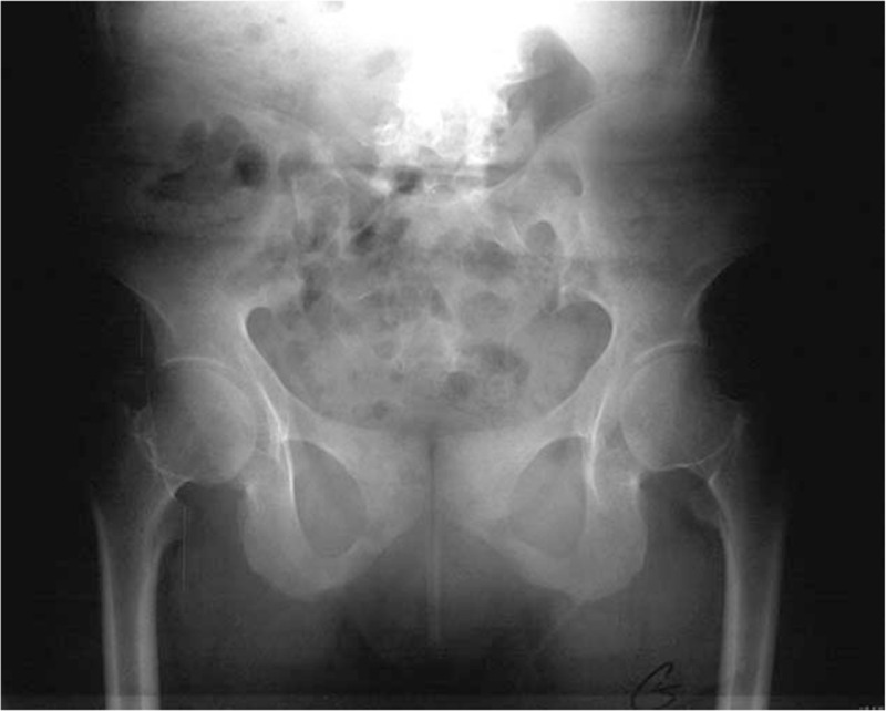
Patient concerns: A 24-year-old girl has history of osteoporosis since her early childhood, associated with multiple axial and appendicular fractures and scoliosis. Recently she presented with episodes of severe back pain, spinal rigidity/stiffness with total loss of spine biomechanics.
Diagnoses: She was provisionally diagnosed as having osteogenesis imperfecta and was investigated for COL1A1/A2 mutations which have been proven to be negative. Autosomal recessive type of osteogenesis imperfecta was proposed as well, no mutations have been encountered. A homozygous for CTSA gene mutation, the gene associated with Galactosialidosis was identified via whole exome sequencing (Next-Generation Sequencing projects) has been identified.
Interventions: Early in her life she had a history of frequent fractures of the long bones since she was 4 years which was followed by vertebral fractures at the age of 12 years. She manifested lower serum 25OH-D levels and were associated with lower LS-aBMD Z-scores with higher urinary bone turnover indexes (urinary NTX/Cr).
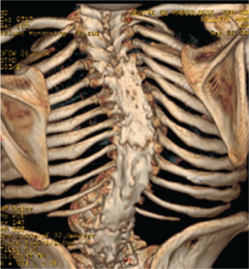
Outcomes: Lysosomal storage diseases (LSD) have a strong correlation with the development of osteoporosis. LSD causes skeletal abnormalities results from a lack of skeletal remodeling and ossification abnormalities owing to abnormal deposition of GAGs (impaired degradation of glycosaminoglycans ) in bone and cartilage. 3D reconstruction CT scan of the spine showed diffuse hyperostosis of almost the entire spine (begins at the level of T4- extending downwards to involve the whole thoraco-lumbar and upper part of the sacrum) with total diffuse fusion of the pedicles, the transverse and articular processes, the laminae and the spinous processes.
Lessons: This is the first clinical report of adult patient with a history of osteoporosis and fractures with the late diagnosis of Galactosialidosis. Osteogenesis imperfecta (autosomal dominant and recessive) were the first given diagnoses which proven negative. The pathophysiology of the spine ankylosis in our current patient and its correlation with LSD, antiresorptive medications, vitamin D3 and supplemental calcium is not fully understood. Therefore, further studies are needed to elucidate this sort of correlation.
Conflict of interest statement
The authors declare no conflict of interests.
Figures


Similar articles
-
Skeletal clinical characteristics of osteogenesis imperfecta caused by haploinsufficiency mutations in COL1A1.J Bone Miner Res. 2013 Sep;28(9):2001-7. doi: 10.1002/jbmr.1942. J Bone Miner Res. 2013. PMID: 23529829
-
Fracture of the spine in patients with ankylosis due to diffuse skeletal hyperostosis: clinical and imaging findings.AJR Am J Roentgenol. 1994 Apr;162(4):899-904. doi: 10.2214/ajr.162.4.8141015. AJR Am J Roentgenol. 1994. PMID: 8141015
-
Osteoporotic vertebral fractures during pregnancy: be aware of a potential underlying genetic cause.J Clin Endocrinol Metab. 2014 Apr;99(4):1107-11. doi: 10.1210/jc.2013-3238. Epub 2014 Jan 13. J Clin Endocrinol Metab. 2014. PMID: 24423337
-
Osteogenesis imperfecta.Nat Rev Dis Primers. 2017 Aug 18;3:17052. doi: 10.1038/nrdp.2017.52. Nat Rev Dis Primers. 2017. PMID: 28820180 Review.
-
Diffuse idiopathic skeletal hyperostosis.Eur J Radiol. 1998 May;27 Suppl 1:S7-11. doi: 10.1016/s0720-048x(98)00036-9. Eur J Radiol. 1998. PMID: 9652495 Review.
Cited by
-
Lung volumetry of osteogenesis imperfecta type 3 subjects is not correlated with thoracic scoliosis and anthropometric data.Orphanet J Rare Dis. 2025 Jun 2;20(1):265. doi: 10.1186/s13023-025-03797-y. Orphanet J Rare Dis. 2025. PMID: 40457460 Free PMC article.
References
-
- McMaster JM. Congenital Scoliosis. The Pediatr Spine: Principles and Practice (chapter 9). 1994;New York: Raven Press, 228.
-
- Resnick D, Shaul RS, Robins JM. Diffuse idiopathic skeletal hyperostosis (DISH): Forestier's disease with extraspinal manifestations. Radiology 1975;115:13–24. - PubMed
-
- Zammarchi E, Donati MA, Morrone A, et al. Early-infantile galactosialidosis: clinical, biochemical, and molecular observations in a new patient. Am J Med Genet 1996;64:453–8. - PubMed
Publication types
MeSH terms
Substances
Supplementary concepts
LinkOut - more resources
Full Text Sources
Other Literature Sources
Medical
Miscellaneous
