Revision surgery after rod breakage in a patient with occipitocervical fusion: A case report
- PMID: 29642217
- PMCID: PMC5908617
- DOI: 10.1097/MD.0000000000010441
Revision surgery after rod breakage in a patient with occipitocervical fusion: A case report
Abstract
Rationale: Rod breakage after occipitocervical fusion (OCF) has never been described in a patient who has undergone surgery for basilar invagination (BI) and atlantoaxial dislocation (AAD). Here, we present an unusual but significant case of revision surgery to correct this complication.
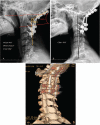
Patient concerns: A 32-year-old female presented with neck pain, unstable leg motion in walking, and also BI with AAD. Her first surgery was planned to correct these conditions and for fusion at the occipital junction (C3-4) using a screw-rod system. At the 31-month follow-up after her first operation, the patient complained of severe neck pain and limitation of motion, suggesting rod breakage.
Diagnoses: Rod breakage after occipitocervical fusion for BI and AAD.
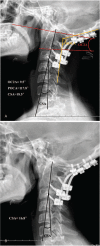
Interventions: The patient underwent reoperation for replacement of the broken rods, adjustment of the occipitocervical angle, maintenance of the bone graft bed, and fusion.
Outcomes: At follow-up, the hardware was found to be in good condition, with no significant loss of cervical lordosis. At the 37-month follow-up after her second operation, the patient was doing better and continuing to recover.
Lessons: We concluded that nonideal choice of occipitocervical angle may play an important role in rod breakage; however, an inadequate bone graft and poor postoperative fusion may also contribute to implant failure.
Conflict of interest statement
The authors report no conflicts of interest.
Figures


Similar articles
-
Outcome of Surgery for Congenital Craniovertebral Junction Anomalies with Atlantoaxial Dislocation/Basilar Invagination: A Retrospective Study of 94 Patients.World Neurosurg. 2021 Feb;146:e313-e322. doi: 10.1016/j.wneu.2020.10.082. Epub 2020 Oct 20. World Neurosurg. 2021. PMID: 33096283
-
Treatment of primary basilar invagination by cervical traction and posterior instrumented reduction together with occipitocervical fusion.Spine (Phila Pa 1976). 2011 Sep 1;36(19):1528-31. doi: 10.1097/BRS.0b013e3181f804ff. Spine (Phila Pa 1976). 2011. PMID: 21270707
-
Surgical treatment for upper cervical deformity with atlantoaxial joint dislocation using individualized 3D printing occipitocervical fusion instrument: A case report and literature review.Medicine (Baltimore). 2021 Mar 26;100(12):e25202. doi: 10.1097/MD.0000000000025202. Medicine (Baltimore). 2021. PMID: 33761705 Free PMC article. Review.
-
Occipitocervical fusion combined with 3-dimensional navigation and 3-dimensional printing technology for the treatment of atlantoaxial dislocation with basilar invagination: A case report.Medicine (Baltimore). 2020 Jan;99(5):e18983. doi: 10.1097/MD.0000000000018983. Medicine (Baltimore). 2020. PMID: 32000432 Free PMC article.
-
Occipitocervical fusion by Luque loop rod instrumentation in Down syndrome.J Pediatr Orthop. 1995 Jul-Aug;15(4):539-42. doi: 10.1097/01241398-199507000-00026. J Pediatr Orthop. 1995. PMID: 7560051 Review.
Cited by
-
Transoral release to realign postoperative loss of reduction following occipitocervical fixation for congenital basilar invagination.Spine Deform. 2021 Jul;9(4):1197-1205. doi: 10.1007/s43390-021-00303-9. Epub 2021 Feb 15. Spine Deform. 2021. PMID: 33590408
-
The importance of stabilisation in enabling bone fusion demonstrated by successful revision of failed occipitocervical fusion using patient-specific atlantoaxial joint spacers: a case report.J Spine Surg. 2025 Jun 27;11(2):354-362. doi: 10.21037/jss-24-157. Epub 2025 Jun 10. J Spine Surg. 2025. PMID: 40621374 Free PMC article.
-
Unexpected Neurological Deterioration Following Occipitocervical Fusion Surgery: A Case Report.Cureus. 2025 Jan 28;17(1):e78129. doi: 10.7759/cureus.78129. eCollection 2025 Jan. Cureus. 2025. PMID: 40018501 Free PMC article.
References
-
- Matsunaga S, Ijiri K, Koga H. Results of a longer than 10-year follow-up of patients with rheumatoid arthritis treated by occipitocervical fusion. Spine 2000;25:1749–53. - PubMed
-
- Matsunaga S, Sakou T, Onishi T, et al. Prognosis of patients with upper cervical lesions caused by rheumatoidarthritis: comparison of occipitocervical fusion between c1 laminectomy and nonsurgical management. Spine 2003;28:1581–7. - PubMed
-
- Lee SC, Chen JF, Lee ST. Clinical experience with rigid occipitocervical fusion in the management of traumatic upper cervical spinal instability. J Clin Neurosci 2006;13:193–8. - PubMed
-
- Bhatia R, Desouza RM, Bull J, et al. Rigid occipitocervical fixation: indications, outeonles, and complications in the modern era. J Neurosurg Spine 2013;18:333–9. - PubMed
Publication types
MeSH terms
LinkOut - more resources
Full Text Sources
Other Literature Sources
Medical
Miscellaneous

