

Nano-Graphene Oxide Functionalized Bioactive Poly(lactic acid) and Poly(ε-caprolactone) Nanofibrous Scaffolds
- PMID: 29642421
- PMCID: PMC5951450
- DOI: 10.3390/ma11040566
Nano-Graphene Oxide Functionalized Bioactive Poly(lactic acid) and Poly(ε-caprolactone) Nanofibrous Scaffolds
Abstract
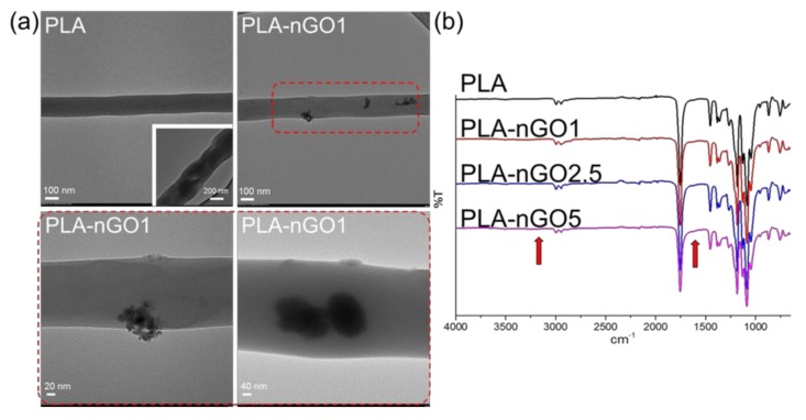
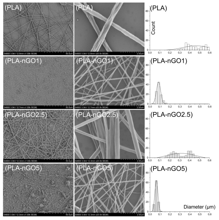
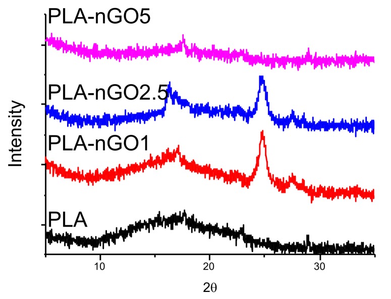
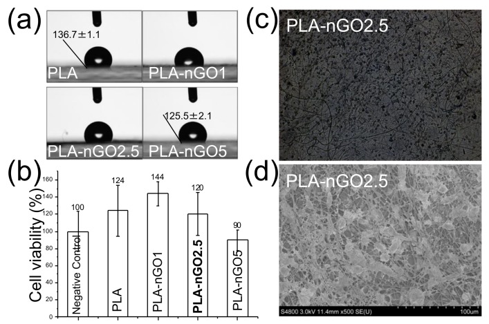
A versatile and convenient way to produce bioactive poly(lactic acid) (PLA) and poly(ε-caprolactone) (PCL) electrospun nanofibrous scaffolds is described. PLA and PCL are extensively used as biocompatible scaffold materials for tissue engineering. Here, biobased nano graphene oxide dots (nGO) are incorporated in PLA or PCL electrospun scaffolds during the electrospinning process aiming to enhance the mechanical properties and endorse osteo-bioactivity. nGO was found to tightly attach to the fibers through secondary interactions. It also improved the electrospinnability and fiber quality. The prepared nanofibrous scaffolds exhibited enhanced mechanical properties, increased hydrophilicity, good cytocompatibility and osteo-bioactivity. Therefore, immense potential for bone tissue engineering applications is anticipated.
Keywords: PCL; PLA; biomineralization; graphene oxide; mechanical properties; scaffold.
Conflict of interest statement
The authors declare no conflict of interest.
Figures


Similar articles
-
Poly (ε-caprolactone)-based electrospun nano-featured substrate for tissue engineering applications: a review.Prog Biomater. 2021 Jun;10(2):91-117. doi: 10.1007/s40204-021-00157-4. Epub 2021 Jun 2. Prog Biomater. 2021. PMID: 34075571 Free PMC article. Review.
-
Advances in Electrospun Poly(ε-caprolactone)-Based Nanofibrous Scaffolds for Tissue Engineering.Polymers (Basel). 2024 Oct 10;16(20):2853. doi: 10.3390/polym16202853. Polymers (Basel). 2024. PMID: 39458681 Free PMC article. Review.
-
The surface grafting of graphene oxide with poly(ethylene glycol) as a reinforcement for poly(lactic acid) nanocomposite scaffolds for potential tissue engineering applications.J Mech Behav Biomed Mater. 2016 Jan;53:403-413. doi: 10.1016/j.jmbbm.2015.08.043. Epub 2015 Sep 8. J Mech Behav Biomed Mater. 2016. PMID: 26409231
-
Enhancement of hydrophilicity, biocompatibility and biodegradability of poly(ε-caprolactone) electrospun nanofiber scaffolds using poly(ethylene glycol) and poly(L-lactide-co-ε-caprolactone-co-glycolide) as additives for soft tissue engineering.J Biomater Sci Polym Ed. 2020 Sep;31(13):1648-1670. doi: 10.1080/09205063.2020.1769799. Epub 2020 Jun 7. J Biomater Sci Polym Ed. 2020. PMID: 32402230
-
Fabrication and characterization of novel ethyl cellulose-grafted-poly (ɛ-caprolactone)/alginate nanofibrous/macroporous scaffolds incorporated with nano-hydroxyapatite for bone tissue engineering.J Biomater Appl. 2019 Mar;33(8):1128-1144. doi: 10.1177/0885328218822641. Epub 2019 Jan 16. J Biomater Appl. 2019. PMID: 30651055
Cited by
-
Graphene Nanoplatelets for the Development of Reinforced PLA-PCL Electrospun Fibers as the Next-Generation of Biomedical Mats.Polymers (Basel). 2020 Jun 21;12(6):1390. doi: 10.3390/polym12061390. Polymers (Basel). 2020. PMID: 32575840 Free PMC article.
-
Surface Modification of Poly(lactic-co-glycolic acid) Microspheres with Enhanced Hydrophilicity and Dispersibility for Arterial Embolization.Materials (Basel). 2019 Jun 18;12(12):1959. doi: 10.3390/ma12121959. Materials (Basel). 2019. PMID: 31216635 Free PMC article.
-
Recent Progress on Bio-Based Polyesters Derived from 2,5-Furandicarbonxylic Acid (FDCA).Polymers (Basel). 2022 Feb 6;14(3):625. doi: 10.3390/polym14030625. Polymers (Basel). 2022. PMID: 35160613 Free PMC article. Review.
-
Porous Silk Fibroin Microspheres Sustainably Releasing Bioactive Basic Fibroblast Growth Factor.Materials (Basel). 2018 Jul 25;11(8):1280. doi: 10.3390/ma11081280. Materials (Basel). 2018. PMID: 30044408 Free PMC article.
-
Biological and physiochemical studies of electrospun polylactid/polyhydroxyoctanoate PLA/P(3HO) scaffolds for tissue engineering applications.RSC Adv. 2023 Aug 11;13(34):24112-24128. doi: 10.1039/d3ra03021k. eCollection 2023 Aug 4. RSC Adv. 2023. PMID: 37577093 Free PMC article.
References
-
- Nejatian T., Khurshid Z., Zafar M.S., Najeeb S., Zohaib S., Mazafari M., Hopkinson L., Sefat F. Biomaterials for Oral and Dental Tissue Engineering. Woodhead Publishing; Cambridge, UK: 2017. 5—dental biocomposites; pp. 65–84.
LinkOut - more resources
Full Text Sources
Other Literature Sources
