. 2018 Apr 7;11(4):569.
doi: 10.3390/ma11040569.

MitoQ Loaded Chitosan-Hyaluronan Composite Membranes for Wound Healing

Affiliations

MitoQ Loaded Chitosan-Hyaluronan Composite Membranes for Wound Healing

Tamer M Tamer et al. Materials (Basel). .

Abstract

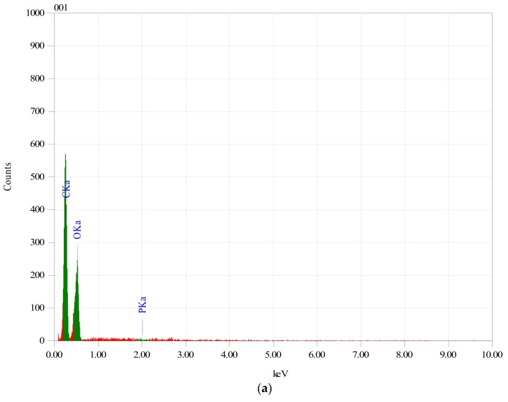
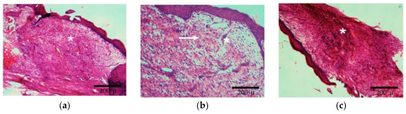
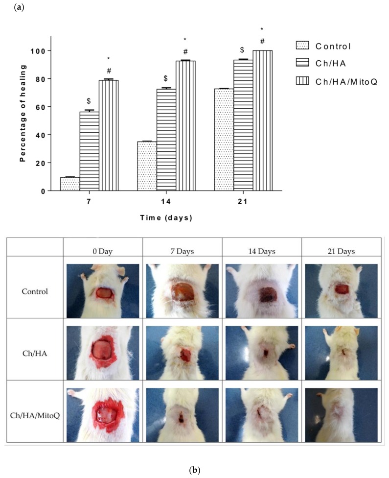
Two self-associating biopolymers, namely chitosan (Ch) and a high-molar-mass hyaluronan (HA), were used to prepare membranes with the aim to protect and to enhance the healing of injured skin. A mitochondrially-targeted antioxidant-MitoQ-was incorporated into the mixture of biopolymers prior to their self-association. These three-component membranes were evaluated in detail utilising surface roughness measurements, contact angle measurements, hemocompatibility, and thrombogenicity analyses. Furthermore, in vivo application of Ch/HA/MitoQ membranes was assessed on injured rabbit and rat skin utilizing histological methods. The results showed that the prepared thrombogenic Ch/HA/MitoQ membranes had higher roughness, which allowed for greater surface area for tissue membrane interaction during the healing processes, and lower cytotoxicity levels than controls. MitoQ-loaded composite membranes displayed superior healing properties in these animal models compared to control membranes.

Keywords: chitosan; hyaluronan; mitochondrially-targeted antioxidant; skin wounds.


Conflict of interest statement

The authors declare no conflict of interest.

Figures

Figure 1
Figure 1
EDX spectrum of (a) Ch/HA and (b) Ch/HA/MitoQ membranes.
Figure 1
Figure 1
EDX spectrum of (a) Ch/HA and (b) Ch/HA/MitoQ membranes.
Figure 2
Figure 2
SEM images of (a)Ch/HA and (b)Ch/HA/MitoQ membranes.
Figure 3
Figure 3
Profiles of the wound closures in rabbits, when the wound was not treated (control), the wound treated with the Ch/HA membrane and the Ch/HA/MitoQ membrane. The asterisk (*) indicates a significant difference at p ≤ 0.05 between the control and Ch/HA/MitoQ. The mark (#) indicates a significant difference between the Ch/HA and Ch/HA/MitoQ membranes at p ≤ 0.05. The mark ($) indicates a significant difference between the control and the Ch/HA membrane at p ≤ 0.05, n = 6 (a, b).
Figure 4
Figure 4
Histology of skin wounds in rabbits after 15 days of treatment. (a)Control: star—vascular granular tissue; (b)Ch/HA membrane: arrow—likely site of activated fibroblasts with formation of collagen fibres (it is acknowledged that a Masson trichrome or immunostaining is required to accurately identify these entities), star—newly-formed veins; (c)Ch/HA/MitoQ membrane: star—nonspecific granular tissue in the initial stage with enhanced cellularity.
Figure 5
Figure 5
Profiles of the wound closures in rats, when the wound was not treated (control), the wound treated with the Ch/HA membrane, and the Ch/HA/MitoQ membrane; and the asterisk (*) indicates a significant difference at p ≤ 0.05 between the control and Ch/HA/MitoQ). The mark (#) indicates a significant difference between the Ch/HA and Ch/HA/MitoQ membranes at p ≤ 0.05. The mark ($) indicates a significant difference between the control and the Ch/HA membrane at p ≤ 0.05, n = 5 (a, b).
Figure 6
Figure 6
Histology of wounds treated with control (gauze), Ch/HA, and Ch/HA/MitoQ membranes at the 7th, 14th, and 21st day after the injury. Epi: Epidermis; K: keratinous layers; IC: possible site of inflammatory cells; BV: blood vessels; HF: hair follicles; SG: sebaceous glands.

References

    1. Lobo V., Patil A., Phatak A., Chandra N. Free radicals, antioxidants and functional foods: Impact on human health. Pharmacogn. Rev. 2010;4:118–126. doi: 10.4103/0973-7847.70902. - DOI - PMC - PubMed
    1. Kowaltowski A.J., de Souza-Pinto N.C., Castilho R.F., Vercesi A.E. Mitochondria and reactive oxygen species. Free Radic. Biol. Med. 2009;47:333–343. doi: 10.1016/j.freeradbiomed.2009.05.004. - DOI - PubMed
    1. Stowe D.F., Camara A.K.S. Mitochondrial reactive oxygen species production in excitable cells: Modulators of mitochondrial and cell function. Antioxid. Redox Signal. 2009;11:1373–1414. doi: 10.1089/ars.2008.2331. - DOI - PMC - PubMed
    1. Guo C., Sun L., Chen X., Zhang D. Oxidative stress, mitochondrial damage and neurodegenerative diseases. Neural Regen. Res. 2013;8:2003–2014. - PMC - PubMed
    1. Valachova K., Banasova M., Topolska D., Sasinkova V., Juranek I., Collins M.N., Soltes L. Influence of tiopronin, captopril and levamisole therapeutics on the oxidative degradation of hyaluronan. Carbohydr. Polym. 2015;134:516–523. doi: 10.1016/j.carbpol.2015.07.029. - DOI - PubMed

LinkOut - more resources