Review
. 2018 Apr 10;19(4):1146.
doi: 10.3390/ijms19041146.

Signals of Systemic Immunity in Plants: Progress and Open Questions

Affiliations
Review

Signals of Systemic Immunity in Plants: Progress and Open Questions

Attila L Ádám et al. Int J Mol Sci. .

Abstract

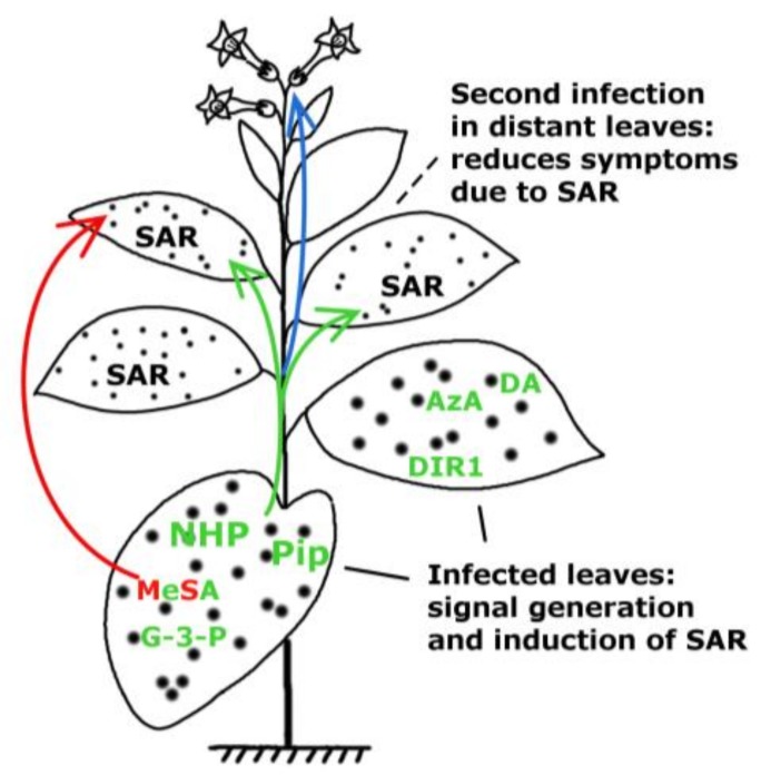
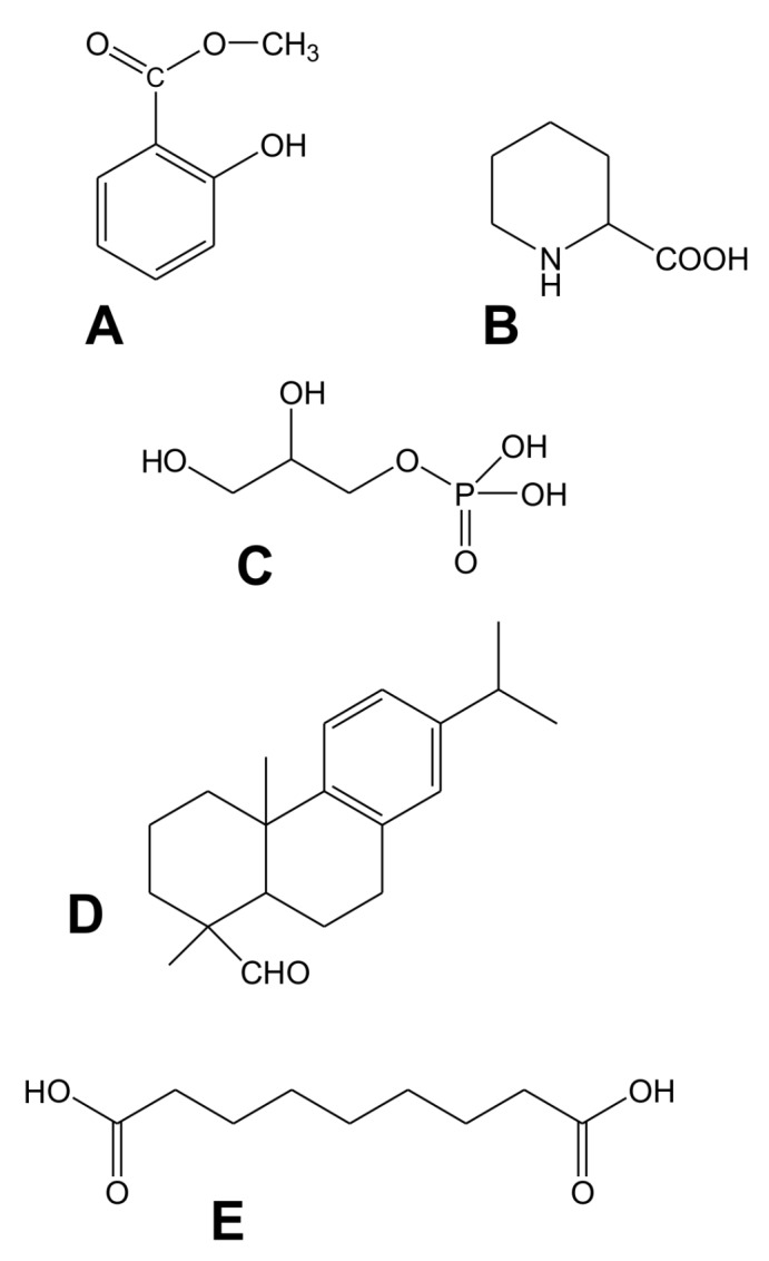
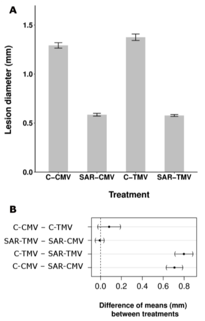
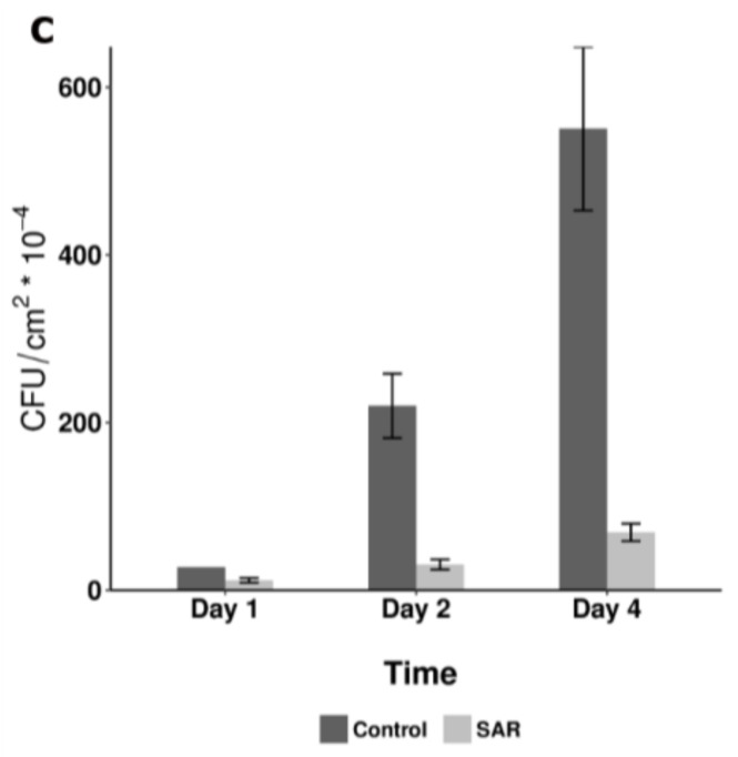
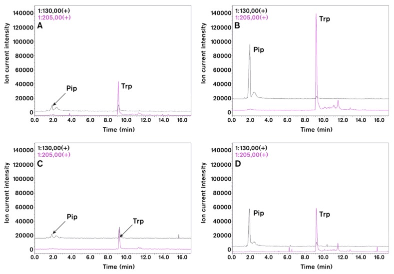
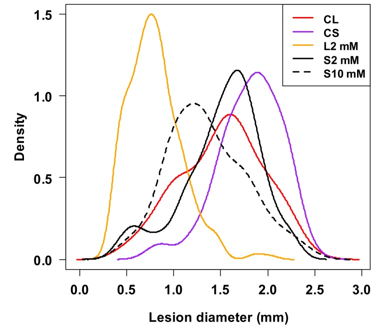
Systemic acquired resistance (SAR) is a defence mechanism that induces protection against a wide range of pathogens in distant, pathogen-free parts of plants after a primary inoculation. Multiple mobile compounds were identified as putative SAR signals or important factors for influencing movement of SAR signalling elements in Arabidopsis and tobacco. These include compounds with very different chemical structures like lipid transfer protein DIR1 (DEFECTIVE IN INDUCED RESISTANCE1), methyl salicylate (MeSA), dehydroabietinal (DA), azelaic acid (AzA), glycerol-3-phosphate dependent factor (G3P) and the lysine catabolite pipecolic acid (Pip). Genetic studies with different SAR-deficient mutants and silenced lines support the idea that some of these compounds (MeSA, DIR1 and G3P) are activated only when SAR is induced in darkness. In addition, although AzA doubled in phloem exudate of tobacco mosaic virus (TMV) infected tobacco leaves, external AzA treatment could not induce resistance neither to viral nor bacterial pathogens, independent of light conditions. Besides light intensity and timing of light exposition after primary inoculation, spectral distribution of light could also influence the SAR induction capacity. Recent data indicated that TMV and CMV (cucumber mosaic virus) infection in tobacco, like bacteria in Arabidopsis, caused massive accumulation of Pip. Treatment of tobacco leaves with Pip in the light, caused a drastic and significant local and systemic decrease in lesion size of TMV infection. Moreover, two very recent papers, added in proof, demonstrated the role of FMO1 (FLAVIN-DEPENDENT-MONOOXYGENASE1) in conversion of Pip to N-hydroxypipecolic acid (NHP). NHP systemically accumulates after microbial attack and acts as a potent inducer of plant immunity to bacterial and oomycete pathogens in Arabidopsis. These results argue for the pivotal role of Pip and NHP as an important signal compound of SAR response in different plants against different pathogens.

Keywords: Arabidopsis; N-hydroxypipecolic acid; SAR signalling; azelaic acid; glycerol-3-phosphate; light dependent signalling; methyl salicylate; pipecolic acid; salicylic acid; spectral distribution of light; tobacco.


Conflict of interest statement

The authors declare no conflict of interest. The founding sponsors had no role in the design of the study; in the collection, analyses, or interpretation of data; in the writing of the manuscript, and in the decision to publish the results.

Figures

Figure 1
Figure 1
Development of systemic acquired resistance (SAR). Putative SAR signal molecules: methyl salicylate (MeSA), lipid transfer protein DIR1 (DEFECTIVE IN INDUCED RESISTANCE1), dehydroabietinal (DA), glycerol-3-phosphate (G3P) or G3P-dependent factor, azelaic acid (AzA), pipecolic acid (Pip) and N-hydroxypipecolic acid (NHP) move from the infected organ (leaves) to pathogen-free distant parts of the plant where they induce SAR against biotrophic and hemibiotrophic pathogens (note the limited symptom expression in the distant, systemic leaves indicated by small spots). Pip and NHP are highlighted. Green and red arrows indicate the movement of signal molecules via phloem transport or the air (putative volatile compounds for inplant or interplant airborne signals), respectively. Blue arrow indicates transgenerational SAR where the epigenetic information is inherited and present in the next generation (re-drawn and modified from [28]). For chemical structures of signal compounds see Figure 2.
Figure 2
Figure 2
Chemical structures of signal compounds participating in SAR induction. (A) Phenolic compound, methyl-salicylate; (B) Non-protein amino acid, pipecolic acid; (C) Glycerol-3-phosphate; (D) Geranylgeranyl diphosphate derived dehydroabietinal; (E) Dicarboxylic acid, azelaic acid.
Figure 3
Figure 3
Aspecific feature of SAR induction in tobacco (N. tabacum cv. Xanthi nc). TMV induced SAR response was effective not only against TMV and another viral (cucumber mosaic virus, CMV necrotic strain) (A,B) but bacterial (Pseudomonas syringae pv. tabaci) (C) pathogens as well. SAR was induced by TMV infection as described earlier [31] and 10 days later the 5th and 6th leaves were challenged by CMV (SAR-CMV) and TMV (SAR-TMV) as compared to controls (C-CMV and C-TMV) (half leaves of each case) (A) or by bacterial suspension (105 CFU mL−1) (C) in triplicates per treatments. (B) Multiple comparison of means of selected treatments in (A). Dots represent the difference of the estimated means between treatments, brackets flank the 95% confidence intervals. The difference is considered significant if the confidence interval does not contain the 0, represented by a vertical dashed line [31]. (C) The SAR induction was also checked by TMV challenge inoculation (showing 69.6% significant inhibition of TMV lesion size). The TMV induced SAR caused time-dependent and significant (generalized linear model) decrease of bacterial multiplication
Figure 3
Figure 3
Aspecific feature of SAR induction in tobacco (N. tabacum cv. Xanthi nc). TMV induced SAR response was effective not only against TMV and another viral (cucumber mosaic virus, CMV necrotic strain) (A,B) but bacterial (Pseudomonas syringae pv. tabaci) (C) pathogens as well. SAR was induced by TMV infection as described earlier [31] and 10 days later the 5th and 6th leaves were challenged by CMV (SAR-CMV) and TMV (SAR-TMV) as compared to controls (C-CMV and C-TMV) (half leaves of each case) (A) or by bacterial suspension (105 CFU mL−1) (C) in triplicates per treatments. (B) Multiple comparison of means of selected treatments in (A). Dots represent the difference of the estimated means between treatments, brackets flank the 95% confidence intervals. The difference is considered significant if the confidence interval does not contain the 0, represented by a vertical dashed line [31]. (C) The SAR induction was also checked by TMV challenge inoculation (showing 69.6% significant inhibition of TMV lesion size). The TMV induced SAR caused time-dependent and significant (generalized linear model) decrease of bacterial multiplication
Figure 4
Figure 4
Effect of TMV (A,B) and CMV (C,D) inoculations on selected amino acids of tobacco leaves (4 days after viral inoculation). HPLC-MS chromatograms of pipecolic acid (Pip, m/z = 130) and tryptophan (Trp, m/z = 205) in virus infected leaves (B,D) as compared to control plants (A,C). Leaf samples (about 200 mg) were extracted with acetonitrile:water (6:4 v/v in 1.5 mL). This condition supports amino acid extraction but limits protein solubilization. HPLC-ESI-MS analyses were carried out with a Shimadzu LCMS-2020 (Shimadzu Co., Kyoto, Japan) analytical system. Chromatographic separations were performed on a SunShell C18 packed column (2.6 µm, 3.0 mm × 100 mm) by using gradient elution (solvent A, 0.1% formic acid solution; solvent B, 0.1% formic acid in acetonitrile) under non-derivatised conditions. Ions for MS detection were obtained by positive mode of electrospray ionization (ESI). Identity of peaks were also checked by co-chromatography of samples with authentic standards (Sigma-Aldrich Co. St. Louis, MI, USA).
Figure 5
Figure 5
Effect of local (CL and L2) and systemic (CS, S2 and S10) pipecolic acid (d-l-Pip) treatments on TMV lesion size distribution by using Kernel density estimation [31,32] (2 days after Pip infiltration). L2, S2 and S10: local (L, leaf 6) and systemic (S, leaf 7) effects of 2 and 10 mM d-l-Pip treatments as compared to corresponding local control (CL) and systemic control (CS) leaves (triplicate per treatments). In a multiple comparison test [31], both concentration of Pip treatment resulted in significant decrease of TMV lesion size. The highest difference was caused by local treatment (L2). The effect of systemic treatment with 10 mM Pip was more pronounced (and significantly different) than with 2 mM Pip

Similar articles

Cited by

References

    1. Winter P.S., Bowman C.E., Villani P.J., Dolan T.E., Hauck N.R. Systemic acquired resistance in moss: Further evidence for conserved defence mechanisms in plants. PLoS ONE. 2014;9:e101880. doi: 10.1371/journal.pone.0101880. - DOI - PMC - PubMed
    1. Ross A.F. Systemic acquired resistance induced by localized virus infections in plants. Virology. 1961;14:340–358. doi: 10.1016/0042-6822(61)90319-1. - DOI - PubMed
    1. Luna E., Bruce T.J., Roberts M.R., Flors V., Ton J. Next-generation systemic acquired resistance. Plant Physiol. 2012;158:844–853. doi: 10.1104/pp.111.187468. - DOI - PMC - PubMed
    1. Sánchez A.L., Stassen J.H., Furci L., Smith L.M., Ton J. The role of DNA (de)methylation in immune responsiveness of Arabidopsis. Plant J. 2016;88:361–374. doi: 10.1111/tpj.13252. - DOI - PMC - PubMed
    1. Conrath U. Molecular aspects of defence priming. Trends Plant Sci. 2011;16:524–531. doi: 10.1016/j.tplants.2011.06.004. - DOI - PubMed

LinkOut - more resources