. 2019 Feb;26(2):250-258.
doi: 10.1177/1933719118768692. Epub 2018 Apr 11.

Regulation of Cell Cycle Regulatory Proteins by MicroRNAs in Uterine Leiomyoma

Affiliations

Regulation of Cell Cycle Regulatory Proteins by MicroRNAs in Uterine Leiomyoma

Tsai-Der Chuang et al. Reprod Sci. 2019 Feb.

Abstract

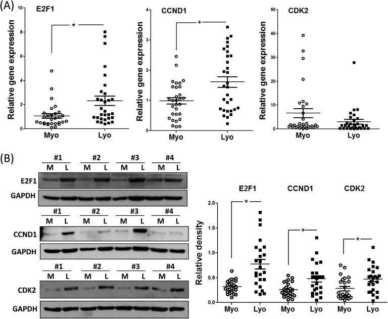
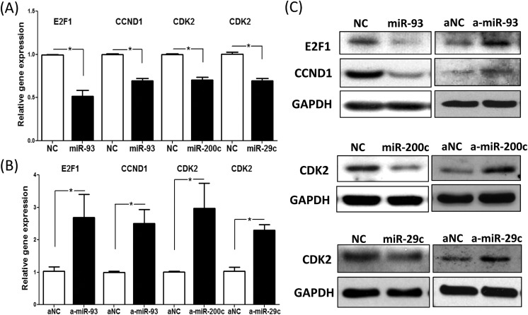
The objective of this study was to determine whether miR-93, miR-29c, and miR-200c, which we previously reported to be downregulated in leiomyomas, target cell cycle regulatory proteins that influence cell proliferation. Based on TargetScan algorithm 3 cell cycle regulatory proteins namely, E2F transcription factor 1 (E2F1), Cyclin D1 (CCND1) and CDK2 which were predicted to be targets of these miRNAs were further analyzed. In 30 hysterectomy specimens, we found the expression of E2F1 and CCND1 messenger RNA (mRNA) was increased in leiomyoma as compared to matched myometrium, with no significant changes in CDK2 mRNA levels. There was a significant increase in the abundance of all 3 proteins in leiomyoma in comparison with matched myometrium. Using luciferase reporter assay, we demonstrated E2F1 and CCND1 are targets of miR-93 and CDK2 is a target of miR-29c and miR-200c. We confirmed these findings through transfection studies in which transfection of primary leiomyoma cells with miR-93 resulted in a significant decrease in the expression of E2F1 and CCND1 mRNA and protein levels, whereas knockdown of miR-93 had the opposite effect. Similarly, overexpression of miR-29c and miR-200c in leiomyoma cells inhibited the expression of CDK2 protein and mRNA, whereas knockdown of this microRNAs (miRNA) had the opposite effect. Transfection of miR-29c, miR-200c, and miR-93 in primary leiomyoma cells resulted in a time-dependent inhibition of cell proliferation and cell motility. These results collectively indicate that the 3 miRNAs known to be downregulated in fibroid tumors are critical in regulation of cell proliferation because of their effects on 3 key cell cycle regulatory proteins, which are overexpressed in uterine leiomyomas.

Keywords: Leiomyoma; cell proliferation; fibroids; microRNA.


Conflict of interest statement

Declaration of Conflicting Interests: The author(s) declared no potential conflicts of interest with respect to the research, authorship, and/or publication of this article.

Figures

Figure 1.
Figure 1.
A, Relative expression of E2F1, CCND1, and E2F1 messenger RNA (mRNA) in leiomyoma (Lyo) and matched myometrium (Myo; n = 30). B, Representative immunoblots for E2F1, CCND1, and cyclin-dependent kinase 2 (CDK2) in paired myometrium (M) and leiomyoma (L). Bar graph shows relative protein band densities (N = 24) in myometrium (Myo) and leiomyoma (Lyo). Results are presented as mean ± SEM and analyzed using paired Student t test. *P < .05.
Figure 2.
Figure 2.
A, The sequence alignment of seed regions of microRNAs (miRNAs) with their target genes at the 3′-UTR region. B, Relative luciferase activity in isolated leiomyoma smooth muscle cells (LSMCs) cotransfected with Renilla and firefly luciferase reporter carrying a 3′-UTR fragment of E2F1, CCND1, cyclin-dependent kinase 2 (CDK2), pre-miR-93, pre-miR-200c, pre-miR-29c, or control oligonucleotides (NC) for 48 hours. Relative luciferase activity is presented as the ratio of Firefly:Renilla as compared to NC, which was independently set as 1. Results are presented as mean ± SEM of at least 3 independent experiments with P values (*P < .05) indicated by corresponding lines.
Figure 3.
Figure 3.
Quantitative RT-PCR analysis of E2F1, CCND1, and cyclin-dependent kinase 2 (CDK2) messenger RNA (mRNA) expression in leiomyoma smooth muscle cells (LSMCs) following transfection with pre-miR-93 (miR-93, for E2F1 and CCND1), pre-miR-200c (miR-200c, for CDK2), pre-miR-29c (miR-29c, for CDK2), and control pre-miR oligonucleotides (NC) (A) or anti-miR-93 (a-miR-93, for E2F1 and CCND1), anti-miR-200c (a-miR-200c, for CDK2), anti-miR-29c (a-miR-29c, for CDK2), and control anti-miR oligonucleotides (aNC) (B) for 72 hours. Results are presented as mean ± SEM of at least 3 independent experiments with P values (*P < .05) indicated by corresponding lines. C, Western blot analysis of E2F1, CCND1, and CDK2 following transfection of LSMCs with control pre-miR oligonucleotides (NC), pre-miR-93 (miR-93, for E2F1 and CCND1), pre-miR-200c (miR-200c, for CDK2), pre-miR-29c (miR-29c, for CDK2) or control anti-miR oligonucleotides (aNC), anti-miR-93 (a-miR-93, for E2F1 and CCND1), anti-miR-200c (a-miR-200c, for CDK2), anti-miR-29c (a-miR-29c, for CDK2) for 96 hours. Results are representative of at least 3 independent cell preparations.
Figure 4.
Figure 4.
A, The rate of cell proliferation was determined after 4 days transfection of pre-miR-93, pre-miR-200c, pre-miR-29c, and control pre-miR oligonucleotides (NC) in leiomyoma smooth muscle cells (LSMCs) by MTT assay on the indicated days, with culture media changed every 2 days. Results are presented as mean ± SEM of 6 independent experiments. *P < .05. B and C, Photomicrographs of LSMCs transfected with pre-miR-93 (B), pre-miR-29c (C), pre-miR-200c (C) or the corresponding negative controls (NC) for 72 hours. The LSMC motility was determined after the biocompatible gels were removed for 21 hours. Motility assays were performed, in triplicates, using 3 independent cell preparations. D, Quantification of cell motility was analyzed by ImageJ and shown as mean ± SEM with P values (*P < .05) as compared to NC.

Similar articles

Cited by

References

    1. Moravek MB, Bulun SE. Endocrinology of uterine fibroids: steroid hormones, stem cells, and genetic contribution. Curr Opin Obstet Gynecol. 2015;27(4):276–283. - PMC - PubMed
    1. Catherino WH, Eltoukhi HM, Al-Hendy A. Racial and ethnic differences in the pathogenesis and clinical manifestations of uterine leiomyoma. Semin Reprod Med. 2013;31(5):370–379. - PMC - PubMed
    1. Segars JH, Parrott EC, Nagel JD, et al. Proceedings from the Third National Institutes of Health International Congress on Advances in Uterine Leiomyoma Research: comprehensive review, conference summary, and future recommendations. Hum Reprod Update. 2014;20(3):309–333. - PMC - PubMed
    1. Tsibris JC, Segars J, Coppola D, et al. Insights from gene arrays on the development and growth regulation of uterine leiomyomata. Fertil Steril. 2002;78(1):114–121. - PMC - PubMed
    1. Huntzinger E, Izaurralde E. Gene silencing by microRNAs: contributions of translational repression and mRNA decay. Nat Rev Genet. 2011;12(2):99–110. - PubMed

Publication types