. 2018 Apr 11;15(1):106.
doi: 10.1186/s12974-018-1140-6.

Activation of melanocortin receptor 4 with RO27-3225 attenuates neuroinflammation through AMPK/JNK/p38 MAPK pathway after intracerebral hemorrhage in mice

Affiliations

Activation of melanocortin receptor 4 with RO27-3225 attenuates neuroinflammation through AMPK/JNK/p38 MAPK pathway after intracerebral hemorrhage in mice

Shengpan Chen et al. J Neuroinflammation. .

Abstract

Background: Neuroinflammation plays an important role in the pathogenesis of intracerebral hemorrhage (ICH)-induced secondary brain injury. Activation of melanocortin receptor 4 (MC4R) has been shown to elicit anti-inflammatory effects in many diseases. The objective of this study was to explore the role of MC4R activation on neuroinflammation in a mouse ICH model and to investigate the contribution of adenosine monophosphate-activated protein kinase (AMPK)/c-Jun N-terminal kinase (JNK)/p38 mitogen-activated protein kinase (p38 MAPK) pathway in MC4R-mediated protection.

Methods: Adult male CD1 mice (n = 189) were subjected to intrastriatal injection of bacterial collagenase or sham surgery. The selective MC4R agonist RO27-3225 was administered by intraperitoneal injection at 1 h after collagenase injection. The specific MC4R antagonist HS024 and selective AMPK inhibitor dorsomorphin were administered prior to RO27-3225 treatment to elucidate potential mechanism. Short- and long-term neurobehavioral assessments, brain water content, immunofluorescence staining, and western blot were performed.

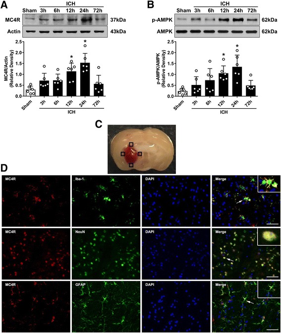
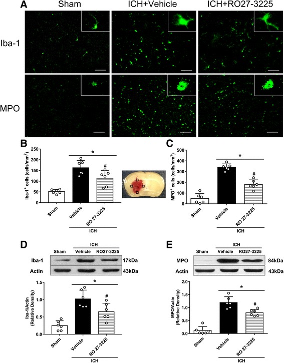
Results: The expression of MC4R and p-AMPK increased after ICH with a peak at 24 h. MC4R was expressed by microglia, neurons, and astrocytes. Activation of MC4R with RO27-3225 improved the neurobehavioral functions, decreased brain edema, and suppressed microglia/macrophage activation and neutrophil infiltration after ICH. RO27-3225 administration increased the expression of MC4R and p-AMPK while decreasing p-JNK, p-p38 MAPK, TNF-α, and IL-1β expression, which was reversed with inhibition of MC4R and AMPK.

Conclusions: Our study demonstrated that activation of MC4R with RO27-3225 attenuated neuroinflammation through AMPK-dependent inhibition of JNK and p38 MAPK signaling pathway, thereby reducing brain edema and improving neurobehavioral functions after experimental ICH in mice. Therefore, the activation of MC4R with RO27-3225 may be a potential therapeutic approach for ICH management.

Keywords: Brain edema; Intracerebral hemorrhage; Melanocortin receptor 4; Neuroinflammation; RO27-3225.


Conflict of interest statement

Ethics approval and consent to participate

All animal experiments were approved by the Institutional Animal Care and Use Committee at Loma Linda University. The study followed the Health’s Guide for the Care and Use of Laboratory Animals (National Research Council) and complied with the ARRIVE guidelines for reporting in vivo experiments.

Competing interests

The authors declare that they have no competing interests.

Publisher’s Note

Springer Nature remains neutral with regard to jurisdictional claims in published maps and institutional affiliations.

Figures

Fig. 1
Fig. 1
Expression profile of MC4R and p-AMPK after ICH. a Representative western blot bands of time course and quantitative analyses of MC4R expression in the ipsilateral hemisphere after ICH. b Representative western blot bands of time course and quantitative analyses of phosphorylated AMPK expression in the ipsilateral hemisphere after ICH. *p < 0.05 vs sham. Error bars are represented as mean ± SD. One-way ANOVA, Tukey’s test, n = 6 per group. c Brain sample with schematic illustration showing the four areas in the perihematomal region (indicated by black squares) from where the images were taken for immunofluorescence staining (pictures are shown in panel d). d Representative images of co-localization of MC4R (red) with microglia/macrophage (Iba-1, green), neurons (NeuN, green), and astrocytes (GFAP, green) in the perihematomal area at 24 h after ICH. Nuclei were stained with DAPI (blue). Scale bar = 50 μm, n = 2
Fig. 2
Fig. 2
The effects of RO27-3225 on neurobehavioral outcomes and brain water content after ICH. a Modified Garcia test, b forelimb placement test, c corner turn test, and d brain water content at 24 h after ICH. e Modified Garcia test, f forelimb placement test, g corner turn test, and h brain water content at 72 h after ICH. *p < 0.05 vs. sham, #p < 0.05 vs. ICH + vehicle. Error bars are represented as mean ± SD. One-way ANOVA, Tukey’s test, n = 6 per group. RO RO27-3225, Ipsi-BG ipsilateral basal ganglia, Ipsi-CX ipsilateral cortex, Cont-BG contralateral basal ganglia, Cont-CX contralateral cortex, Cerebel cerebellum
Fig. 3
Fig. 3
The effects of RO27-3225 on microglia/macrophage activation and neutrophil infiltration after ICH. a Representative images of immunofluorescence staining of Iba-1 (green) and MPO (green) in the perihematomal area at 24 h after ICH. Brain sample with schematic illustration shows the four areas (indicated by black squares) used for Iba-1 and MPO-positive cell counting in the perihematomal region. b, c Quantitative analyses of Iba-1 and MPO-positive cells in the perihematomal area at 24 h after ICH. d, e Representative western blot bands and quantitative analyses of Iba-1 and MPO protein levels in the ipsilateral hemisphere at 24 h after ICH. *p < 0.05 vs. sham, #p < 0.05 vs. ICH + vehicle. Error bars are represented as mean ± SD. One-way ANOVA, Tukey’s test, n = 6 per group
Fig. 4
Fig. 4
The effects of RO27-3225 on long-term neurobehavioral outcomes after ICH. a Foot fault test and b Rotarod test in the first, second, and third week after ICH. c Escape latency and d swim distance of Morris water maze on days 21 to 25 after ICH. e Typical traces and f probe quadrant duration of Morris water maze on day 25 after ICH. *p < 0.05 vs. sham, #p < 0.05 vs. ICH + vehicle. Error bars are represented as mean ± SD. Two-way repeated measures ANOVA, Tukey’s test (ad), and one-way ANOVA, Tukey’s test (f), n = 8 per group
Fig. 5
Fig. 5
MC4R antagonist and AMPK inhibitor reversed the effects of RO27-3225 on neurobehavioral outcomes after ICH. a Modified Garcia test, b forelimb placement test, c corner turn test, and d the effects of RO27-3225 on body weight loss at 24 h after ICH. *p < 0.05 vs. sham, #p < 0.05 vs. ICH + vehicle, @p < 0.05 vs. ICH + RO27-3225 + HS024, and &p < 0.05 vs. ICH + RO27-3225 + DMSO. Error bars are represented as mean ± SD. One-way ANOVA, Tukey’s test, n = 6 per group
Fig. 6
Fig. 6
The effects of RO27-3225 and MC4R antagonist HS024 on the expression of MC4R and its downstream signaling proteins. a Representative western blot bands. bg Quantitative analyses of MC4R, phosphorylated AMPK, phosphorylated JNK, phosphorylated p38 MAPK, TNF-α, and IL-1β in the ipsilateral hemisphere at 24 h after ICH. *p < 0.05 vs. sham, #p < 0.05 vs. ICH + vehicle, and @p < 0.05 vs. ICH + RO27-3225 + HS024. Error bars are represented as mean ± SD. One-way ANOVA, Tukey’s test, n = 6 per group
Fig. 7
Fig. 7
The effects of RO27-3225 and AMPK inhibitor dorsomorphin on the expression of MC4R and its downstream signaling proteins. a Representative western blot bands. bg Quantitative analyses of MC4R, phosphorylated AMPK, phosphorylated JNK, phosphorylated p38 MAPK, TNF-α, and IL-1β in the ipsilateral hemisphere at 24 h after ICH. *p < 0.05 vs. sham, #p < 0.05 vs. ICH + vehicle, and &p < 0.05 vs. ICH + RO27-3225 + DMSO. Error bars are represented as mean ± SD. One-way ANOVA, Tukey’s test, n = 6 per group

References

    1. Macellari F, Paciaroni M, Agnelli G, Caso V. Neuroimaging in intracerebral hemorrhage. Stroke. 2014;45:903–908. doi: 10.1161/STROKEAHA.113.003701. - DOI - PubMed
    1. Ziai WC. Hematology and inflammatory signaling of intracerebral hemorrhage. Stroke. 2013;44:S74–S78. doi: 10.1161/STROKEAHA.111.000662. - DOI - PMC - PubMed
    1. Wilkinson DA, Pandey AS, Thompson BG, Keep RF, Hua Y, Xi G. Injury mechanisms in acute intracerebral hemorrhage. Neuropharmacology. 2017; - PMC - PubMed
    1. Zhou Y, Wang Y, Wang J, Anne Stetler R, Yang QW. Inflammation in intracerebral hemorrhage: from mechanisms to clinical translation. Prog Neurobiol. 2014;115:25–44. doi: 10.1016/j.pneurobio.2013.11.003. - DOI - PubMed
    1. Chen S, Yang Q, Chen G, Zhang JH. An update on inflammation in the acute phase of intracerebral hemorrhage. Transl Stroke Res. 2015;6:4–8. doi: 10.1007/s12975-014-0384-4. - DOI - PubMed

MeSH terms