. 2018 Oct 2;138(14):1446-1462.
doi: 10.1161/CIRCULATIONAHA.117.033249.

Defective Base Excision Repair of Oxidative DNA Damage in Vascular Smooth Muscle Cells Promotes Atherosclerosis

Affiliations

Defective Base Excision Repair of Oxidative DNA Damage in Vascular Smooth Muscle Cells Promotes Atherosclerosis

Aarti Shah et al. Circulation. .

Abstract

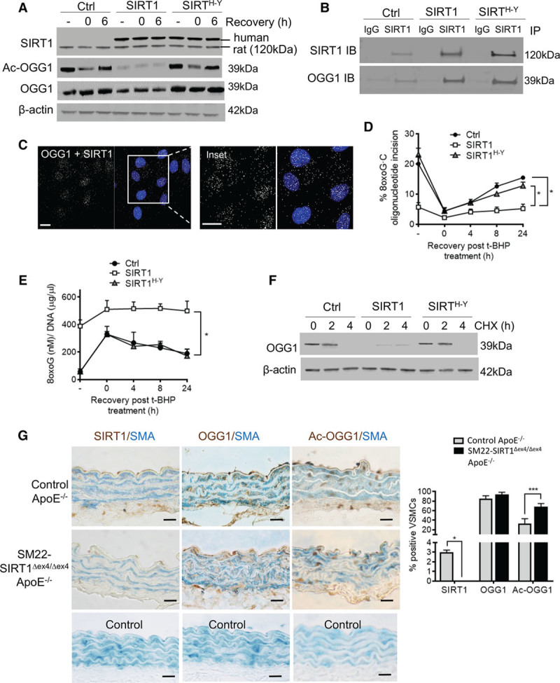
Background: Atherosclerotic plaques demonstrate extensive accumulation of oxidative DNA damage, predominantly as 8-oxoguanine (8oxoG) lesions. 8oxoG is repaired by base excision repair enzymes; however, the mechanisms regulating 8oxoG accumulation in vascular smooth muscle cells (VSMCs) and its effects on their function and in atherosclerosis are unknown.

Methods: We studied levels of 8oxoG and its regulatory enzymes in human atherosclerosis, the mechanisms regulating 8oxoG repair and the base excision repair enzyme 8oxoG DNA glycosylase I (OGG1) in VSMCs in vitro, and the effects of reducing 8oxoG in VSMCs in atherosclerosis in ApoE-/- mice.

Results: Human plaque VSMCs showed defective nuclear 8oxoG repair, associated with reduced acetylation of OGG1. OGG1 was a key regulatory enzyme of 8oxoG repair in VSMCs, and its acetylation was crucial to its repair function through regulation of protein stability and expression. p300 and sirtuin 1 were identified as the OGG1 acetyltransferase and deacetylase regulators, respectively, and both proteins interacted with OGG1 and regulated OGG1 acetylation at endogenous levels. However, p300 levels were decreased in human plaque VSMCs and in response to oxidative stress, suggesting that reactive oxygen species-induced regulation of OGG1 acetylation could be caused by reactive oxygen species-induced decrease in p300 expression. We generated mice that express VSMC-restricted OGG1 or an acetylation defective version (SM22α-OGG1 and SM22α-OGG1K-R mice) and crossed them with ApoE-/- mice. We also studied ApoE-/- mice deficient in OGG1 (OGG1-/-). OGG1-/- mice showed increased 8oxoG in vivo and increased atherosclerosis, whereas mice expressing VSMC-specific OGG1 but not the acetylation mutant OGG1K-R showed markedly reduced intracellular 8oxoG and reduced atherosclerosis. VSMC OGG1 reduced telomere 8oxoG accumulation, DNA strand breaks, cell death and senescence after oxidant stress, and activation of proinflammatory pathways.

Conclusions: We identify defective 8oxoG base excision repair in human atherosclerotic plaque VSMCs, OGG1 as a major 8oxoG repair enzyme in VSMCs, and p300/sirtuin 1 as major regulators of OGG1 through acetylation/deacetylation. Reducing oxidative damage by rescuing OGG1 activity reduces plaque development, indicating the detrimental effects of 8oxoG on VSMC function.

Keywords: DNA damage; DNA glycosylases; atherosclerosis; oxidative stress; vascular diseases.


Figures

Figure 1.
Figure 1.
Human plaque VSMCs show defective nuclear BER activity and reduced acetyl-OGG1 expression. A, Base excision repair (BER) assay measuring incision of a fluorescently labeled 8-oxoguanine (8oxoG.C) oligonucleotide in nuclear and cytoplasmic fractions of cultured plaque and normal aortic vascular smooth muscle cells (VSMCs). Representative gel (Left) and quantification (Right) are shown. Incision product is denoted by an arrow (n=4). B, Quantitative polymerase chain reaction analysis of 8oxoG DNA glycosylase I (OGG1) expression in cultured plaque and normal aortic VSMCs (n=4). C, Western blot of OGG1 and acetylated OGG1 (Ac-OGG1) in plaque and normal aortic VSMCs (n=4). D, Immunohistochemistry for 8oxoG, Ac-OGG1, or OGG1 (brown) in sections of human plaques and normal aorta (n=10). Sections are also costained for α-smooth muscle actin (SMA; blue). Insets show high-power views of outlined areas. Negative controls using isotype-matched antibodies and quantification are shown below. Scale bars, 25 µm. All graphical data are mean±SEM. *P<0.05, **P<0.01, Student t test.
Figure 2.
Figure 2.
OGG1 is the major BER enzyme for 8oxoG, and its activity is regulated by acetylation. A, 8-Oxoguanine (8oxoG) DNA glycosylase I (OGG1) protein expression in control or OGG1 exon1 or exon7 CRISPR knockout rat vascular smooth muscle cell lines (n=4). B, Base excision repair (BER) assay analysis and (C) 8oxoG intracellular levels measured by ELISA in control (Ctrl), OGG1Exon1KO, or OGG1Exon7KO rat vascular smooth muscle cells (VSMCs) at untreated baseline (-) or after tert-butyl hydroperoxide (t-BHP) treatment for 1 hour (t=0) and after t-BHP removal (4-, 8-, 24-hour recovery; n=5). D, Western blot of OGG1, acetylated OGG1 (Ac-OGG1), or myc tag in rat VSMCs expressing the empty vector (Ctrl), human OGG1 (OGG1), or acetylation mutant OGG1 (OGG1K-R). Immunoblotting for OGG1 shows a lower band for endogenous rat OGG1 and a higher band for exogenous human OGG1 (n=4). E, BER assay analysis and (F) 8oxoG intracellular levels measured by ELISA in Ctrl, OGG1, or OGG1K-R cells at untreated baseline (-) or after t-BHP treatment for 1 hour (t=0) and after t-BHP removal (4-, 8-, 24-hour recovery; n=4). All graphical data are mean±SEM. *P<0.05, **P<0.01, ***P<0.001, 1-way ANOVA (Bonferroni post hoc).
Figure 3.
Figure 3.
p300 is downregulated in plaque VSMCs and regulates OGG1 acetylation and BER. A, Western blot of p300 expression in cultured human plaque or normal aortic vascular smooth muscle cells (VSMCs; n=4). B, Western blot of 8-oxoguanine (8oxoG) DNA glycosylase I (OGG1), acetylated (Ac-) OGG1, and p300 levels in human VSMCs after 1 hour of tert-butyl hydroperoxide (t-BHP) treatment and 0- to 24-hour recovery (n=4). C, Immunoprecipitation (IP) of human vascular smooth muscle cell lysates with an anti-p300 antibody analyzed by immunoblotting (IB) with anti-OGG1 and anti-p300 antibodies (n=3). D, Proximity ligation assay of OGG1-p300 interaction with rabbit anti-OGG1 and mouse anti-p300 antibodies (n=3). Scale bar, 20µm. E, Western blot for Ac-OGG1 and Ac-histone 4 (H4) in VSMCs expressing OGG1 or OGG1K-R with or without CTPB treatment (10 μmol/L; n=3). F, Quantification of base excision repair(BER) assay in OGG1 or OGG1K-R VSMCs either untreated (-) or after 1 hour of t-BHP (0) or 6-hour recovery with/without CTPB (n=3). Representative gel is shown in Figure IVa in the online-only Data Supplement. G, Western blot of OGG1 expression after cycloheximide (CHX) treatment (0–4 hours) in OGG1 or OGG1K-R VSMCs with/without the p300 activator CTPB (n=3). H, Western blot of OGG1 expression after CHX treatment (0–4 hours) in control cells with/without CTPB with/without the proteasomal degradation inhibitor MG132 (10 μmol/L; n=3). Scale bars, 25 µm. All graphical data are mean±SEM. *P<0.05, Student t test.
Figure 4.
Figure 4.
SIRT1 binds OGG1 and regulates deacetylation of OGG1 in vitro and in vivo. A, Western blot of total sirtuin 1 (SIRT1), acetylated (Ac-) 8-oxoguanine (8oxoG) DNA glycosylase I (OGG1), and OGG1 expression in rat vascular smooth muscle cells (VSMCs) expressing the empty vector (control [Ctrl]), human SIRT1 (SIRT1), or deacetylase defective mutant SIRTH364Y cells after 1 hour of tert-butyl hydroperoxide (t-BHP) treatment and after 0- to 6-hour recovery (n=3). Immunoblotting for SIRT1 shows a lower band for endogenous rat SIRT1 and a higher band for exogenous human SIRT1. B, Immunoprecipitation (IP) of human vascular smooth muscle cell lysates with an anti-SIRT1 antibody analyzed by immunoblotting (IB) with anti-SIRT1 and anti-OGG1 antibodies (n=3). C, Proximity ligation assay of OGG1-SIRT1 interaction with rabbit anti-OGG1 and mouse anti-SIRT1 antibodies (n=3). D, Quantification of base excision repairassay in Ctrl, SIRT1, or SIRT1H-Y VSMCs either untreated (-) or after 1 hour of t-BHP (0) or 0- to 24-hour recovery (n=3). Representative gel is shown in Figure Vb in the online-only Data Supplement. E, 8oxoG intracellular levels measured by ELISA in control, SIRT1, or SIRT1H-Y VSMCs after 1 hour of t-BHP treatment and after 0- to 24-hour recovery (n=3). F, Western blot of OGG1 expression after cycloheximide (CHX; 0–4 hours) treatment of control, SIRT1, or SIRT1H-Y VSMCs (n=3). G, Immunohistochemistry for SIRT1, OGG1, or Ac-OGG1 (brown) in aortas from control ApoE−/− or SIRT1−/−/ApoE−/− mice (n=10). Tissue sections were also costained for α-smooth muscle actin (SMA; blue). Negative control sections with isotype-matched antibodies are shown below. Scale bar, 25 µm. All graphical data are mean±SEM. *P<0.05, ***P<0.001, 1-way ANOVA (Bonferroni post hoc).
Figure 5.
Figure 5.
OGG1 regulates 8oxoG expression in VSMCs in vivo and BER. A, Western blot of 8-oxoguanine (8oxoG) DNA glycosylase I (OGG1), acetylated (Ac-) OGG1, and myc tag in vascular smooth muscle cells (VSMCs) cultured from wild-type (control), OGG1−/−, SM22α-OGG1, or SM22α-OGG1K-R mouse aortas (n=4). B, 8oxoG intracellular levels measured by ELISA in control, OGG1−/−, SM22α-OGG1, or SM22α-OGG1K-R VSMCs (n=4). C, Base excision repair(BER) assay quantification in control, OGG1−/−, SM22α-OGG1, or SM22α-OGG1K-R VSMCs treated with tert-butyl hydroperoxide (t-BHP) for 1 hour and recovered for up to 24 hours (n=4). All graphical data are mean±SEM. *P<0.05, 1-way ANOVA (Bonferroni post hoc).
Figure 6.
Figure 6.
Effects of 8-oxoguanine DNA glycosylase I (OGG1) on plaque development and morphology in vivo. A, Representative images and quantification of en face preparations of descending aortas from control ApoE−/− (n=12), OGG1−/− ApoE−/− (n=14), SM22α-OGG1 ApoE−/− (n=12), and SM22α-OGG1K-R ApoE−/− (n=13) mice stained with Oil Red O. Scale bar, 2 mm. B, Hematoxylin-eosin (H&E) and Masson trichrome immunohistochemistry of control ApoE−/− (n=12), OGG1−/− ApoE−/− (n=14), SM22α-OGG1 ApoE−/− (n=12), and SM22α-OGG1K-R ApoE−/− (n=13) mouse aortic roots at 22 weeks after fat feeding from 8 to 22 weeks. Quantification of percent plaque size, plaque area, core area, and cap area (micrometers squared). Scale bar, 200 µm. All graphical data are mean±SEM. **P<0.01, ***P<0.001, 1-way ANOVA (Bonferroni post hoc).
Figure 7.
Figure 7.
8-Oxoguanine (8oxoG) DNA glycosylase I (OGG1) regulates DNA strand breaks, cell senescence, apoptosis, and inflammasome pathways. A, Comet assay with quantification for cultured vascular smooth muscle cells (VSMCs) from wild-type control, OGG1−/−, SM22α-OGG1, or SM22α-OGG1K-R mice treated with tert-butyl hydroperoxide (t-BHP) for 1 hour and recovered for 6 hours (n=3). Scale bar, 100 µm. UT indicates untreated. B, Chromatin immunoprecipitation–quantitative polymerase chain reaction using primers to a telomeric region on 8oxoG-bound chromatin from control, OGG1−/−, SM22α-OGG1, or SM22α-OGG1K-R mouse VSMCs (n=3). Data are shown as fold-change over immunoglobulin G (IgG). C, Percent VSMCs expressing senescence-associated β-galactosidase activity (SaβG) from wild-type control, OGG1−/−, SM22α-OGG1, or SM22α-OGG1K-R mice (n=3). D, Apoptosis assayed by annexin V and propidium iodide staining of mouse cells with/without t-BHP treatment by flow cytometry (n=3). E, Relative expression of transcripts for inflammatory cytokines and inflammasome-associated components in aortic arch tissue from control ApoE−/−, OGG1−/− ApoE−/−, SM22α-OGG1 ApoE−/−, and SM22α-OGG1K-R ApoE−/− mice after fat feeding (n=4). All graphical data are mean±SEM. *P<0.05, **P<0.01, ***P<0.001, 1-way ANOVA (Bonferroni post hoc).
Figure 8.
Figure 8.
Model of 8-oxoguanine (8oxoG) DNA glycosylase I (OGG1) regulation in VSMCs in atherosclerosis. Ac indicated acetylated; BER, base excision repair; IL, interleukin; Lys, lysine; and SIRT1, sirtuin 1.

Comment in

Similar articles

Cited by

References

    1. Neeley WL, Essigmann JM. Mechanisms of formation, genotoxicity, and mutation of guanine oxidation products. Chem Res Toxicol. 2006;19:491–505. doi: 10.1021/tx0600043. - PubMed
    1. Grollman AP, Moriya M. Mutagenesis by 8-oxoguanine: an enemy within. Trends Genet. 1993;9:246–249. - PubMed
    1. Hoeijmakers JH. Genome maintenance mechanisms for preventing cancer. Nature. 2001;411:366–374. doi: 10.1038/35077232. - PubMed
    1. Vartanian V, Lowell B, Minko IG, Wood TG, Ceci JD, George S, Ballinger SW, Corless CL, McCullough AK, Lloyd RS. The metabolic syndrome resulting from a knockout of the NEIL1 DNA glycosylase. Proc Natl Acad Sci USA. 2006;103:1864–1869. doi: 10.1073/pnas.0507444103. - PMC - PubMed
    1. Martinet W, Knaapen MW, De Meyer GR, Herman AG, Kockx MM. Oxidative DNA damage and repair in experimental atherosclerosis are reversed by dietary lipid lowering. Circ Res. 2001;88:733–739. - PubMed

Publication types

MeSH terms