. 2018 Apr 24;115(17):E3905-E3913.
doi: 10.1073/pnas.1716164115. Epub 2018 Apr 11.

Widespread changes in transcriptome profile of human mesenchymal stem cells induced by two-dimensional nanosilicates

Affiliations

Widespread changes in transcriptome profile of human mesenchymal stem cells induced by two-dimensional nanosilicates

James K Carrow et al. Proc Natl Acad Sci U S A. .

Abstract

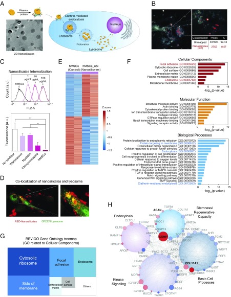
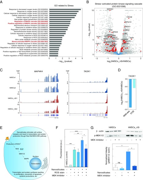
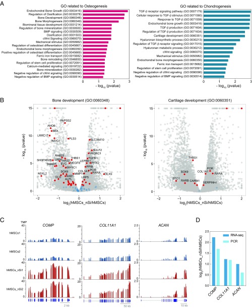
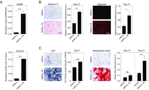
Two-dimensional nanomaterials, an ultrathin class of materials such as graphene, nanoclays, transition metal dichalcogenides (TMDs), and transition metal oxides (TMOs), have emerged as a new generation of materials due to their unique properties relative to macroscale counterparts. However, little is known about the transcriptome dynamics following exposure to these nanomaterials. Here, we investigate the interactions of 2D nanosilicates, a layered clay, with human mesenchymal stem cells (hMSCs) at the whole-transcriptome level by high-throughput sequencing (RNA-seq). Analysis of cell-nanosilicate interactions by monitoring changes in transcriptome profile uncovered key biophysical and biochemical cellular pathways triggered by nanosilicates. A widespread alteration of genes was observed due to nanosilicate exposure as more than 4,000 genes were differentially expressed. The change in mRNA expression levels revealed clathrin-mediated endocytosis of nanosilicates. Nanosilicate attachment to the cell membrane and subsequent cellular internalization activated stress-responsive pathways such as mitogen-activated protein kinase (MAPK), which subsequently directed hMSC differentiation toward osteogenic and chondrogenic lineages. This study provides transcriptomic insight on the role of surface-mediated cellular signaling triggered by nanomaterials and enables development of nanomaterials-based therapeutics for regenerative medicine. This approach in understanding nanomaterial-cell interactions illustrates how change in transcriptomic profile can predict downstream effects following nanomaterial treatment.

Keywords: 2D nanomaterials; RNA-seq; human mesenchymal stem cells; nanosilicates; whole-transcriptome sequencing.


Conflict of interest statement

Conflict of interest statement: J.K.C. and A.K.G. are coauthors on US Patent Application No. WO2017112802 A1 published on June 29, 2017 (US Provisional Patent Application No. 62/270,403 filed on December 21, 2015).

Figures

Fig. 1.
Fig. 1.
Biophysical interaction of nanosilicates and hMSCs. (A) Two-dimensional nanosilicates electrostatically bind to proteins from biological fluids and are subsequently internalized by cells via surface-mediated endocytosis. (B) Hyperspectral imaging indicating distribution of nanosilicates throughout the cell body following endocytosis. The image was captured from transverse section of cell body. (C) Flow cytometry analysis of rhodamine-tagged nanosilicates demonstrate dose-dependent cellular uptake. The nanosilicates were primarily internalized via clathrin-mediated process (chlorpromazine) as opposed to macropinocytosis (wortmannin) or caveolar-mediated (nystatin). **P < 0.01; ***P < 0.001. (D) LAMP1 staining (green) for lysosomal membranes further tracks nanosilicates (red) following endocytosis. (E) Row-scaled z-scores of quantile normalized gene expression [in log2(RPKM)] of >4,000 genes following treatment with nanosilicates (padjust < 0.05, red, up-regulated: 1,897 genes; blue, down-regulated: 2,171 genes). (F) Significant GO terms of associated biological processes, cellular components, and molecular functions from differentially regulated genes (P < 0.05). Terms related to biological process and cellular components indicate strong biophysical interactions between cells and nanosilicates. (G) Clustering of significant 244 cellular component gene ontology (GO) terms into broader cellular component categories. (H) Gene network displaying interconnected genetic targets after nanosilicate treatment with high degrees of expression and statistical significance (red, up-regulated; blue, down-regulated; size increases with significance).
Fig. 2.
Fig. 2.
Nanosilicates lead to stress-induced MAPK signaling. (A) Nanosilicate treatment results in activation of stress-related response. A list of significant GO terms related to stress after nanosilicate treatment indicate signal propagation via MAPK/ERK signaling pathways. (B) The majority of genes involved in stress-activated kinase signaling cascade (GO:0031098) undergo a significant differential expression. (C) The change in gene expression profile of MAP4K4 and TAOK1 (aligned reads normalized by total library size). (D) Comparison of TAOK1 gene expression obtained from RNA-seq was validated using qRT-PCR. (E) Nanosilicates trigger a stress-responsive kinase cascade (Ras–Raf–MEK–ERK pathways), leading to changes in reactive oxygen species (ROS) production and subsequent RNA transcription and protein synthesis. (F) Flow-cytometric analysis was performed to measure the stress-responsive kinase cascade, by measuring ROS production with a ROS-sensitive fluorescent reporter dye. Experiments were performed in the presence or absence of a MAPK inhibitor. A significant increase in ROS-mediated fluorescent signal is observed upon exposure to nanosilicate, and this is abrogated after treatment with the MAPK inhibitor. *P < 0.05; **P < 0.01; ***P < 0.001; ****P < 0.0001. (G) Production of p-MEK1/2 was determined using Western blot in presence of nanosilicates and MEK inhibitor, establishing the role of nanosilicate in MAPK/ERK signaling. *P < 0.05. ns, not significant.
Fig. 3.
Fig. 3.
Transcriptomic analysis elucidates nanosilicate-induced bioactivity. (A) GO terms related to osteogenesis and chondrogenesis indicate nanosilicate-induced hMSC differentiation. (B) Significant gene expression changes in genes involved in bone development (GO:0060348) and cartilage development (GO:0060351). (C) Gene expression profile of COMP, COL11A1, and ACAN, demonstrating up-regulation due to nanosilicate treatment (aligned reads normalized by total library size). (D) Differential gene expression from RNA-seq was validated using qRT-PCR, indicating similar trend.
Fig. 4.
Fig. 4.
Nanosilicate-induced hMSCs differentiation. (A) Western blot showing production of COL1A1 and COMP after exposure to nanosilicates for 7 d in normal media. (B) The effect of nanosilicates on production of GAGs was determined by safranin O and aggrecan staining after culturing hMSCs in chondro-conductive media for 21 d. (C) The effect of nanosilicates on osteogenic differentiation was determined by ALP activity and formation of mineralized matrix after culturing hMSCs in osteo-conductive media for 21 d. *P < 0.05; ***P < 0.001; ****P < 0.0001. ns, not significant.

Similar articles

Cited by

References

    1. Butler SZ, et al. Progress, challenges, and opportunities in two-dimensional materials beyond graphene. ACS Nano. 2013;7:2898–2926. - PubMed
    1. Chen Y, Tan C, Zhang H, Wang L. Two-dimensional graphene analogues for biomedical applications. Chem Soc Rev. 2015;44:2681–2701. - PubMed
    1. Chimene D, Alge DL, Gaharwar AK. Two-dimensional nanomaterials for biomedical applications: Emerging trends and future prospects. Adv Mater. 2015;27:7261–7284. - PubMed
    1. Jiang W, Kim BYS, Rutka JT, Chan WCW. Nanoparticle-mediated cellular response is size-dependent. Nat Nanotechnol. 2008;3:145–150. - PubMed
    1. Kerativitayanan P, Carrow JK, Gaharwar AK. Nanomaterials for engineering stem cell responses. Adv Healthc Mater. 2015;4:1600–1627. - PubMed

Publication types

MeSH terms