

Regulator of calcineurin-2 is a centriolar protein with a role in cilia length control
- PMID: 29643119
- PMCID: PMC5992583
- DOI: 10.1242/jcs.212258
Regulator of calcineurin-2 is a centriolar protein with a role in cilia length control
Abstract
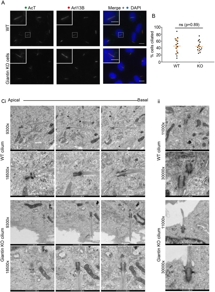
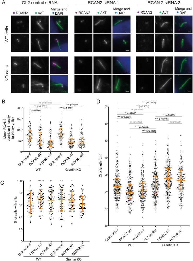
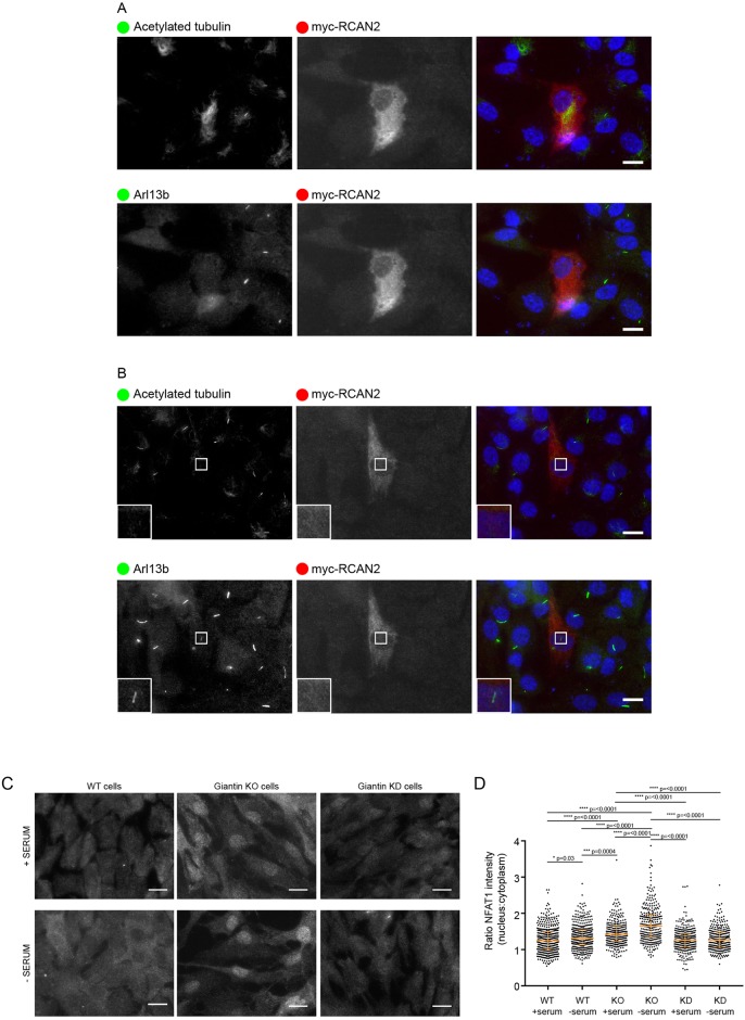
Almost every cell in the human body extends a primary cilium. Defective cilia function leads to a set of disorders known as ciliopathies, which are characterised by debilitating developmental defects that affect many tissues. Here, we report a new role for regulator of calcineurin 2 (RCAN2) in primary cilia function. It localises to centrioles and the basal body and is required to maintain normal cilia length. RCAN2 was identified as the most strongly upregulated gene from a comparative RNAseq analysis of cells in which expression of the Golgi matrix protein giantin had been abolished by gene editing. In contrast to previous work where we showed that depletion of giantin by RNAi results in defects in ciliogenesis and in cilia length control, giantin knockout cells generate normal cilia after serum withdrawal. Furthermore, giantin knockout zebrafish show increased expression of RCAN2. Importantly, suppression of RCAN2 expression in giantin knockout cells results in the same defects in the control of cilia length that are seen upon RNAi of giantin itself. Together, these data define RCAN2 as a regulator of cilia function that can compensate for the loss of giantin function.
Keywords: Calcineurin; Cilia; Giantin; Golgi; RCAN2.
© 2018. Published by The Company of Biologists Ltd.
Conflict of interest statement
Competing interestsThe authors declare no competing or financial interests.
Figures

Similar articles
-
A role for the Golgi matrix protein giantin in ciliogenesis through control of the localization of dynein-2.J Cell Sci. 2013 Nov 15;126(Pt 22):5189-97. doi: 10.1242/jcs.131664. Epub 2013 Sep 17. J Cell Sci. 2013. PMID: 24046448 Free PMC article.
-
The Golgi matrix protein giantin is required for normal cilia function in zebrafish.Biol Open. 2017 Aug 15;6(8):1180-1189. doi: 10.1242/bio.025502. Biol Open. 2017. PMID: 28546340 Free PMC article.
-
A ciliopathy complex builds distal appendages to initiate ciliogenesis.J Cell Biol. 2021 Sep 6;220(9):e202011133. doi: 10.1083/jcb.202011133. Epub 2021 Jul 9. J Cell Biol. 2021. PMID: 34241634 Free PMC article.
-
Primary cilia biogenesis and associated retinal ciliopathies.Semin Cell Dev Biol. 2021 Feb;110:70-88. doi: 10.1016/j.semcdb.2020.07.013. Epub 2020 Jul 31. Semin Cell Dev Biol. 2021. PMID: 32747192 Free PMC article. Review.
-
Scoring a backstage pass: mechanisms of ciliogenesis and ciliary access.J Cell Biol. 2012 Jun 11;197(6):697-709. doi: 10.1083/jcb.201111146. J Cell Biol. 2012. PMID: 22689651 Free PMC article. Review.
Cited by
-
Regulator of calcineurin 3 as a novel predictor of diagnosis and prognosis in pan-cancer.Croat Med J. 2024 Aug 31;65(4):356-372. doi: 10.3325/cmj.2024.65.356. Croat Med J. 2024. PMID: 39219199 Free PMC article.
-
Systematic Discovery of Short Linear Motifs Decodes Calcineurin Phosphatase Signaling.Mol Cell. 2020 Jul 16;79(2):342-358.e12. doi: 10.1016/j.molcel.2020.06.029. Epub 2020 Jul 8. Mol Cell. 2020. PMID: 32645368 Free PMC article.
-
Regulator of Calcineurin (RCAN): Beyond Down Syndrome Critical Region.Mol Cells. 2020 Aug 31;43(8):671-685. doi: 10.14348/molcells.2020.0060. Mol Cells. 2020. PMID: 32576715 Free PMC article. Review.
-
The factory, the antenna and the scaffold: the three-way interplay between the Golgi, cilium and extracellular matrix underlying tissue function.Biol Open. 2023 Feb 15;12(2):bio059719. doi: 10.1242/bio.059719. Epub 2023 Feb 21. Biol Open. 2023. PMID: 36802341 Free PMC article. Review.
-
Zebrafish as an Emerging Model for Osteoporosis: A Primary Testing Platform for Screening New Osteo-Active Compounds.Front Endocrinol (Lausanne). 2019 Jan 29;10:6. doi: 10.3389/fendo.2019.00006. eCollection 2019. Front Endocrinol (Lausanne). 2019. PMID: 30761080 Free PMC article. Review.
References
-
- Asante D., Maccarthy-Morrogh L., Townley A. K., Weiss M. A., Katayama K., Palmer K. J., Suzuki H., Westlake C. J. and Stephens D. J. (2013). A role for the Golgi matrix protein giantin in ciliogenesis through control of the localization of dynein-2. J. Cell Sci. 126, 5189-5197. 10.1242/jcs.131664 - DOI - PMC - PubMed
Publication types
MeSH terms
Substances
Grants and funding
- BB/N000420/1/BB_/Biotechnology and Biological Sciences Research Council/United Kingdom
- 21937/VAC_/Versus Arthritis/United Kingdom
- BB/L014181/1/BB_/Biotechnology and Biological Sciences Research Council/United Kingdom
- 21211/VAC_/Versus Arthritis/United Kingdom
- MR/K018019/1/MRC_/Medical Research Council/United Kingdom
LinkOut - more resources
Full Text Sources
Other Literature Sources
Molecular Biology Databases
