Review
. 2018 Apr 11;82(2):e00068-17.
doi: 10.1128/MMBR.00068-17. Print 2018 Jun.

Fungal Morphogenesis, from the Polarized Growth of Hyphae to Complex Reproduction and Infection Structures

Affiliations
Review

Fungal Morphogenesis, from the Polarized Growth of Hyphae to Complex Reproduction and Infection Structures

Meritxell Riquelme et al. Microbiol Mol Biol Rev. .

Abstract

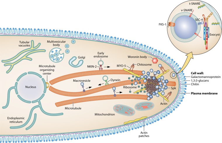
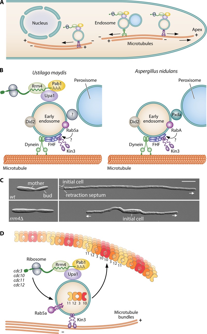
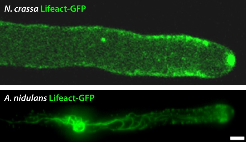
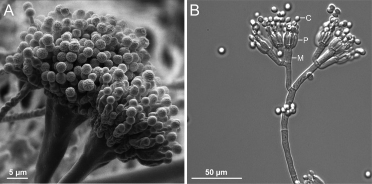
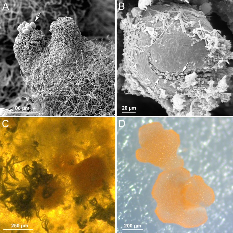
Filamentous fungi constitute a large group of eukaryotic microorganisms that grow by forming simple tube-like hyphae that are capable of differentiating into more-complex morphological structures and distinct cell types. Hyphae form filamentous networks by extending at their tips while branching in subapical regions. Rapid tip elongation requires massive membrane insertion and extension of the rigid chitin-containing cell wall. This process is sustained by a continuous flow of secretory vesicles that depends on the coordinated action of the microtubule and actin cytoskeletons and the corresponding motors and associated proteins. Vesicles transport cell wall-synthesizing enzymes and accumulate in a special structure, the Spitzenkörper, before traveling further and fusing with the tip membrane. The place of vesicle fusion and growth direction are enabled and defined by the position of the Spitzenkörper, the so-called cell end markers, and other proteins involved in the exocytic process. Also important for tip extension is membrane recycling by endocytosis via early endosomes, which function as multipurpose transport vehicles for mRNA, septins, ribosomes, and peroxisomes. Cell integrity, hyphal branching, and morphogenesis are all processes that are largely dependent on vesicle and cytoskeleton dynamics. When hyphae differentiate structures for asexual or sexual reproduction or to mediate interspecies interactions, the hyphal basic cellular machinery may be reprogrammed through the synthesis of new proteins and/or the modification of protein activity. Although some transcriptional networks involved in such reprogramming of hyphae are well studied in several model filamentous fungi, clear connections between these networks and known determinants of hyphal morphogenesis are yet to be established.

Keywords: cell wall; cytoskeleton; fungal development; hyphal morphogenesis; polarity.


Figures

FIG 1
FIG 1
VSC (vesicle supply center) model for hyphal morphogenesis. (A) The “hyphoid,” a perfect hyphal shape. The hyphoid curve is a geometric function derived from a computer-simulated secretory process, where growth units (vesicles) emanating from a forward-moving source (the VSC) extend the cell surface in a sharply polarized manner. When analyzed mathematically, the process yielded the hyphoid equation y = x cot (x V/N), where N is the amount of cell wall-building vesicles produced per unit of time and V is the rate of advancement of the VSC; when plotted on Cartesian coordinates, the function generates a unique curve that follows closely the actual profile of regular hyphae (Adapted from reference .) (B) Displacement and advancement of the VSC from its concentric position in a spore generate a germinating tube. (C) The formation and advancement of a new VSC at a subapical hyphal region generate a lateral branch.
FIG 2
FIG 2
Architecture of a fungal hyphal tip. Different populations of vesicles concentrate at the Spitzenkörper. Examples of the main organelles of the secretory pathway and the cytoskeleton are displayed. The circle shows a macrovesicle carrying β-1,3-glucan synthase and the protein complexes required for vesicle fusion with the plasma membrane.
FIG 3
FIG 3
Cell end marker proteins determine the interplay between the microtubule and the actin cytoskeleton in A. nidulans. (A) Scheme of cell end markers transported at the MT plus end and delivered to the apical membrane. The prenylated TeaR proteins are probably delivered with vesicles. The motor protein KipA transports TeaA and probably other tip proteins toward the MT plus end. (B) Differential interference contrast images of wild-type, ΔteaA, and ΔteaR strains. ΔteaA strains exhibited zigzag and ΔteaR strains curved hyphae. (C) Monomeric red fluorescent protein 1 (mRFP1)-TeaA or GFP-TeaR localizes to one point at the tip and along the tip membrane. (D) Series of PALM images of an mEosFP-TeaR-expressing hypha (5-min time interval). Cell profiles are shown in different line styles. The right column shows overlays of PALM images from two time points (top, 0 and 5 min; middle, 5 and 10 min), and overlays of outlines reveal growth regions coinciding with TeaR cluster locations. (Panels A to C are modified from reference with permission; panel D is modified from reference .)
FIG 4
FIG 4
The microtubule cytoskeleton. (A) Microtubule cytoskeleton and nuclear organization in Neurospora crassa and Aspergillus nidulans. Bars = 5 μm. (Courtesy of Rosa M. Ramírez Cota; reprinted with permission.) (B) Microtubule-organizing centers in A. nidulans. Shown is a germling with two mitotic spindles. Microtubules were stained with GFP-tagged alpha tubulin, and the SPBs were labeled with mRFP-tagged ApsB. The right fluorescent pictures show mitotic and astral microtubules emanating from the SPBs (labeled with GFP-Spa10) and cytoplasmic microtubules emanating from a septal MTOC. (C) Schemes of a SPB and a sMTOC.
FIG 5
FIG 5
mRNA transport. (A) Scheme of MT-based transport in fungal hyphae. (B) Transport of endosomes in Ustilago maydis and Aspergillus nidulans. (C) Rrm4 is important for efficient polar growth. (D) RNA transport ensures septin gradient formation. (Panels A and B [left] are modified from reference ; panel C is modified from reference ; panel D is adapted from reference with permission.)
FIG 6
FIG 6
Inositol pyrophosphate-regulated cell morphogenesis in the fission yeast Schizosaccharomyces pombe. The transition from mono- to bipolar growth (NETO [new end take off]) requires inositol pyrophosphate generated by the S. pombe Asp1 protein, as does the switch from single-celled yeast growth to invasive pseudohyphal growth (dimorphic switch).
FIG 7
FIG 7
Actin organization at hyphal apical and subapical regions in N. crassa and A. nidulans. Bar = 5 μm. (Courtesy of L. Quintanilla and B. D. Shaw; reprinted with permission.)
FIG 8
FIG 8
The activity of the transcription factor BrlA results in different conidiophore morphological patterns in Aspergillus nidulans (scanning electron micrograph) (image by G. H. Braus) (A) and Penicillium chrysogenum (light microscopic image) (image by U. Kück) (B). The P. chrysogenum conidiophore shows a simpler structure, lacking a multinucleated vesicle and producing fewer metulae (M), phialides (P), and conidia (C).
FIG 9
FIG 9
Fruiting bodies from diverse ascomycetes. (A) Perithecia from Sordaria macrospora. (Image by U. Kück.) (B) Cleistothecium from Aspergillus nidulans. (Image by G. H. Braus.) (C) Cleistothecia from Penicillium chrysogenum. (Image by U. Kück.) (D) Apothecium from Pyronema confluens (adapted from reference 446). Panels A and B show scanning electron micrographs, and panels C and D show light microscopic images. Arrows in panels A and B indicate ascospores that are actively discharged from the S. macrospora perithecium, while ascospores from A. nidulans are distributed when the mature fruiting body disintegrates.
FIG 10
FIG 10
Hierarchical regulatory network controlling different cell types of Ustilago maydis. During the fusion of two nonpathogenic, yeast-like sporidia, the binding of peptide pheromones to the pheromone receptor (Pra) activates a MAP kinase (MAPK) cascade, and environmental signals feed via a so-far-unidentified receptor into a cAMP-dependent protein kinase (PKA) pathway. Both pathways converge at Prf1, the central transcription factor required for mating-dependent gene expression. Prf1 is additionally regulated at the transcriptional level by the transcription factors Rop1 and Hap2, both of which are thought to be targets of the MAPK cascade. The phosphorylation of Prf1 results in the induction of the bE and bW genes; after the fusion of two sporidia, bE and bW form a heterodimeric transcription factor that is the master regulator to induce filamentous growth and pathogenic development. The central node for gene regulation at the onset of pathogenic development is the transcription factor Rbf1. rbf1 gene expression is already induced by Prf1 prior to cell fusion, but expression is boosted after cell fusion and the formation of the filamentous dikaryon by the bE/bW heterodimer. Rbf1 is sufficient to trigger the expression of all genes required for plant penetration. Among the Rbf1-induced genes are the transcription factors Biz1 and Hdp2, both of which are absolutely required for plant infection. Hdp2 and Biz1 are highly expressed at the later stages of in planta development, while rbf1 expression is barely detectable within the plant. biz1 and hdp2 are also regulated by physical cues on the plant surface via the plasma membrane receptor Sho1/Msb2. Two transcription factors specifically expressed during the biotrophic phase are Fox1, which is thought to contribute to the regulation of several effector genes, and Ros1, a regulator for the late developmental steps during spore formation.
FIG 11
FIG 11
The injury response in Trichoderma. (A) A colony of T. atroviride (IMI206040) growing in the dark was damaged with a cookie mold and photographed 48 h later (right). An undamaged control is also shown (left). (B) Microscopic changes observed upon injury. One hour after injury, hyphae were stained with lactophenol cotton blue and examined under a light microscope. Arrows indicate newly formed hypha. (C) Illustration of the regeneration and conidiation processes of T. atroviride after injury. Upon damage, ATP is released (exogenous ATP [eATP]), triggering an increase in the level of cytosolic calcium required for regeneration (E. Medina-Castellanos, J. M. Villalobos-Escobedo, M. Riquelme, N. D. Read, C. Abreu-Goodger, and A. Herrera-Estrella, unpublished data). ROS and oxylipins (Oxy) play an important role in cell differentiation during conidiophore formation in response to injury. (Adapted from reference .)
None
None
None
None
None
None
None
None
None
None
None
None
None

Similar articles

Cited by

References

    1. Fisher MC, Henk DA, Briggs CJ, Brownstein JS, Madoff LC, McCraw SL, Gurr SJ. 2012. Emerging fungal threats to animal, plant and ecosystem health. Nature 484:186–194. doi:10.1038/nature10947. - DOI - PMC - PubMed
    1. Kupferschmidt K. 2012. Attack of the clones. Science 337:636–638. doi:10.1126/science.337.6095.636. - DOI - PubMed
    1. Cardwell KF, Desjardins A, Henry SH, Munkvold G, Robens J. 2001. Mycotoxins: the cost of achieving food security and food quality. APSnet Feature doi:10.1094/APSnetFeature-2001-0901. - DOI
    1. Woolhouse M, Gaunt E. 2007. Ecological origins of novel human pathogens. Crit Rev Microbiol 33:231–242. doi:10.1080/10408410701647560. - DOI - PubMed
    1. Bartnicki-Garcia S, Lippman E. 1969. Fungal morphogenesis: cell wall construction in Mucor rouxii. Science 165:302–304. doi:10.1126/science.165.3890.302. - DOI - PubMed

Publication types