. 2018 Jun 8;293(23):8843-8860.
doi: 10.1074/jbc.M117.807503. Epub 2018 Apr 11.

Structural control of caspase-generated glutamyl-tRNA synthetase by appended noncatalytic WHEP domains

Affiliations

Structural control of caspase-generated glutamyl-tRNA synthetase by appended noncatalytic WHEP domains

Dalia Halawani et al. J Biol Chem. .

Abstract

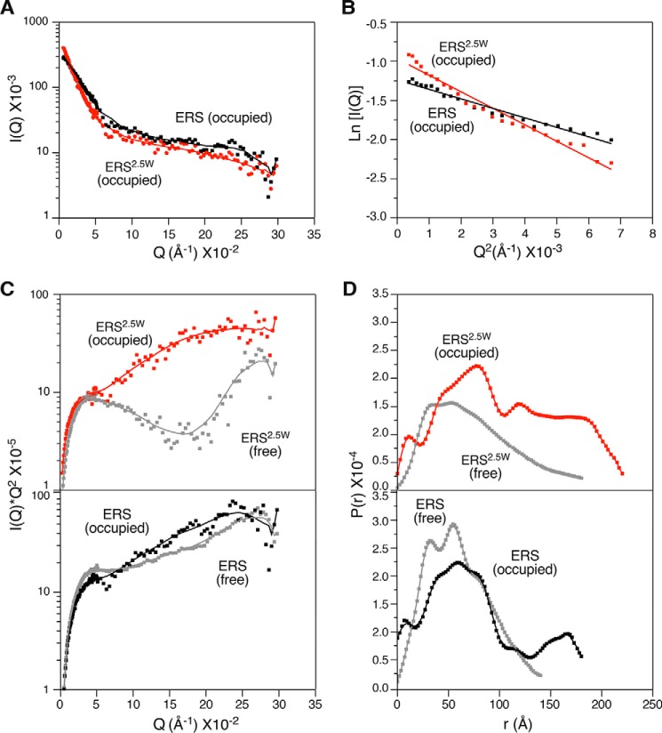
Aminoacyl-tRNA synthetases are ubiquitous, evolutionarily conserved enzymes catalyzing the conjugation of amino acids onto cognate tRNAs. During eukaryotic evolution, tRNA synthetases have been the targets of persistent structural modifications. These modifications can be additive, as in the evolutionary acquisition of noncatalytic domains, or subtractive, as in the generation of truncated variants through regulated mechanisms such as proteolytic processing, alternative splicing, or coding region polyadenylation. A unique variant is the human glutamyl-prolyl-tRNA synthetase (EPRS) consisting of two fused synthetases joined by a linker containing three copies of the WHEP domain (termed by its presence in tryptophanyl-, histidyl-, and glutamyl-prolyl-tRNA synthetases). Here, we identify site-selective proteolysis as a mechanism that severs the linkage between the EPRS synthetases in vitro and in vivo Caspase action targeted Asp-929 in the third WHEP domain, thereby separating the two synthetases. Using a neoepitope antibody directed against the newly exposed C terminus, we demonstrate EPRS cleavage at Asp-929 in vitro and in vivo Biochemical and biophysical characterizations of the N-terminally generated EPRS proteoform containing the glutamyl-tRNA synthetase and most of the linker, including two WHEP domains, combined with structural analysis by small-angle neutron scattering, revealed a role for the WHEP domains in modulating conformations of the catalytic core and GSH-S-transferase-C-terminal-like (GST-C) domain. WHEP-driven conformational rearrangement altered GST-C domain interactions and conferred distinct oligomeric states in solution. Collectively, our results reveal long-range conformational changes imposed by the WHEP domains and illustrate how noncatalytic domains can modulate the global structure of tRNA synthetases in complex eukaryotic systems.

Keywords: EPRS; aminoacyl-tRNA synthetase; aminoacylation; biophysics; caspase; glutamyl-prolyl-tRNA synthetase; microscale thermophoresis; neoepitope; neutron scattering; protein conformation; small-angle neutron scattering; transfer RNA (tRNA).


Conflict of interest statement

The authors declare that they have no conflicts of interest with the contents of this article

Figures

Figure 1.
Figure 1.
926DQVD929 is a functional and evolutionarily conserved caspase-cleavage site in EPRS. A, domain organization of human EPRS and two caspase-generated fragments, ERS2.5W and PRS0.5W. Arrows indicate predicted caspase cleavage sites. Epitopes for domain- or tag-specific antibodies are indicated. Sequence and localization of the caspase cleavage site, DQVD929, are shown. Peptide used for neoepitope antibody generation is underlined. B, quantitative Western blot analysis of transfected HEK293F lysates treated with recombinant caspase-3 (Casp3) in the presence or absence of Z-VAD (20 μm) and probed with domain- or tag-specific EPRS antibodies. C, quantitative Western blot analysis of EPRSWT- or EPRSD929A-transfected HEK293F lysates treated with recombinant caspase-3 in the presence or absence of Z-VAD (20 μm). D, multiple sequence alignment of the region containing the caspase-cleavage site in EPRS. Blue, yellow, and magenta denote the P1 aspartic acid and conserved and similar amino acid substitutions, respectively. Mammalian species are indicated in blue. E, domain organization of EPRS in multiple species and of ears-1 and pars-1 in C. elegans. Open arrowheads indicate the conserved caspase-cleavage site.
Figure 2.
Figure 2.
Catalytic activity of ERS2.5W and PRS0.5W. A and B, aminoacylation activity of purified recombinant ERS2.5W (A) and PRS0.5W (B). Purified ERS2.5W and PRS0.5W on Coomassie-stained SDS-polyacrylamide gel are shown at the right panel.
Figure 3.
Figure 3.
Interaction of caspase-generated EPRS fragments, ERS2.5W and PRS0.5W, with MSC components. A, EPRS and truncated domains expressed in HEK293F cells with theoretical molecular mass and MSC association. B, immunoblot (IB) analysis of immunoprecipitated (IP) EPRS, or truncated variants, probed with antibodies for FLAG tag and MSC components.
Figure 4.
Figure 4.
Detection of EPRS cleavage at Asp-929 in vitro and in vivo using neoepitope antiserum. A, immunoblot (IB) of lysates of N2a cells transfected with vector, FLAG-tagged ERS2.5W, or full-length EPRS probed with neoepitope serum with or without blocking peptide (left panel) or with anti-linker antibody (right panel). B, immunoblot of lysates from HEK293F cells transfected with vector, FLAG-tagged ERS2.5W, or EPRS before (left panel) or after (right panel) immunoprecipitation (IP) using neoepitope serum. C, Coomassie stain of FLAG-tagged EPRS purified by pulldown from HEK293F cells (left panel). Immunoblot of purified wildtype (WT) EPRS or D929A mutant after caspase cleavage (right panel). Immunoblot was Ponceau-stained and probed with neoepitope serum. D, immunoblot of lysates from TNFα-treated HT29 cells co-incubated with either vehicle or CHX in the presence or absence of Z-VAD (40 μm) for up to 16 h. Specific (arrow) and nonspecific (NS) immunoreactivities are indicated. E, immunofluorescence micrographs of human colon tissue detected with neoepitope or preimmune serum (green) and DAPI nuclear stain (blue) at 5× (upper panel) or 20× (lower panel) magnification. Lamina propria (LP) and colonic crypt (crypt) are indicated. Scale bar represents 100 μm.
Figure 5.
Figure 5.
In vivo detection of ERS2.5W. Immunofluorescence micrographs of human tissue microarray detected with neoepitope serum (green) and DAPI (blue) are shown. Scale bars represent 100 μm.
Figure 6.
Figure 6.
Solution structures of ERS2.5W and ERS in the ligand-free state as determined by SANS. A, size-exclusion chromatography of recombinant ERS2.5W (red) and ERS (black). Molecular mass standards are indicated on top. Coomassie-stained gel of purified recombinant ERS2.5W and ERS (insets). B, Coomassie stain analysis of recombinant ERS2.5W (left panel) and ERS (right panel) before and after BS3 cross-linking; monomer (arrow) and oligomer (*) species are indicated. C, scattering intensity (I) as a function of scattering vector (Q) for ERS2.5W (red) and ERS (black). D, Guinier plot of ERS2.5W (red) and ERS (black). E, overlay of Kratky plots of scattering data comparing ERS2.5W (red) and ERS (black) showing altered domain structure. F, overlay of pair-distance distribution function, P(r), of ERS2.5W (red) and ERS (black) showing distinct conformational features.
Figure 7.
Figure 7.
Ab initio modeling of WHEP-dependent conformational rearrangement of the catalytic core and appended GST–C domain. A–D, conformers of ERS2.5W (A, green), ERS (B, orange), and overlays (C and D) constructed from minimal ensemble search of 16 possible conformers derived using DAMMIN ab initio modeling of SANS data. Three rotational displays are visualized using PyMOL. Measurements of maximal vertical and horizontal dimensions are indicated by double-headed arrows.
Figure 8.
Figure 8.
Appended WHEP domains alter the conformational state of the synthetase core and appended GST–C domain. A, ERS2.5W and ERS domain organization with indicated localization of tryptophan residues and antibody-binding epitopes. B, spectra of intrinsic protein fluorescence of ERS2.5W, ERS, or buffer only with peak fluorescence wavelength indicated at 341 nm. C, quantification of peak intrinsic fluorescence of ERS2.5W, ERS, or buffer only from triplicate measurements ± S.D. Asterisk indicates statistical significance at p < 0.001. D, quantitative Western blotting of ERS2.5W (upper panel) or ERS (lower panel) subjected to trypsin digestion for the indicated times using antibodies directed against the His-tag (green) or the ERS domain (red). Overlay of the green and red immunoblots (IB) (merged) identifies protein fragments with dual immunoreactivity for the His-tag and ERS domain antibodies (yellow). E and F, quantification of full-length protein remaining after trypsin digestion at the indicated time points as detected by the His epitope antibody (E) or ERS domain antibody (F) for ERS2.5W (red) or ERS (black). A.U., arbitrary units.
Figure 9.
Figure 9.
Ligand-dependent conformational changes in ERS2.5W and ERS determined by SANS. A, scattering intensity as a function of scattering vector (Q) for ERS2.5W (red) and ERS (black) in the presence of ligands. B, Guinier plots of ERS2.5W (red) and ERS (black) in the presence of ligands. C, overlay of Kratky plots of scattering data showing changes in the conformational state of ERS2.5W (top panel) or ERS (bottom panel) in response to ligand binding. D, overlay of pair-distance distribution function, P(r), showing changes in the conformational state of ERS2.5W (top) or ERS (bottom) in response to ligand binding. Red and black traces indicate ERS2.5W and ERS, respectively, in ligand-occupied states (occupied); gray traces show ligand-free states (free) of both proteins.
Figure 10.
Figure 10.
WHEP domains facilitate ERS2.5W self-association in response to ligand binding. A, schematic of MST experimental design. Fluorescently-labeled protein remains monomeric in the absence of ligand and self-associates in the presence of ligands (ATP + l-Glu). Titration of unlabeled protein in MST results in binding and formation of labeled/unlabeled hetero-oligomers. Ligand-induced self-association impedes hetero-oligomerization, resulting in an apparent higher dissociation constant (KD) and a smaller fraction of bound labeled protein. B and D, change in thermophoretic mobility of labeled ERS2.5W (B) or ERS (D) in response to titration of unlabeled protein. C and E, fraction of bound labeled ERS2.5W (C) or ERS (E) as a function of unlabeled protein titration. F, change in MST-determined thermophoretic mobility of labeled ERS2.5W (red) and ERS (black) in response to ATP, tRNA, or l-Glu.
Figure 11.
Figure 11.
Structural arrangement of the glutamyl-tRNA synthetase as a bent rod is a shared feature of prokaryotic and eukaryotic synthetases. A and B, superposition of the surface structure of Thermus thermophilus ERS (blue) on SANS-derived structures of human ERS2.5W (green) (A) and ERS (orange) (B) in three rotational displays as visualized by PyMOL.

Similar articles

Cited by

References

    1. Schimmel P. (1987) Aminoacyl tRNA synthetases: general scheme of structure-function relationships in the polypeptides and recognition of transfer RNAs. Annu. Rev. Biochem. 56, 125–158 10.1146/annurev.bi.56.070187.001013 - DOI - PubMed
    1. Ibba M., and Soll D. (2000) Aminoacyl-tRNA synthesis. Annu. Rev. Biochem. 69, 617–650 10.1146/annurev.biochem.69.1.617 - DOI - PubMed
    1. Ko Y. G., Kim E. Y., Kim T., Park H., Park H. S., Choi E. J., and Kim S. (2001) Glutamine-dependent antiapoptotic interaction of human glutaminyl-tRNA synthetase with apoptosis signal-regulating kinase 1. J. Biol. Chem. 276, 6030–6036 10.1074/jbc.M006189200 - DOI - PubMed
    1. Park M. C., Kang T., Jin D., Han J. M., Kim S. B., Park Y. J., Cho K., Park Y. W., Guo M., He W., Yang X. L., Schimmel P., and Kim S. (2012) Secreted human glycyl-tRNA synthetase implicated in defense against ERK-activated tumorigenesis. Proc. Natl. Acad. Sci. U.S.A. 109, E640–E647 10.1073/pnas.1200194109 - DOI - PMC - PubMed
    1. Yannay-Cohen N., Carmi-Levy I., Kay G., Yang C. M., Han J. M., Kemeny D. M., Kim S., Nechushtan H., and Razin E. (2009) LysRS serves as a key signaling molecule in the immune response by regulating gene expression. Mol. Cell 34, 603–611 10.1016/j.molcel.2009.05.019 - DOI - PubMed

Publication types