. 2018 Apr 11;285(1876):20180051.
doi: 10.1098/rspb.2018.0051.

Microbial decay analysis challenges interpretation of putative organ systems in Cambrian fuxianhuiids

Affiliations

Microbial decay analysis challenges interpretation of putative organ systems in Cambrian fuxianhuiids

Jianni Liu et al. Proc Biol Sci. .

Abstract

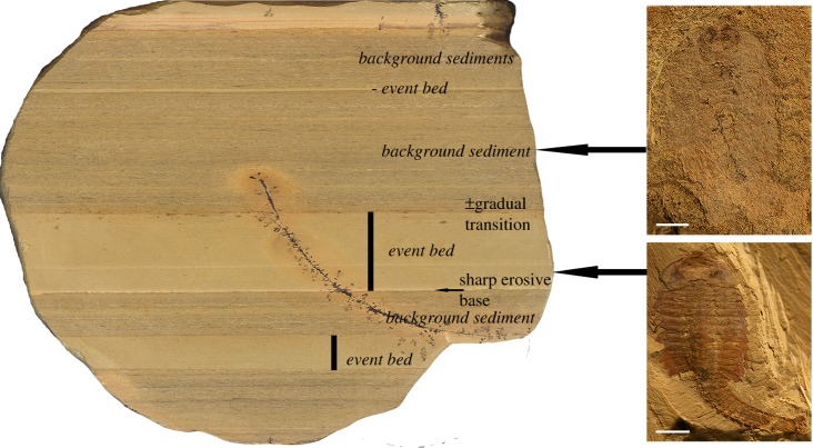
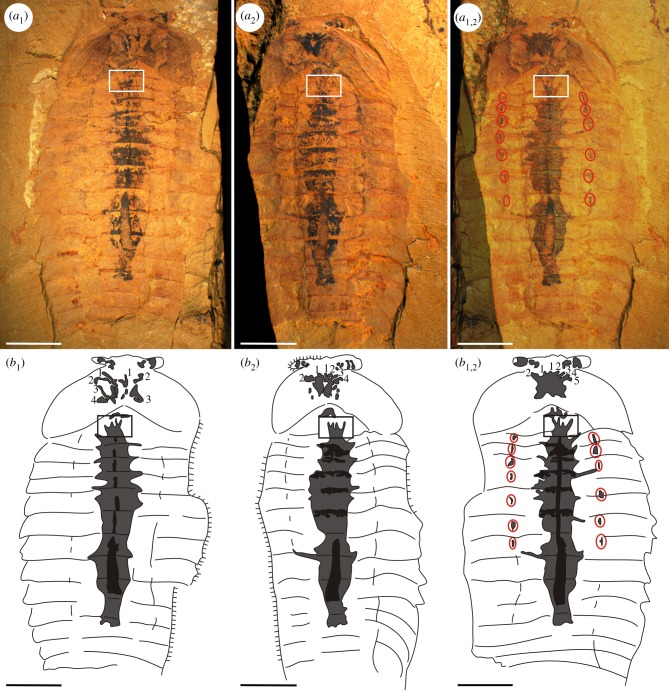
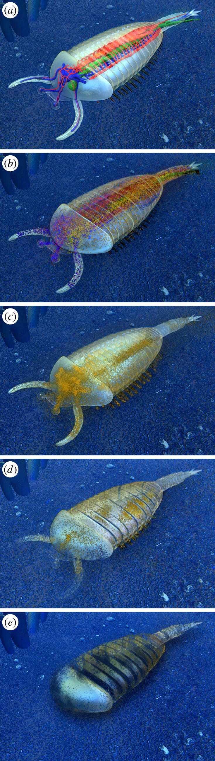
The Chengjiang fossil Lagerstätte (Cambrian Stage 3) from Yunnan, southern China is renowned for its soft-tissue preservation. Accordingly structures in fuxianhuiids, radiodontans and great appendage arthropods have been interpreted as the nervous and cardiovascular systems, including brains, hearts and blood vessels. That such delicate organ systems survive the fossilization process seems remarkable; given that this mode of preservation involves major taphonomic changes, such as flattening, microbial degradation, chemical alteration and replacement. Here, we document a range of taphonomic preservation states in numerous articulated individuals of Fuxianhuia protensa We suggest that organic (partly iron mineral-replaced) bulbous structures in the head region, previously interpreted as brain tissue, along with sagittally located organic strands interpreted as part of the cardiovascular system or as nerve cords, may be better explained as microbial biofilms that developed following decomposition of the intestine, muscle and other connective tissues, forming halos surrounding the original organic remains.

Keywords: Cambrian; Chengjiang fossil Lagerstätte; cardiovascular system; microbial biofilms; nervous tissue.


Conflict of interest statement

We have no competing interests.

Figures

Figure 1.
Figure 1.
Event beds (EBs) and background beds (BGBs) belonging to the Chengjiang fossil Lagerstätte in a vertical section of the mid-Yu'anshan Formation mudstones. The characteristics of EBs are: 1. fine claystone probably of distant tempestites or nepheloid layer; 2. total organic carbon (TOC) (0.11–0.21%) slightly lower than in BGBs, but the preserved TOC does not allow strong early diagenetic sulphate reduction; 3. beds homogeneous or with upward grading with a sharp erosional base; 4. thanatocenosis indicating instantaneous embedding (day level), parautochthonous, >90% articulated, often with ‘soft-tissue’ preservation, partly 3D preservation and oblique embedding of carcasses; 5. diversity higher than in adjacent BGB; 6. depositional time represented in EBs is in the range of hours to days; and 7. decay rate in organic remains is lower because of oxidant depletion, but some decay due to endogenous gut bacteria. BGBs characterized by: 1. claystone with higher content of silt; 2. TOC (0.18–0.48%) slightly higher than in EBs, with abundant faecal strings and organic hash beds laminated with its base gradually from underlying EBs; 3. fossil remains slightly time-averaged, majority are disarticulated shell fragments, articulated remains very rare, often partly destroyed or with a spongy appearance and mostly without ‘soft-tissue’ preservation; 4. diversity lower than in adjacent EBs; 5. time represented in a millimetric lamina is in the range of some years; and 6. decay rate in organic remains high by aerobic respiration and microbial decomposition. Scale bar in fossils is 5 mm. (Online version in colour.)
Figure 2.
Figure 2.
Fuxianhuia protensa from an ‘event bed’ (EB) and a ‘background bed’ (BGB) of the Chengjiang fossil Lagerstätte, showing typical differences in preservation. (a) EB fossil (ELI-JS721) showing excellent preservation of the external anatomy (e.g. the head shield, trunk tergites, antennae and stalked eyes). (b) BGB fossil (ELI-JS327A) preserving only remains of the gut and very faint segmentation in the partly decayed cuticle. Scale bar equals 5 mm. (Online version in colour.)
Figure 3.
Figure 3.
Enlargement of the head of Fuxianhuia protensa from the Chengjiang fossil Lagerstätte, showing different shapes of the ‘brain’. (a) Fossil (ELI-HY030A) with quadrate-shaped ‘brain’. (b) Fossil (ELI-HY030B) with upside down triangular-shaped ‘brain’. Note the part and counterpart of one specimens with different shapes of the ‘brain’ (which is created by split artefacts). (c) Fossil (ELI-JS050A) with trapezoidal-shaped ‘brain’. (d) Fossil (ELI-JS050B) with triangular-shaped ‘brain’. (e) Fossil (ELI-EJ062A) with triangular-shaped ‘brain’. (f) Fossil (ELI-EJ062B) with irregular-shaped ‘brain’, showing some branches. (g) Fossil (ELI-EJ067B) with rectangular-shaped ‘brain’. (h) Fossil (ELI-JS0087A) with butterfly-shaped ‘brain’. (i) Fossil (ELI-EJ231A) with rounded ‘brain’. (j) Fossil (ELI-JS469B2) with sub-rounded ‘brain’. (k) Fossil (ELI-EJ129A) with oval-shaped ‘brain’. (l) Fossil (ELI-EJ264B) with bifid ‘brain’. Scale bar, 1 mm. (Online version in colour.)
Figure 4.
Figure 4.
‘Brain-like’ structures in specimens of Fuxianhuia protensa revealing different shapes and branching morphologies. (a,a1) ELI-EJ0072A with eight branches on the left, four branches on the right. (b,b1) ELI-EJ0076A with six branches on the left, four branches on the right. (c,c1) ELI-CG752 with four branches on the left, three branches on the right. (d,d1) ELI-EJ264B with two branches on the left, four branches on the right. (e,e1) ELI-TLP0004B, round ‘brain-like’ structure with only one branch on the right. (f,f1) ELI-JS354B with one branch on the left, two branches on the right. (g,g1) ELI-JS327B, ‘brain-like’ structure almost filling the head shield without branches. (h,h1) ELI-JS359A, large ‘brain-like’ structure with four branches on the left and five branches on the right. Scale bar in (a,b,e,f,g) is 2.5 mm; (c,d,h) is 2 mm.
Figure 5.
Figure 5.
Fuxianhuia protensa EJ264A, B from the Chengjiang fossil Lagerstätte. Note the different appearance of the ‘brain-like’ structures, which were created by splitting artefacts, and the gut (black) is surrounded by a rounded organic halo (dark grey) in the thoracic region. (a1) ELI-EJ264A, ‘brain-like’ structure with four branches on the left and three branches on the right. (a2) ELI-EJ264B, ‘brain-like’ structure with two branches on the left and four branches on the right. (a1,2) Combined image from both halves of the specimen, showing the complete biofilm forming the ‘brain-like’ structure which differs in appearance from either the part or counterpart. (b1,b2,b1,2) Camera lucida drawings of (a1,a2,a1,2). Box marks some spinose projections of an additional bulbous structure in the foregut-midgut transition, which are similar to more frontally positioned structures previously interpreted as ‘nerves’ emerging from a ‘brain’. Red circles mark spot-like organic remains (b1,2) at the positions of apodemes, where stronger connective tissue may have existed. Scale bar equals 5 mm.
Figure 6.
Figure 6.
Five reconstructed decay stages of Fuxianhuia protensa from the Chengjiang fossil Lagerstätte with fossil examples and their explanatory drawings. (a) Decay stage 1 (EBs), specimen ELI-EJ0072A: external cuticle is well-preserved and small microbial biofilms occur in the head and gut (black part in a1). (b) Decay stage 2 (EBs), specimen ELI-EJ0076A: external cuticle is still well-preserved, but the gut has been ruptured and displaced (black part in b1) and the development of a microbial halo in the mid-thorax is common (grey part in b1). (c) Decay stage 3 (BGBs), specimen ELI-JS516: external cuticle is obscured and the gut mostly degraded, the microbial communities strongly radiate laterally in the muscles and connective tissues of head and thorax (black part in c1; the box marks a bulbous structure in the midgut region strongly resembling the morphology of those in the head region previously interpreted as ‘brain’ and ‘nervous tissues’). (d) Decay stage 4 (BGBs), specimen ELI-JS327B: cuticles of the trunk tergites, head shield, eyes and antennae have also begun to decompose, microbial biofilm occupies nearly the entire body cavity and head shield; labile tissues have been fully decomposed, microbes metabolize the more durable connective tissue (black part in d1). (e) Decay stage 5 (BGBs), specimen JS354B, with strongly decayed external cuticle and internal biofilms shrunken in size and fragmented into small or particulate regions (black part in e1). Red circles mark spot-like organic remains at the positions of apodemes, where stronger connective tissue may have existed.
Figure 7.
Figure 7.
The bulbous structure in the mid gut region of Fuxianhuia protensa, which shows a comparable morphology to the ‘brain-like’ structures in the head region. (ae) The bulbous structures in the mid gut of specimens ELI-EJ203, EJ073A, JS403A, JS516, JS630A. (fj) The complete specimens of ELI-EJ203, EJ073A, JS403A, JS516, JS630A. Box marks the region of the bulbous structure in the body. Scale bars: (ae) is 1 mm; (fj) is 5 mm. (Online version in colour.)
Figure 8.
Figure 8.
Taphonomical decay model, corresponding to the five decay stages shown in figure 6. Note: Decay stage 0 (undecayed organism) is not preserved in the Chengjiang fossil Lagerstätte.

Similar articles

Cited by

References

    1. Chen JY, Ramsköld L, Zhou GQ. 1994. Evidence for monophyly and arthropod affinity of Cambrian giant predators. Science 264, 1304–1308. (10.1126/science.264.5163.1304) - DOI - PubMed
    1. Chen JY, Edgecombe GD, Ramskold L, Zhou GQ. 1995. Head segmentation in early Cambrian Fuxianhuia: implications for arthropod evolution. Science 268, 1339–1342. (10.1126/science.268.5215.1339) - DOI - PubMed
    1. Hou XG, Bergström J. 1997. Arthropods of the Lower Cambrian Chengjiang fauna southwest China. Fossils Strata 45, 1–116.
    1. Shu DG, et al. 1999. Lower Cambrian vertebrates from south China. Nature 402, 42–46. (10.1038/46965) - DOI
    1. Shu DG, Conway Morris S, Han J, Chen L, Zhang XL, Zhang ZF, Liu HQ, Li Y, Liu JN. 2001. Primitive deuterostomes from the Chengjiang Lagerstätte (Lower Cambrian, China). Nature 414, 419–424. (10.1038/35106514) - DOI - PubMed

Publication types

LinkOut - more resources