. 2018 Apr;15(141):20170889.
doi: 10.1098/rsif.2017.0889.

Nerve regeneration in the cephalopod mollusc Octopus vulgaris: label-free multiphoton microscopy as a tool for investigation

Affiliations

Nerve regeneration in the cephalopod mollusc Octopus vulgaris: label-free multiphoton microscopy as a tool for investigation

Pamela Imperadore et al. J R Soc Interface. 2018 Apr.

Abstract

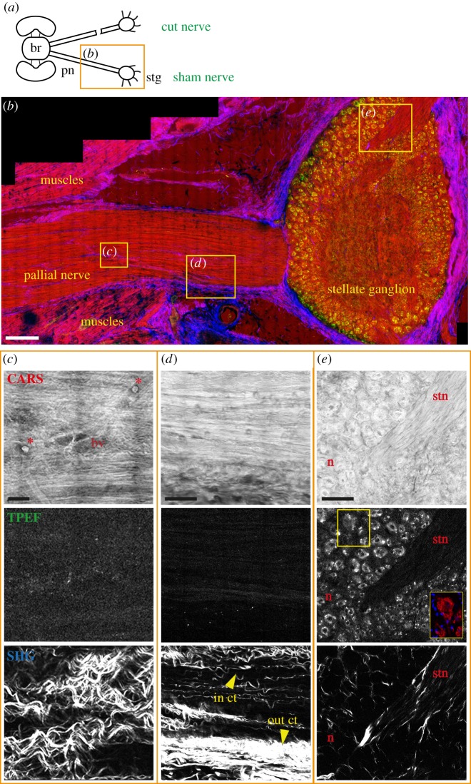
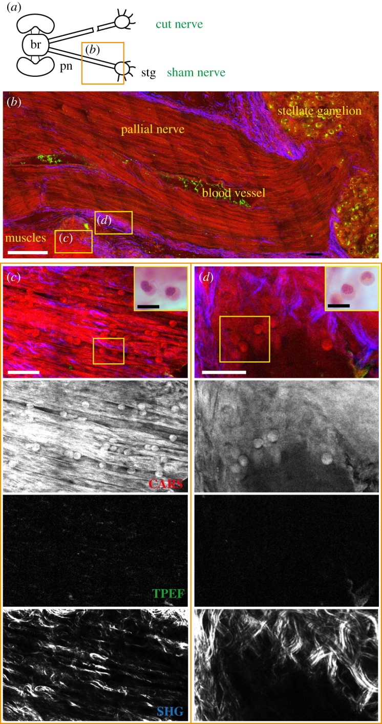
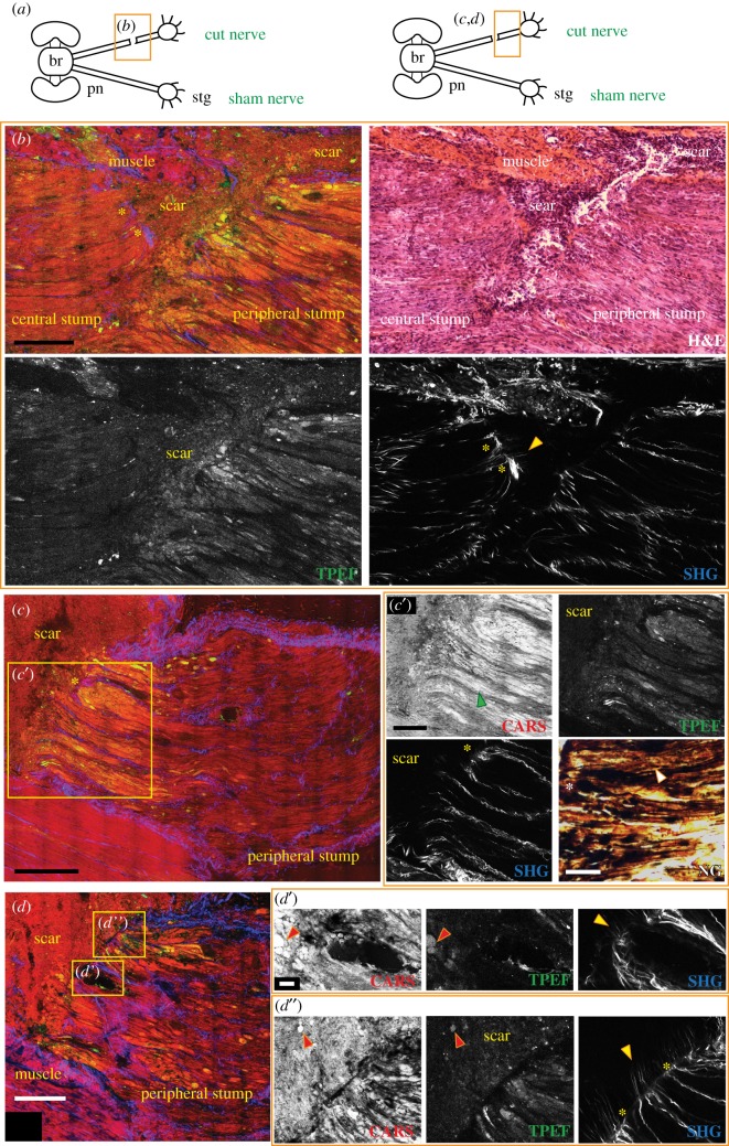
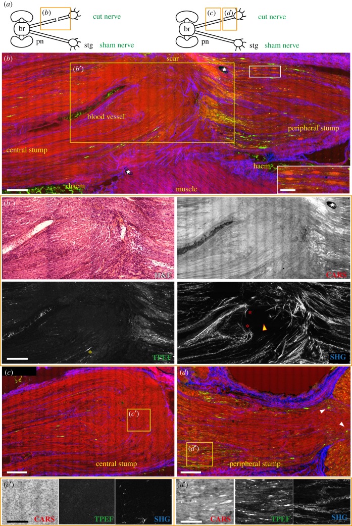
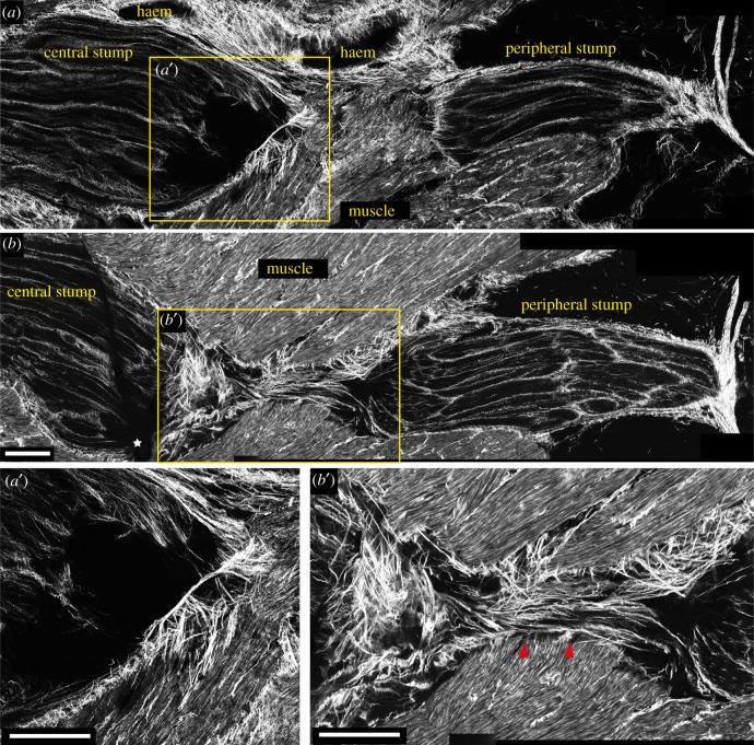
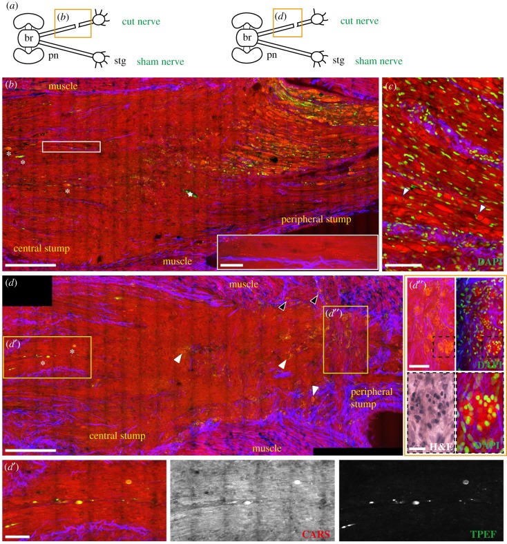
Octopus and cephalopods are able to regenerate injured tissues. Recent advancements in the study of regeneration in cephalopods appear promising encompassing different approaches helping to decipher cellular and molecular machinery involved in the process. However, lack of specific markers to investigate degenerative/regenerative phenomena and inflammatory events occurring after damage is limiting these studies. Label-free multiphoton microscopy is applied for the first time to the transected pallial nerve of Octopus vulgaris Various optical contrast methods including coherent anti-Stokes Raman scattering (CARS), endogenous two-photon excited fluorescence (TPEF) and second harmonic generation (SHG) have been used. We detected cells and structures often not revealed with classical staining methods. CARS highlighted the involvement of haemocytes in building up scar tissue; CARS and TPEF facilitated the identification of degenerating fibres; SHG allowed visualization of fibrillary collagen, revealing the formation of a connective tissue bridge between the nerve stumps, likely involved in axon guidance. Using label-free multiphoton microscopy, we studied the regenerative events in octopus without using any other labelling techniques. These imaging methods provided extremely helpful morpho-chemical information to describe regeneration events. The techniques applied here are species-specific independent and should facilitate the comparison among various animal species.

Keywords: CARS; Octopus vulgaris; SHG; TPEF; label-free multiphoton microscopy; nerve regeneration.


Conflict of interest statement

We declare we have no competing interests.

Figures

Figure 1.
Figure 1.
Sham control nerve. (a) Schematic drawing showing the connections between the brain (br) and stellate ganglion (stg) through the right and left pallial nerves (pn). The orange rectangle delimits the area of the nerve shown in the figure. The sham nerve is shown in (b) after multiphoton imaging. CARS signals (red in b) highlighted the structure of axons in the pallial nerve (bd) and the neuropil, neurons (n) and stellar nerves (stn) inside the stellate ganglion (b,e). CARS signal also showed blood vessels (bv) and haemocytes (red asterisks) in the nerve (c). In (b) also muscles appear to give a strong signal in CARS. SHG (blue in b) allowed visualization of the outer connective tissue (out ct) enwrapping the pallial nerve (b,d) and the stellate ganglion (b), and the inner connective tissue (in ct) surrounding bundles of fibres inside the pallial nerve (c,d). TPEF signal (in green) was strongly detected only in the neurons of the stellate ganglion (e). Neurons were identified via acetylated tubulin labelling, visible in red in the yellow box in (e). Glial cells around neurons are not highlighted using multiphoton microscopy, but their nuclei are detected via DAPI counterstain (blue in the yellow box in e). Scale bars: (b) 250 µm, (c) 20 µm, (d,e) 50 µm.
Figure 2.
Figure 2.
Haemocytes infiltrating sham tissues. (a) Schematic drawing showing the connections between the brain (br) and stellate ganglion (stg) through the right and left pallial nerves (pn). The orange rectangle delimits the area of the nerve shown in the figure panel. (b) A blood vessel is detected inside the sham nerve via CARS. Haemocytes were visualized in this vessel and infiltrating the damaged tissues, like muscles (c) and connective tissue (d) injured to expose the nerve, as round spheres with a diameter of 7–12 µm. This is confirmed by H&E staining, which highlighted the classical U-shaped nuclei of haemocytes (yellow box enlargements in c and d). TPEF did not give detectable signal in muscles or connective tissue as shown in single channel images (TPEF in c and d). Connective tissue is detected in the pallial nerve forming axon bundles, enveloping the whole nerve (b). It also gives a strong signal inside the muscle (c) surrounding the nerve. Scale bars: (b) 250 µm, (c) 20 µm, (d) 25 µm, yellow box in (c,d) 10 µm.
Figure 3.
Figure 3.
Injured nerve 3 days post lesion. (a) Schematic drawing showing the connections between the brain (br) and stellate ganglion (stg) through the right and left pallial nerves (pn). The orange rectangle delimits the area of the nerve shown in the figure panel. (b) The lesion site of a nerve 3 days post lesion imaged by multiphoton microscopy and later stained using H&E. In the merged image, the two stumps of the nerve appear to be separated by a scar which also forms in the muscular tissue. The scar is highlighted by CARS and TPEF. Axons in both stumps are seen to degenerate, especially in the peripheral stump characterized by fibres swelling and fragmenting (b,c,c′, CARS and TPEF; see green arrow in c′ CARS) and debris were also detected as round structures inside the scar that forms between the two stumps (highlighted by red arrows in d′ and d″, CARS and TPEF). Degeneration is further confirmed by Nauta–Gygax (NG) staining (c′) where axon tip swelling is marked by white asterisks and small granules of fragmenting axons are highlighted by white arrows. Connective tissue appears to seal around cut axons of both stumps (b,c′, SHG marked with yellow asterisks) but also to stick out straight from them invading the scar (b,d′,d″, SHG, marked with yellow arrows). A blood vessel is also visible in the peripheral stump. It contains haemocytes that are released inside the scar (d′, CARS) and contribute to its formation. Scale bars: (bd) 250 µm, (c′) 125 µm, (c′, NG) 50 µm, (d′,d″) 20 µm.
Figure 4.
Figure 4.
Injured nerve 7 days post lesion. (a) Schematic drawing showing the connections between the brain (br) and stellate ganglion (stg) through the right and left pallial nerves (pn). The orange rectangle delimits the area of the nerve shown in the figure panel. The lesioned nerve 7 days post lesion is shown in (b). The scar has now a limited size, mainly confined to the side. Blood vessels running in the central stump and haemorrhagic areas (haem) around the nerve are visible, especially due to blood giving a strong signal in TPEF (green in b). Degenerating fibres occupy a large area of the peripheral stump, observable via CARS and TPEF. Some of them assume an ovoidal shape (highlighted in the white box enlargement in b). Some degenerating fibres are also visible in the central stump (b′) with TPEF (highlighted by yellow asterisk) well organized in rows. The inner connective tissue grows backwards to seal the cut axons (red asterisks in b′, SHG). Beyond this point, regenerating axons from the stump grow as a bulk of disorganized fibres (c′, CARS) where connective tissue is not present (also marked with yellow arrow in b′, SHG). (d) Degeneration in the peripheral stump involves a large number of fibres (d′, CARS and TPEF) which also get closer to the ganglion (see white arrows in d). White stars in figure (b) indicate artefacts. Scale bars: (b,b′,c,d) 250 µm, (white insert in b) 100 µm, (c′,d′) 50 µm.
Figure 5.
Figure 5.
Connective tissue (SHG signal) in injured nerve 7 days post lesion. (a,b) The outer connective tissue sealed around the cut stumps of the nerve 7 days post lesion, shaping the central stump in a spike-like structure. (a′) Regenerating fibres in the central stump lack inner connective tissue. (b,b′) Shows how close to the muscular tissue underneath the lesioned nerve, connective tissue forms a bridge that fills the gap between the two stumps (marked by red arrows in b′). White stars in figure (b) indicate artefacts. Scale bar (a,a′,b,b′): 250 µm.
Figure 6.
Figure 6.
Injured nerve 14 days post lesion. (a) Schematic drawing showing the connections between the brain (br) and stellate ganglion (stg) through the right and left pallial nerves (pn). The orange rectangle delimits the area of the nerve shown in the figure panel. (b,d) The central and peripheral stumps of a nerve 14 days post lesion. The majority of the fibres in the peripheral stump appear swollen and fragmented, while the few degenerating axons in the central stump are marked with white asterisks (also visible in d′, CARS and TPEF). Small granules, arranged in rows are visible in the central stump (enlargement in the white rectangle in b). In (c) the peripheral stump is counterstained with DAPI, showing that cell nuclei are outside the ovoidal fragments and not infiltrating them. Some of these fragments contain a structure resembling a nucleus (marked with white arrows) which, however, does not stain with DAPI. Drop shaped structures were identified in the lesioned nerve (white arrows in d; enlargement in d″) and muscles (black arrows in d), highlighted by CARS and TPEF (red and green in d). Counterstain with DAPI allowed identification of those structures as cell clusters (d″) surrounded by connective tissue (d″ SHG). H&E staining (black dotted box in d″) slightly stained these structures. White stars in figure (b) indicate artefacts. Scale bars: (b,d) 250 µm, (c) 100 µm, (d′,d″) 50 µm, (e) 20 µm, white rectangle in (b) 25 µm.

Similar articles

Cited by

References

    1. Bely AE, Nyberg KG. 2010. Evolution of animal regeneration: re-emergence of a field. Trends Ecol. Evol. 25, 161–170. ( 10.1016/j.tree.2009.08.005) - DOI - PubMed
    1. Bejjani RE, Hammarlund M. 2012. Neural regeneration in Caenorhabditis elegans. Annu. Rev. Genet. 46, 499–513. ( 10.1146/annurev-genet-110711-155550) - DOI - PMC - PubMed
    1. Brockes JP, Kumar A. 2008. Comparative aspects of animal regeneration. Annu. Rev. Cell Dev. Biol. 24, 525–549. ( 10.1146/annurev.cellbio.24.110707.175336) - DOI - PubMed
    1. Chen Z-L, Yu W-M, Strickland S. 2007. Peripheral regeneration. Annu. Rev. Neurosci. 30, 209–233. ( 10.1146/annurev.neuro.30.051606.094337) - DOI - PubMed
    1. Kumar A, Brockes JP. 2012. Nerve dependence in tissue, organ, and appendage regeneration. Trends Neurosci 35, 691–699. ( 10.1016/j.tins.2012.08.003) - DOI - PubMed

Publication types

MeSH terms

LinkOut - more resources