Review
. 2018 Apr;15(141):20170916.
doi: 10.1098/rsif.2017.0916.

Long-range proton-coupled electron transfer in biological energy conversion: towards mechanistic understanding of respiratory complex I

Affiliations
Review

Long-range proton-coupled electron transfer in biological energy conversion: towards mechanistic understanding of respiratory complex I

Ville R I Kaila. J R Soc Interface. 2018 Apr.

Abstract

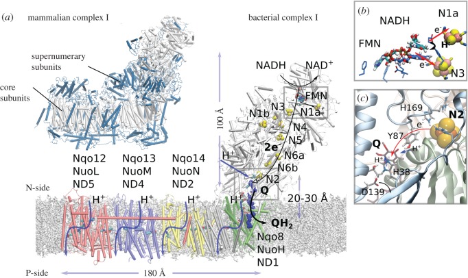
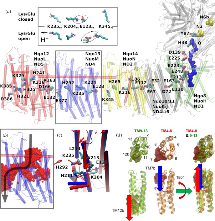
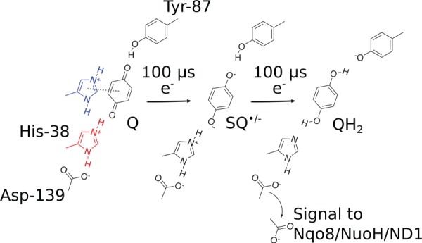
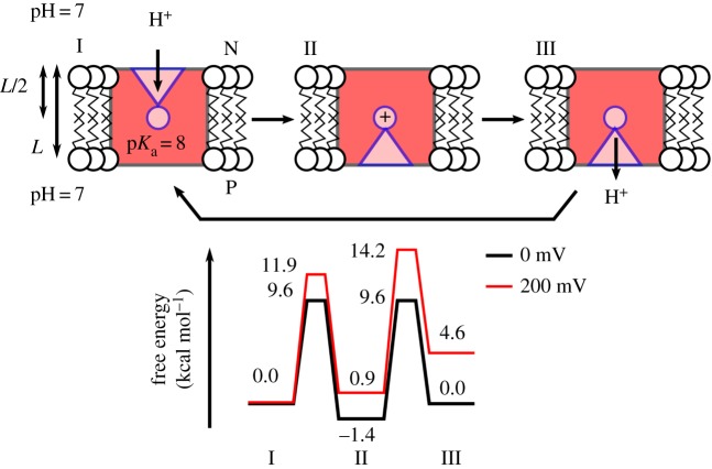
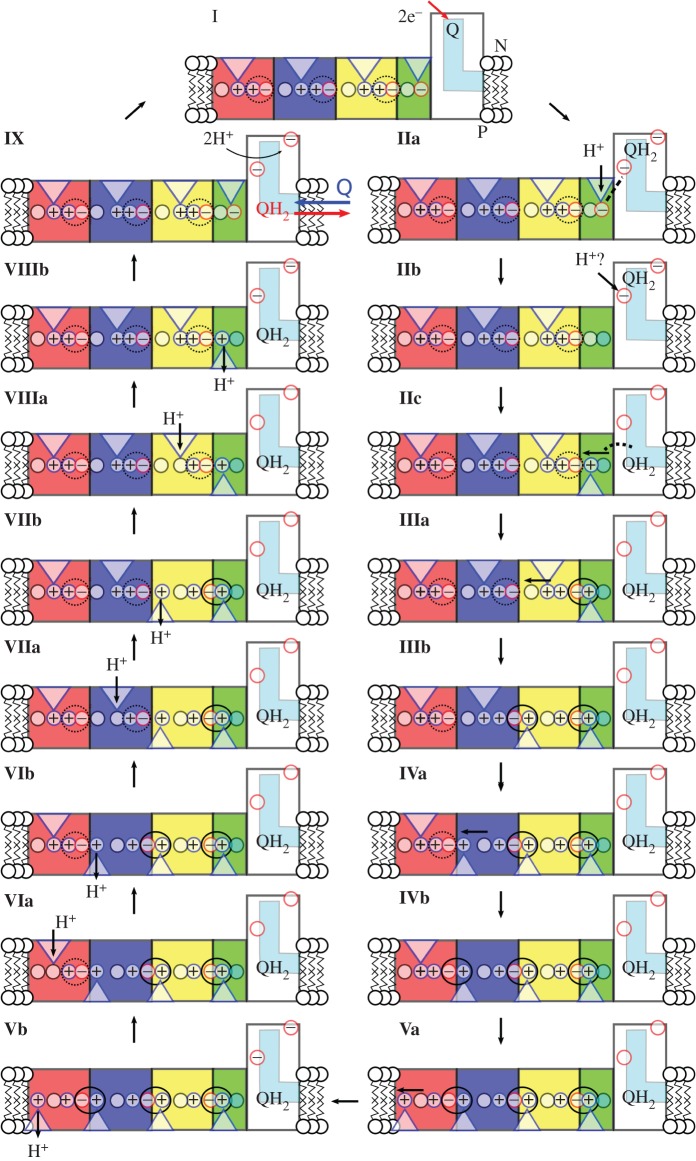
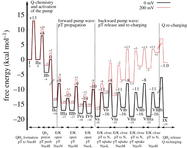
Biological energy conversion is driven by efficient enzymes that capture, store and transfer protons and electrons across large distances. Recent advances in structural biology have provided atomic-scale blueprints of these types of remarkable molecular machinery, which together with biochemical, biophysical and computational experiments allow us to derive detailed energy transduction mechanisms for the first time. Here, I present one of the most intricate and least understood types of biological energy conversion machinery, the respiratory complex I, and how its redox-driven proton-pump catalyses charge transfer across approximately 300 Å distances. After discussing the functional elements of complex I, a putative mechanistic model for its action-at-a-distance effect is presented, and functional parallels are drawn to other redox- and light-driven ion pumps.

Keywords: NADH:ubiquinone oxidoreductase; PCET; bioenergetics; molecular simulations; proton transfer.


Conflict of interest statement

I declare I have no competing interests.

Figures

Figure 1.
Figure 1.
Structure and function of complex I. (a) The hydrophilic domain of complex I catalyses hydride transfer from NADH to FMN that triggers a stepwise electron transfer along a chain of eight FeS centres to quinone (Q), located approximately 20–30 Å above the membrane plane. The quinone-reduction process is coupled to proton pumping across the antiporter-like membrane subunits Nqo14, Nqo13, Nqo12 and Nqo8. The structural model was built based on complex I from Thermus thermophilus (PDB: 4HEA). Inset: the bovine complex I (PDB ID: 5LC5) showing the supernumerary subunits (in blue) that surround the core complex (in white). (b) Close-up of the NADH/FMN site (PDB ID: 3IAM). (c) Close-up of the N2/Q-reduction site, with Q in its stacking conformation with His-38 (see text). The figure was prepared with VMD [15].
Figure 2.
Figure 2.
Structure of the proton-pumping membrane domain in complex I. (a) The membrane domain of complex I showing conserved buried charged/hydrophilic residues. NuoL (red); NuoM (blue); NuoN (yellow); NuoH (green); Q (blue van der Waals representation). Inset: The conformation of the Lys/Glu ion pair (here Lys-204M/Glu-123M) can modulate the pKa of the middle Lys (Lys-235M). (b) Water molecules (in red) establish protonic connectivity in all antiporter-like subunits. The figure shows time-averaged occupancies of water molecules, i.e. water molecules that visit the channel area during the simulation time, in the NuoM subunit based on microsecond MD simulations. (c) Snapshot of structures obtained from MD simulations of open (in blue) and closed (in red) proton channels from the N-side in Nqo13 (NuoM/ND4), showing conformational changes in the broken helix element. (d) The structural symmetry of the antiporter-like subunits with an N-side input channel near broken helix TM7b and output channel near broken helix TM12b. The two five-helical bundles (TM4–8 and TM9-13) are related by rotation and translation symmetry, which is different from the rotation–inversion symmetry found in typical transporters.
Scheme 1.
Scheme 1.
Quinone chemistry in the active site of complex I. The quinone headgroup forms a hydrogen bond with Tyr-87 of Nqo4 (Thermus numbering), and alternates between stacking (in blue) and hydrogen-bonded conformations with His-38 of Nqo4 (in red), which modulates the redox potential of the Q [68]. Electron transfer from NADH along the FeS chain in approximately 100 µs [61] reduces the quinone to a transient anionic semiquinone (SQ•/−) species. The second electron, stored at the FeS chain, e.g. at the N1a centre, is transferred in approximately 100 µs to further reduce the SQ•/− species to the quinol (QH2) by proton transfer from His-38 and Tyr-87. This weakens the electrostatic interactions between His-38 and Asp-139, which is flipped towards the Nqo8/NuoH/ND1 subunit, and in turn triggers the pump machinery [86].
Figure 3.
Figure 3.
Principles for constructing free-energy diagrams for proton transfer across the membrane. The free energies are related to equilibrium constants and rates by equations (7.1)–(7.3), and the effect of a membrane potential is estimated from equation (7.4). The figure shows transfer of a proton to a single group with pKa = 8 across the membrane from pH = 7 (N-side) to pH = 7 (P-side) without and with an external pmf of 200 mV. The model assumes that the proton transfer from the N-side and P-side to the buried proton loading site takes place on 1 µs timescales. The figure shows a passive proton channel, whereas a proton pump would operate with an element that modulates the pKa of the buried site by e.g. an electron transfer site [140].
Figure 4.
Figure 4.
Long-range PCET model for complex I. The schematic figure shows NuoH (in green), NuoN (in yellow), NuoM (in blue), NuoL (in red), Q-channel (in cyan), water connectivity (with blue triangles), and closed and open Lys-Glu ion pairs (marked with dotted and solid black circles, respectively). Central steps: I: Reduction of Q by NADH. IIa: Formation of QH2 by local pT from His-38 and Tyr-87 triggers pKa shift in the NuoH subunit. IIb: Proton uptake by the Glu-quartet in NuoH from the N-side. Reprotonation of His-38/Tyr-87 could also take place already in this state if the redox signal has been transmitted to the NuoH subunit (see §3). IIc: QH2 moves down in the Q-channel, which pushes the ‘NuoH’-proton towards NuoA/J/K/H. IIIa: Protonation of terminal residues in NuoA/J/K/H leads to accumulation of positive charge that opens up the Lys-186N/Glu-112N ion pair. This leads to pT from Lys-186N to Lys-345N, and dehydration of the water contact to the N-side. IIIb: Protonation of the terminal end of NuoN opens up the Lys-204M/Glu-123M-ion pair. IVa: Opening of the NuoM Lys-Glu ion pair lowers the pKa of Lys-235M that leads to pT towards Glu-377M, and dehydration of the water contact to the N-side. IVb: Protonation of the terminal end of the NuoM opens up the Lys-216L/Asp-166L ion pair. Va: Opening of the NuoL Lys-Glu ion pair lowers the pKa of Lys-329L that leads to pT towards Lys-385L, and dehydration of the water contact to the N-side. Vb: Proton release to the P-side from NuoL. VIa: Closure of the Lys-216L/Asp-166L ion pair and re-protonation of Lys-329L from the N-side. VIb: Closure of the NuoL Lys-Glu ion pair destabilizes the proton at the terminal end of the NuoM subunit that is ejected to the P-side. VIIa: Closure of the Lys-204M/Glu-123M ion pair and re-protonation Lys-235M from the N-side. VIIb: Closure of the NuoM Lys-Glu ion pair destabilizes the proton stored at the terminal end of the NuoN subunit that is ejected to the P-side. VIIIa: Closure of the Lys-186N/Glu-112N ion pair and re-protonation Lys-216N from the N-side. VIIIb: Closure of the NuoN Lys-Glu ion pair destabilizes the proton stored at the terminal end of the NuoH subunit that is ejected to the P-side. IX: Exchange of QH2 with an oxidized quinone, and re-protonation of Tyr-87/His-38 from the N-side. Owing to the strong pairwise coupling between the ion-pair conformation in subunit i + 1 and the pKa of the terminal residue in subunit i, the proton release steps are expected to propagate in the backwave process rather than releasing protons in subunits closer to QH2.
Figure 5.
Figure 5.
Qualitative free-energy profiles for the long-range PCET process for complex I estimated at a pmf = 0 mV (in black) and 200 mV (in red). Labelling of states is defined in figure 4. The pKa of the terminal proton acceptor in the antiporter-like subunits were assumed to have a pKa of 9–10 [76,109], and the N- and P-sides have a pH = 7. Quinone transfer along its channel was assumed to release approximately 600 mV (IIb → IIc). The thermodynamic values are estimated at T = 310 K and transition state energies are estimated based on transition state theory using a standard pre-exponential factor. States with dashed lines cannot be determined due to missing data. See main text (§7) for derivation of the free-energy profiles.

References

    1. Reece SY, Nocera DG. 2009. Proton-coupled electron transfer in biology: results from synergistic studies in natural and model systems. Annu. Rev. Biochem. 78, 673–699. ( 10.1146/annurev.biochem.78.080207.092132) - DOI - PMC - PubMed
    1. Kaila VRI, Verkhovsky MI, Wikström M. 2010. Proton-coupled electron transfer in cytochrome oxidase. Chem. Rev. 110, 7062–7081. ( 10.1021/cr1002003) - DOI - PubMed
    1. Hammes-Schiffer S, Stuchebrukhov AA. 2010. Theory of coupled electron and proton transfer reactions. Chem. Rev. 110, 6939–6960. ( 10.1021/cr1001436.) - DOI - PMC - PubMed
    1. Siegbahn PEM, Blomberg MRA. 2010. Quantum chemical studies of proton-coupled electron transfer in metalloenzymes. Chem. Rev. 110, 7040–7061. ( 10.1021/cr100070p) - DOI - PubMed
    1. Cardona T, Sedoud A, Cox N, Rutherford AW. 2012. Charge separation in Photosystem II: a comparative and evolutionary overview. Biochim. Biophys. Acta 1817, 26–43. ( 10.1016/j.bbabio.2011.07.012) - DOI - PubMed

Publication types

MeSH terms