. 2018 Apr 11;10(436):eaan3464.
doi: 10.1126/scitranslmed.aan3464.

The systemic response to surgery triggers the outgrowth of distant immune-controlled tumors in mouse models of dormancy

Affiliations

The systemic response to surgery triggers the outgrowth of distant immune-controlled tumors in mouse models of dormancy

Jordan A Krall et al. Sci Transl Med. .

Abstract

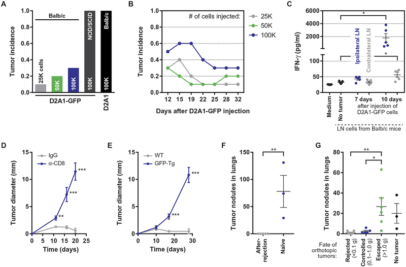
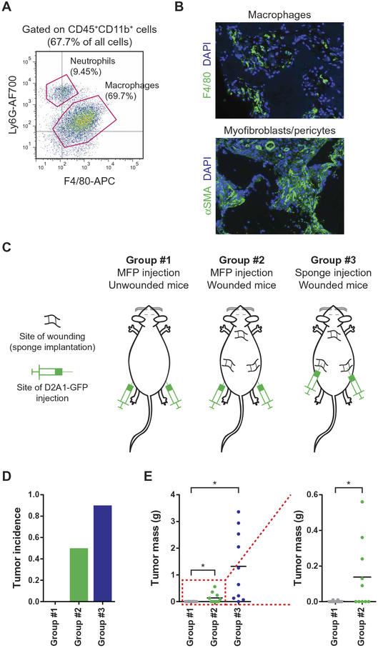
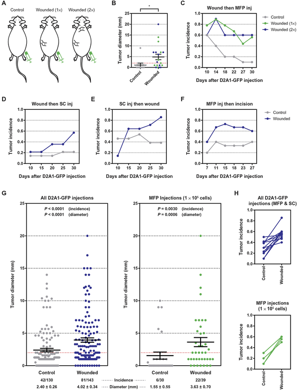
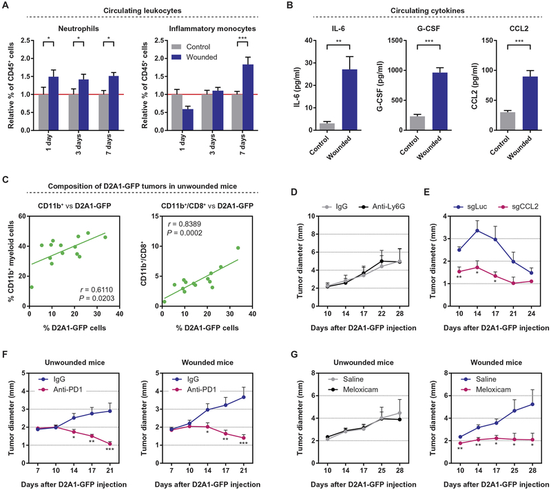
Patients undergoing surgical resection of primary breast tumors confront a risk for metastatic recurrence that peaks sharply 12 to 18 months after surgery. The cause of early metastatic relapse in breast cancer has long been debated, with many ascribing these relapses to the natural progression of the disease. Others have proposed that some aspect of surgical tumor resection triggers the outgrowth of otherwise-dormant metastases, leading to the synchronous pattern of relapse. Clinical data cannot distinguish between these hypotheses, and previous experimental approaches have not provided clear answers. Such uncertainty hinders the development and application of therapeutic approaches that could potentially reduce early metastatic relapse. We describe an experimental model system that definitively links surgery and the subsequent wound-healing response to the outgrowth of tumor cells at distant anatomical sites. Specifically, we find that the systemic inflammatory response induced after surgery promotes the emergence of tumors whose growth was otherwise restricted by a tumor-specific T cell response. Furthermore, we demonstrate that perioperative anti-inflammatory treatment markedly reduces tumor outgrowth in this model, suggesting that similar approaches might substantially reduce early metastatic recurrence in breast cancer patients.


Conflict of interest statement

Competing interests: The authors declare that they have no competing financial interests.

Figures

Fig. 1.
Fig. 1.. The outgrowth of D2A1-GFP tumors in Balb/c mice is restricted by a GFP-specific CD8+ T cell response.
(A) Endpoint tumor incidence, 32 days after the orthotopic injection of 2.5 × 10⁴, 5 × 10⁴, or 1 × 10⁵ D2A1–green fluorescent protein (GFP) cells into syngeneic Balb/c mice; 1 × 10⁵ D2A1-GFP cells injected into immunodeficient nonobese diabetic/severe combined immunodeficient mice; and 1 × 10⁵ unlabeled D2A1 cells injected into Balb/c mice (n = 10 per group). Tumor incidence is reported as the fraction of mice bearing tumors of diameter ≥ 2 mm (see Supplementary Materials and Methods). (B) Fraction of tumor-bearing mice as a function of time after the orthotopic injection of D2A1-GFP cells into Balb/c mice at doses of 2.5 × 10⁴, 5 × 10⁴, or 1 × 10⁵ per injection (n = 8 to 10 per group). (C) Secretion of interferon-γ (IFN-γ) into culture medium during ex vivo coculture of irradiated D2A1-GFP cells with lymph node (LN) cells isolated from the inguinal LNs of tumor-free or D2A1-GFP–bearing Balb/c mice. Data for cells isolated from the ipsilateral (tumor-draining) and contralateral LNs are shown for tumor-bearing mice (n = 5 per group). (D) Tumor diameter after the orthotopic injection of 1 × 10⁵ D2A1-GFP cells into Balb/c mice 1 day after initiating injections of either anti-CD8 antibodies or control immunoglobulin G (IgG) (n = 9 to 10 per group). (E) Tumor diameter after the orthotopic injection of 1 × 10⁵ D2A1-GFP cells into wild-type (WT) or GFP-transgenic (Tg) mice, both on a Balb/c:C57BL/6 F1 background (n = 11 per group). (F) Number of tumor nodules in the lungs after the intravenous injection via the tail vein of 1 × 10⁶ D2A1-GFP cells into Balb/c mice that had previously rejected D2A1-GFP tumors or into naïve mice (n = 3 to 5 per group). (G) Number of tumor nodules in the lungs after the intravenous injection via the tail vein of D2A1-GFP 5 × 10⁵ cells into Balb/c mice bearing D2A1-GFP cells in the mammary fat pad (MFP) at different stages of growth or rejection (n = 3 to 6 per group). For all panels, data are plotted as means ± SEM. P values were calculated using the Mann-Whitney test (*P < 0.05, **P < 0.005, ***P < 0.0005).
Fig. 2.
Fig. 2.. The postsurgical wound-healing response protects local immunogenic tumors from immune-mediated destruction.
(A) Identification of sponge-infiltrating myeloid cells using flow cytometry after staining for the presence of neutrophils (CD45+CD11b+Ly6G+) and macrophages (CD45+CD11b+F4/80+), 7 days after sponge implantation. (B) Immunofluorescence staining of tissue sections containing stroma-infiltrated sponges. Sections were stained with anti-F4/80 (macrophages, top) or anti–α–smooth muscle actin (αSMA) (myofibroblasts and blood vessel–lining pericytes, bottom). Representative images at ×10 magnification are shown for sections isolated 14 days after wounding by subcutaneous sponge implantation. DAPI, 4′,6-diamidino-2-phenylindole. (C to E) Injection of immunogenic D2A1-GFP cells into control or wounded Balb/c mice. (C) Schematic illustrating the experimental design in which 1 × 10⁵ tumor cells were injected into the MFP of control mice (left) or mice bearing a local wound (center) or were injected directly into a site of wound healing (right). The incidence (D) and mass (E) of the resulting tumors were determined 30 days after the injection of tumor cells (n = 10 per group). For all panels, data are plotted as means ± SEM. P values were calculated using the Mann-Whitney test (*P < 0.05).
Fig. 3.
Fig. 3.. The systemic response to surgery triggers the outgrowth of immunogenic tumor cells at distant anatomical sites.
(A to C) Injection of immunogenic D2A1-GFP cells into syngeneic Balb/c mice wounded at distant sites. (A) Schematic illustrating the experimental design in which 1 × 10⁵ D2A1-GFP cells were injected into the MFP of unwounded mice or into mice that had been previously wounded by sponge implantation at one or two distant sites (1× and 2×, respectively). (B) Tumor diameter at the conclusion of the experiment and (C) tumor incidence as a function of time are shown (n = 9 to 10 per group). The dashed red line indicates a tumor diameter of 2 mm, the threshold for tumor incidence (see Materials and Methods). inj, injection. (D to F) Tumor incidence as a function of time for three experiments in which tumors and surgical wounds were inflicted at contralateral sites: (D) mice were wounded by sponge implantation 7 days before the subcutaneous (SC) injection of 1 × 10⁵ tumor cells; (E) mice were wounded by sponge implantation 7 days after the subcutaneous injection of 1 × 10⁵ tumor cells; and (F) mice were wounded by a cutaneous incision 7 days after the orthotopic injection of 2 × 10⁵ tumor cells into the MFP (n = 12 to 14 per group in each experiment). (G) Meta-analysis of tumor diameter and tumor incidence for experiments in which D2A1-GFP cells were injected into unwounded Balb/c mice or into mice surgically wounded at distant anatomical sites. Data are shown for all experiments with D2A1-GFP cells (left) and for experiments in which 1 × 10⁵ D2A1-GFP cells were injected orthotopically into an MFP (right). (H) Linkage plots of tumor incidence for experiments in which D2A1-GFP cells were injected into unwounded mice or into mice surgically wounded at distant anatomical sites. Data sets are the same as in (G). For all panels, data are plotted as means ± SEM. P values were calculated using the Mann-Whitney test (B) (*P < 0.05) or Fisher’s exact test (G).
Fig. 4.
Fig. 4.. Surgery initiates a systemic inflammatory response that triggers the outgrowth of distant immunogenic tumors and can be inhibited by perioperative anti-inflammatory treatment.
(A) Relative proportion of circulating neutrophils and inflammatory (Ly6Chi) monocytes in wounded and control Balb/c mice, 1, 3, and 7 days after surgery. The proportion of each cell type in the circulation was determined as a percentage of CD45+ leukocytes, and the values were normalized to those of control mice on each collection day (n = 4 to 6 per group). (B) Concentrations of interleukin-6 (IL-6), granulocyte colony-stimulating factor (G-CSF), and CCL2 in the circulation of control and wounded Balb/c mice, 24 hours after surgery, as detected by enzyme-linked immunosorbent assay (n = 6 per group). (C) Correlation between the percentage of tumor-infiltrating CD11b+ myeloid cells (left) or the myeloid-to-CD8+ T cell ratio (right) and the percentage of D2A1-GFP cells within orthotopic tumors, 17 days after the injection of tumor cells into unwounded Balb/c mice. (D) Tumor diameter after the subcutaneous injection of 5 × 10⁵ D2A1-GFP cells into Balb/c mice that were subsequently treated with anti-Ly6G or isotype-control antibodies (n = 12 per group). (E) Tumor diameter after the orthotopic injection of 1 × 10⁶ D2A1-GFP–sgLuciferase (sgLuc) or D2A1-GFP–sgCCL2 cells into Balb/c mice (n = 5 per group). (F) Tumor diameter after the orthotopic injection of 1 × 10⁵ D2A1-GFP cells into previously unwounded (left) or wounded (right) Balb/c mice that were subsequently treated with anti-PD1 or isotype-control antibodies (n = 15 mice per group). PD1, programmed cell death protein 1. (G) Tumor diameter after the orthotopic injection of 1 × 10⁵ D2A1-GFP cells into previously unwounded (left) or wounded (right) Balb/c mice treated peri- and postoperatively with saline or meloxicam (n = 15 mice per group). For all panels, data are plotted as means ± SEM. P values were calculated using Student’s t test (A and B) or the Mann-Whitney test (C to G) (*P < 0.05, **P < 0.005, ***P < 0.0005).

Comment in

  • On the cutting edge.
    Harjes U. Harjes U. Nat Rev Cancer. 2018 Jul;18(7):404-405. doi: 10.1038/s41568-018-0017-4. Nat Rev Cancer. 2018. PMID: 29713084 No abstract available.

Similar articles

Cited by

References

    1. Giancotti FG, Mechanisms governing metastatic dormancy and reactivation. Cell 155, 750–764 (2013). - PMC - PubMed
    1. Aguirre-Ghiso JA, Models, mechanisms and clinical evidence for cancer dormancy. Nat. Rev. Cancer 7, 834–846 (2007). - PMC - PubMed
    1. Colleoni M, Sun Z, Price KN, Karlsson P, Forbes JF, Thürlimann B, Gianni L, Castiglione M, Gelber RD, Coates AS, Goldhirsch A, Annual hazard rates of recurrence for breast cancer during 24 years of follow-up: Results from the international breast cancer study group trials I to V. J. Clin. Oncol 34, 927–935 (2016). - PMC - PubMed
    1. Cheng L, Swartz MD, Zhao H, Kapadia AS, Lai D, Rowan PJ, Buchholz TA, Giordano SH, Hazard of recurrence among women after primary breast cancer treatment—A 10-year follow-up using data from SEER-medicare. Cancer Epidemiol. Biomarkers Prev. 21, 800–809 (2012). - PubMed
    1. Pantel K, Brakenhoff RH, Brandt B, Detection, clinical relevance and specific biological properties of disseminating tumour cells. Nat. Rev. Cancer 8, 329–340 (2008). - PubMed

Publication types

Substances