. 2018 Jun 13;92(13):e00430-18.
doi: 10.1128/JVI.00430-18. Print 2018 Jul 1.

Neuronal Subtype Determines Herpes Simplex Virus 1 Latency-Associated-Transcript Promoter Activity during Latency

Affiliations

Neuronal Subtype Determines Herpes Simplex Virus 1 Latency-Associated-Transcript Promoter Activity during Latency

Jorge Ruben Cabrera et al. J Virol. .

Abstract

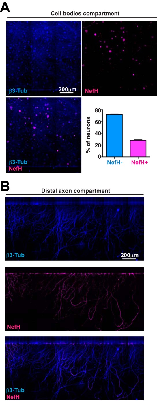
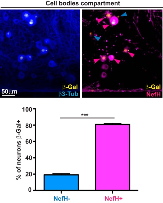
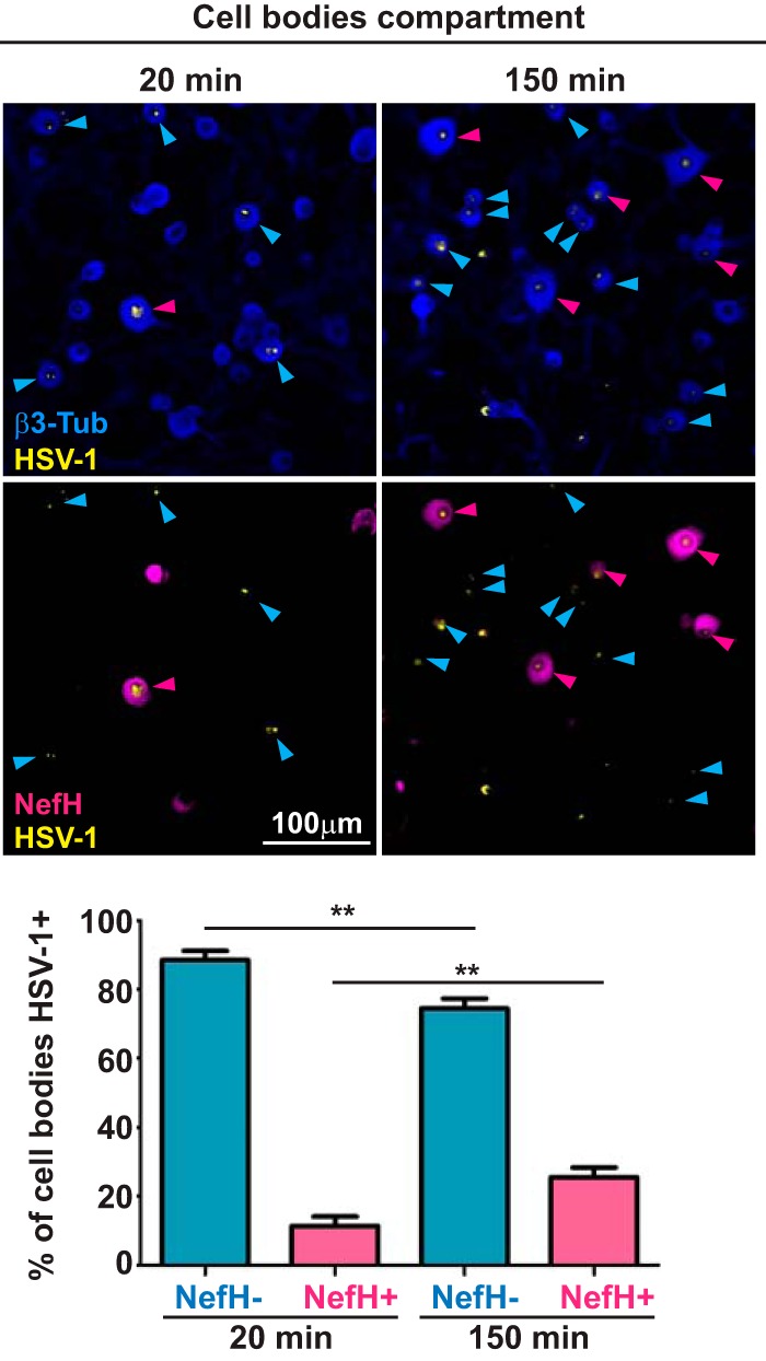
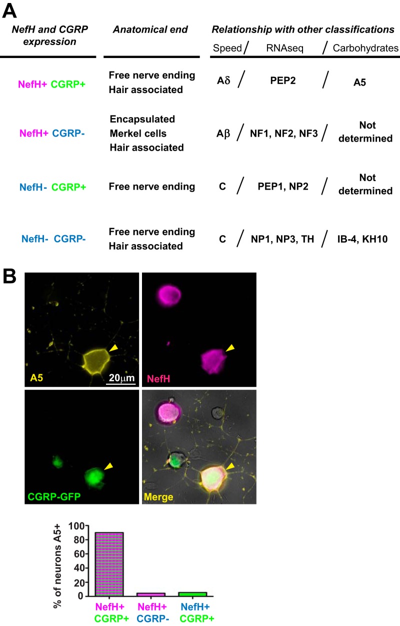
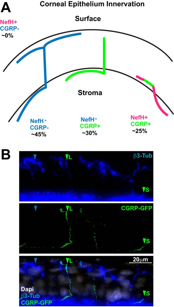
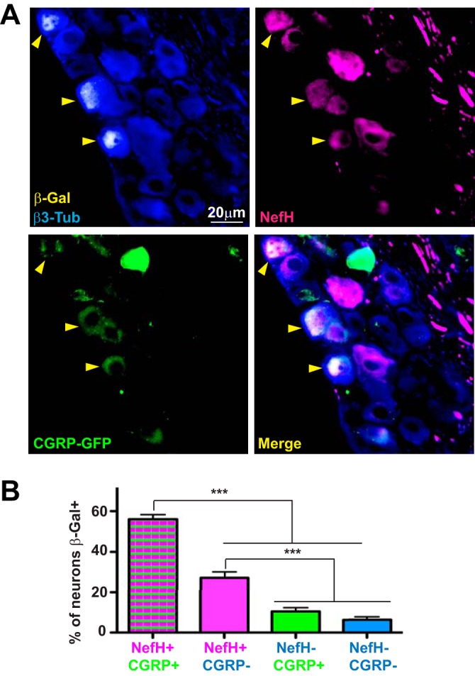
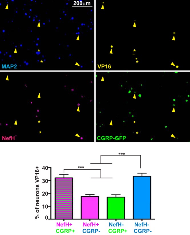
Herpes simplex virus (HSV) latency in neurons remains poorly understood, and the heterogeneity of the sensory nervous system complicates mechanistic studies. In this study, we used primary culture of adult trigeminal ganglion (TG) mouse neurons in microfluidic devices and an in vivo model to examine the subtypes of sensory neurons involved in HSV latency. HSV-infected neurofilament heavy-positive (NefH+) neurons were more likely to express latency-associated transcripts (LATs) than infected neurofilament heavy-negative (NefH-) neurons. This differential expression of the LAT promoter correlated with differences in HSV-1 early infection that manifested as differences in the efficiency with which HSV particles reached the cell body following infection at the distal axon. In vivo, we further identified a specific subset of NefH+ neurons which coexpressed calcitonin gene-related peptide α (NefH+ CGRP+ neurons) as the sensory neuron subpopulation with the highest LAT promoter activity following HSV-1 infection. Finally, an early-phase reactivation assay showed HSV-1 reactivating in NefH+ CGRP+ neurons, although other sensory neuron subpopulations were also involved. Together, these results show that sensory neurons expressing neurofilaments exhibit enhanced LAT promoter activity. We hypothesize that the reduced efficiency of HSV-1 invasion at an early phase of infection may promote efficient establishment of latency in NefH+ neurons due to initiation of the antiviral state preceding arrival of the virus at the neuronal cell body. While the outcome of HSV-1 infection of neurons is determined by a broad variety of factors in vivo, neuronal subtypes are likely to play differential roles in modulating the establishment of latent infection.IMPORTANCE Two pivotal properties of HSV-1 make it a successful pathogen. First, it infects neurons, which are immune privileged. Second, it establishes latency in these neurons. Together, these properties allow HSV to persist for the lifetime of its host. Neurons are diverse and highly organized cells, with specific anatomical, physiological, and molecular characteristics. Previous work has shown that establishment of latency by HSV-1 does not occur equally in all types of neurons. Our results show that the kinetics of HSV infection and the levels of latency-related gene expression differ in certain types of neurons. The neuronal subtype infected by HSV is therefore a critical determinant of the outcome of infection and latency.

Keywords: herpes simplex virus; latency; neurons.


Figures

FIG 1
FIG 1
Trigeminal ganglion neuronal subpopulations are identifiable by neurofilament heavy expression. (A) Representative images from the cell bodies compartment of microfluidic devices containing explanted, polarized, adult mouse TG neurons. Neurons polarizing over 3 days in culture were fixed within the devices and immunostained for β3-tubulin (β3-Tub) (blue; upper left) to demarcate all neurons and for neurofilament heavy (NefH; magenta; upper right) to identify NefH+ neurons. The merged image is shown at the lower left. The graph represents the percentage of NefH versus NefH+ neurons counted from tiles obtained from the central channel in eight replicate chambers. (B) Representative images of TG axons in the distal axon compartment of the microfluidic device depicted in panel A. (Top) β3-Tub (blue); (middle) NefH (magenta); (bottom) merged image.
FIG 2
FIG 2
LAT expression is primarily observed in NefH+ neurons following axonal HSV-1 infection. TG neurons cultured in microfluidic devices for 3 days were infected at the distal axon compartment with KOS/62 (MOI = 10). At 5 dpi, the cultures were fixed and immunostained for expression of the LAT promoter-driven β-galactosidase (β-Gal; yellow), β3-Tub (blue), and NefH (magenta). In the right micrograph, the blue arrowheads point to β-Gal+ NefH neurons and magenta arrowheads point to β-Gal+ NefH+ neurons. The graph represents the percentage of NefH and NefH+ neurons positive for β-Gal (LAT) expression. The threshold for β-Gal positivity was set using mock-infected chambers. Data are for 8 chambers and are from 3 independent experiments. ***, P < 0.001.
FIG 3
FIG 3
HSV-1 early-infection efficiency is lower in NefH+ neurons. TG neurons cultured in microfluidic devices for 3 days were infected synchronously at the distal axon compartment with KOS (MOI = 200). Cultures were fixed and processed at two time points: 20 min (left) and 150 min (right) postadsorption. Immunostaining for β3-Tub (blue) and NefH (magenta) revealed total neurons and NefH+ neurons, respectively. Immunostaining for HSV-1 is in yellow. Blue arrowheads point to NefH-infected neurons. Magenta arrowheads point to NefH+-infected neurons. The graph represents the percentage of NefH and NefH+ cell bodies positive for KOS at the early and late time points. The threshold for KOS detection was set using mock-infected chambers. Data are for 7 chambers for 20 min postadsorption and 7 chambers for 150 min postadsorption from 2 independent experiments. **, P < 0.01.
FIG 4
FIG 4
A5+ TG neurons are mainly NefH+ CGRP+ neurons. (A) TG neuron classification based on NefH and CGRP expression. The four defined subpopulations are compared to other existing classifications: anatomical ending, conduction speed, large-scale single-cell RNA-seq, or carbohydrate expression. The information presented is based on previously published data (18–20, 36, 37). (B) Adult CGRP-GFP mouse TG neurons were explanted onto coverslips and immunostained for the A5 cell surface carbohydrate (yellow) and NefH (magenta). CGRP-GFP neurons were green due to GFP expression from the transgenic mouse. Yellow arrowheads point to an A5+ neuron. Neurons were identified by their morphology under phase contrast. The graph represents the percentage of neurons in each subpopulation that were positive for Α5. Data are for >200 neurons from 2 independent experiments.
FIG 5
FIG 5
The innervation of mouse corneal epithelium is heterogeneous. (A) Overview of mouse corneal epithelial innervation based on previously published data (39–41). (B) A representative z-stack projection of corneal epithelium from adult CGRP-GFP mice. Tissue sections were immunostained for β3-Tub (blue), and CGRP+ neurons were green from GFP expression in the transgenic mouse. The merged image is shown at the bottom (DAPI [4′,6-diamidino-2-phenylindole] is in white). Blue arrowheads point to CGRP FNEs. Green arrowheads point to CGRP+ FNEs. L, long; S, short.
FIG 6
FIG 6
HSV-1 LAT expression is predominantly observed in NefH+ CGRP+ sensory neurons following corneal infection. (A) Representative image of TGs from adult CGRP-GFP mice corneally infected with KOS/62 (1 × 10⁶ PFU/eye) for >21 days. Tissue sections were immunostained for β-Gal (yellow), β3-Tub (blue), and NefH (magenta). CGRP+ neurons were green owing to GFP expression from the transgenic mouse. Yellow arrowheads point to β-Gal+ neurons in which LAT promoter activity is evident. The merged image is shown at the bottom right. (B) The graph represents the percentage of each subpopulation of neurons positive for β-Gal. The threshold for β-Gal detection was set using mock-infected tissue. No fewer than 6 random sections per TG were analyzed. Data are for 12 TGs from 2 independent experiments. ***, P < 0.001. Bar, 20 μm.
FIG 7
FIG 7
HSV-1 is capable of establishing latency in multiple neuronal populations. Representative images of dissociated TG neurons from adult CGRP-GFP mice infected corneally with KOS/62 (1 × 10⁶ PFU/eye) for >21 days. Neurons were seeded without trophic support and in the presence of trichostatin A to ensure full reactivation from all latently infected neurons. Twenty-four hours later, neurons were immunostained for MAP2 to label all neurons (blue), VP16 (yellow), and NefH (magenta). CGRP+ neurons were identified by GFP emission (green). Yellow arrowheads point to VP16+ neurons. Yellow asterisks mark nonspecific staining or infected, syncytial neurons that were excluded from quantitation. The graph represents the percentage of neurons of each subpopulation that were VP16 positive. The threshold for VP16 detection was set using a culture of mock-infected neurons. Data are for 12 TG from 2 independent experiments. ***, P < 0.001.

References

    1. Knipe DM, Howley PM, Cohen JI, Griffin DE, Lamb RA, Martin MA, Racaniello VR, Roizman B (ed). 2013. Fields virology, 6th ed Lippincott Williams & Wilkins, Philadelphia, PA.
    1. Xu F, Sternberg MR, Kottiri BJ, McQuillan GM, Lee FK, Nahmias AJ, Berman SM, Markowitz LE. 2006. Trends in herpes simplex virus type 1 and type 2 seroprevalence in the United States. JAMA 296:964–973. doi:10.1001/jama.296.8.964. - DOI - PubMed
    1. Whitley RJ, Roizman B. 2001. Herpes simplex virus infections. Lancet 357:1513–1518. doi:10.1016/S0140-6736(00)04638-9. - DOI - PubMed
    1. Cunningham AL, Diefenbach RJ, Miranda-Saksena M, Bosnjak L, Kim M, Jones C, Douglas MW. 2006. The cycle of human herpes simplex virus infection: virus transport and immune control. J Infect Dis 194(Suppl):S11–S18. doi:10.1086/505359. - DOI - PubMed
    1. Knipe DM, Cliffe A. 2008. Chromatin control of herpes simplex virus lytic and latent infection. Nat Rev Microbiol 6:211–221. doi:10.1038/nrmicro1794. - DOI - PubMed

Publication types

MeSH terms

Substances

LinkOut - more resources