Review
. 2018;94(4):180-203.
doi: 10.2183/pjab.94.013.

MPF-based meiotic cell cycle control: Half a century of lessons from starfish oocytes

Affiliations
Review

MPF-based meiotic cell cycle control: Half a century of lessons from starfish oocytes

Takeo Kishimoto. Proc Jpn Acad Ser B Phys Biol Sci. 2018.

Abstract

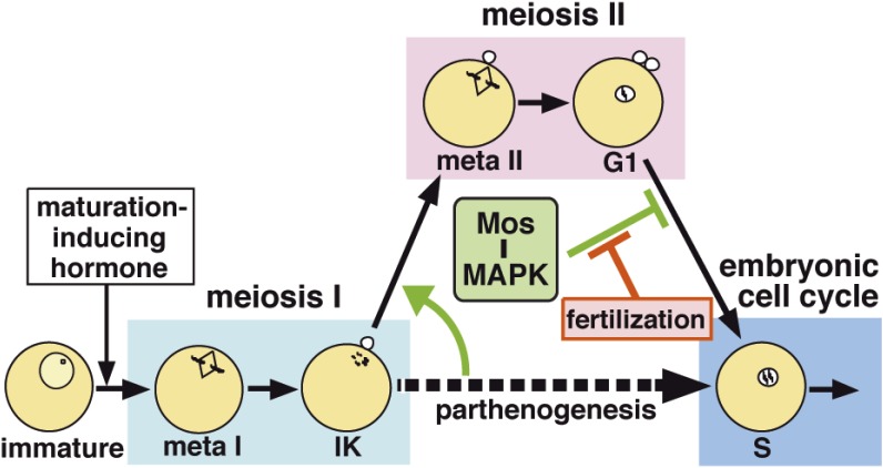
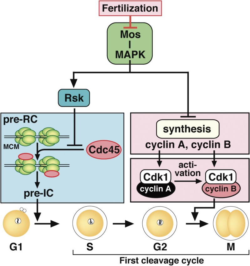
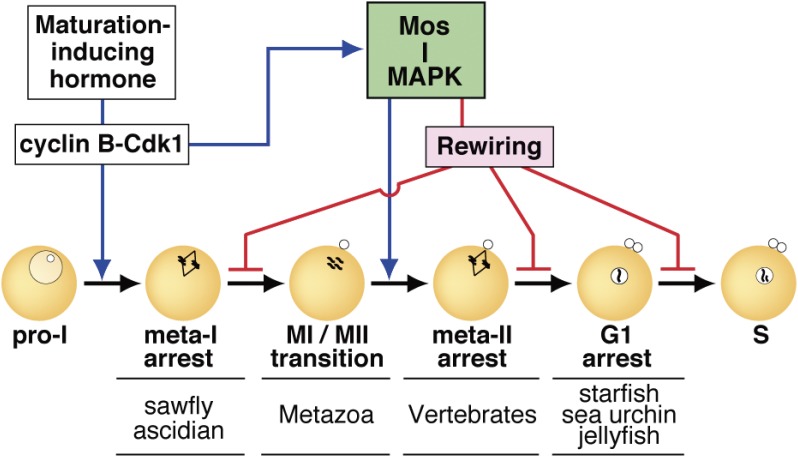
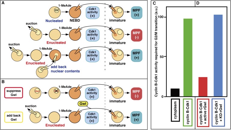
In metazoans that undergo sexual reproduction, genomic inheritance is ensured by two distinct types of cell cycle, mitosis and meiosis. Mitosis maintains the genomic ploidy in somatic cells reproducing within a generation, whereas meiosis reduces by half the ploidy in germ cells to prepare for successive generations. The meiotic cell cycle is believed to be a derived form of the mitotic cell cycle; however, the molecular mechanisms underlying both of these processes remain elusive. My laboratory has long studied the meiotic cell cycle in starfish oocytes, particularly the control of meiotic M-phase by maturation- or M phase-promoting factor (MPF) and the kinase cyclin B-associated Cdk1 (cyclin B-Cdk1). Using this system, we have unraveled the molecular principles conserved in metazoans that modify M-phase progression from the mitotic type to the meiotic type needed to produce a haploid genome. Furthermore, we have solved a long-standing enigma concerning the molecular identity of MPF, a universal inducer of M-phase both in mitosis and meiosis of eukaryotic cells.

Keywords: M-phase; MPF; cell cycle; cyclin B-Cdk1; meiosis; oocyte.


Figures

Figure 1.
Figure 1.
Hormonal control of oocyte maturation and demonstration of MPF. (A) In the endocrine control of oocyte maturation, starfish GSS (released from the nervous system) or frog GTH (released from the pituitary) functions as the first substance acting on ovarian follicles. The second substance, maturation-inducing hormone (starfish 1-MeAde or frog progesterone), is produced by and released from follicles, and acts on the oocyte surface. Based on these, a third substance that is responsible for oocyte maturation was hypothesized in the oocyte cytoplasm, and subsequently designated as maturation-promoting factor (MPF) upon its demonstration as shown in B. (B) MPF, the third substance, was demonstrated by cytoplasmic transfer from maturation-inducing hormone-treated maturing oocytes into untreated immature oocytes, which in turn undergo maturation (upper box). At the same time, the MPF activity was shown not to decrease through multiple successive transfers into immature oocytes in which de novo protein synthesis was prevented (lower box). This was called the “amplification” of MPF, implying that the inactive form of MPF is present in immature oocytes and that it can be autocatalytically activated by the active form of MPF. GV, germinal vesicle (oocyte nucleus).
Figure 2.
Figure 2.
Activation of the cyclin B-Cdk1 complex at entry into M-phase and its subsequent inactivation at exit from M-phase. Cdc2 was renamed as cyclin-dependent kinase 1 (Cdk1) in the designation of the Cdk family in 1991.78) (A) At the G2/M-phase border, Myt1/Wee1 (which phosphorylates cyclin B-associated Cdk1 on Thr14 and Tyr15 for inhibition) surpasses Cdc25 (which dephosphorylates cyclin B-associated Cdk1 on the sites phosphorylated by Myt1/Wee1 for activation), and accordingly, cyclin B-Cdk1 remains in an inactive form, in which all three sites on Cdk1 are phosphorylated (indicated by the circled P). At the transition into M-phase, the initial activator (which is different depending on the cell type) first tips the balance between Myt1/Wee1 and Cdc25 activities to trigger the initial activation of cyclin B-Cdk1. Subsequently, active cyclin B-Cdk1 directly phosphorylates Cdc25 and Myt1/Wee1 for activation and inhibition, respectively, and hence the Myt1/Wee1-Cdc25 balance is further reversed via an autoactivation loop, leading to the robust and full autoregulatory activation of cyclin B-Cdk1. Details on the autoactivation loop are shown in Fig. 9. (B) At exit from M-phase, active cyclin B-Cdk1 activates an E3 ligase, the anaphase-promoting complex/cyclosome (APC/C), leading to poly-ubiquitination of Cdk1-associated cyclin B. Subsequently, the proteasome recognizes the poly-ubiquitin chain, dissociates poly-ubiquitinated cyclin B from Cdk1, and then proteolyses cyclin B, resulting in inactivation of cyclin B-Cdk1. U, ubiquitin; E1, ubiquitin-activating enzyme; E2, ubiquitin-conjugating enzyme.
Figure 3.
Figure 3.
Dynamics of cell cycle regulators and their choreographers in starfish meiotic and early cleavage cycles. (A) Fully grown immature starfish oocytes are arrested at prophase of meiosis I (MI), which is equivalent to G2-phase in somatic cells. This arrest is characterized by the presence of a large nucleus called the germinal vesicle (GV). Once these immature oocytes are isolated into seawater and treated with 1-MeAde (1-methyladenine), the starfish maturation-inducing hormone, meiosis resumes as hallmarked by GV breakdown (GVBD), followed by two consecutive M-phases, MI and meiosis II (MII), without an intervening S-phase. After the completion of MII, mature haploid eggs arrest at G1-phase unless fertilization occurs. Starfish oocytes are fertilizable throughout the meiotic cell cycle (even at prophase I or G1-phase), whereas physiological fertilization possibly occurs in late MI. After the completion of MII, fertilized eggs start to undergo cleavage cycles consisting of alternating S- and M-phases. The cell cycle dynamics of various regulators are schematically shown in B–D. meta I and meta II, metaphase of MI and MII, respectively; Ik, interkinesis period; 1PB and 2PB, the first and second polar body; 1CL, the first cleavage. (B) Cyclin B protein is already present in immature oocytes. By contrast, cyclin A protein (and Wee1 protein shown in D) is undetectable in immature oocytes and starts to accumulate near the end of MI (shaded areas). After meiotic resumption, the protein levels of cyclins A and B cycle along with the cell cycle, peaking at each metaphase. The Cdk1 level remains constant throughout the entire process. Accordingly, Cdk1 activity is represented by cyclin B-Cdk1 in MI, and largely by both cyclin B-Cdk1 and cyclin A-Cdk1 in and after MII. (C, D) In contrast to cyclins, the protein levels of Cdc25, Myt1, Greatwall kinase (Gwl), mitogen-activated protein kinase (MAPK), polo-like kinase 1 (Plk1), and Aurora (Aur) remain constant throughout the entire process. All of Gwl, Plk1, Aur, and MAPK are activated downstream of cyclin B-Cdk1 at meiotic resumption. The meiotic cycles are characterized by the unique dynamics in activities of MAPK and Plk1, and hence Cdc25 and Myt1 during the MI to MII transition and after the completion of MII (during G1-phase arrest) in unfertilized eggs (dotted lines). In contrast, Gwl activity correlates with cyclin B-Cdk1 activity. PP2A-B55 and PP1 activities are assumed to roughly mirror those of cyclin B-Cdk1 and Gwl. See the text for details.
Figure 4.
Figure 4.
Signaling pathway leading to the activation of cyclin B-Cdk1 at the meiotic G2/M-phase transition in starfish oocytes. This pathway consists of the initial activation of cyclin B-Cdk1 and its subsequent autoregulatory activation, as shown in Fig. 2A. The initial activation pathway may be characteristic of the starfish oocyte system, whereas the autoregulatory activation pathway is largely conserved. The putative 1-MeAde receptor on the oocyte surface couples with heterotrimeric G-protein, from which the Gβγ complex is released to cause the initial activation of cyclin B-Cdk1 via two parallel pathways. In one pathway, Gβγ activates phosphoinositide 3-kinase (PI3K) to produce phosphatidylinositol 3,4,5-triphosphate (PIP3), depending on which Akt/protein kinase B (PKB) is activated. Akt/PKB directly phosphorylates Myt1 and Cdc25 for downregulation and upregulation, respectively. In the other pathway, Gβγ along with PI3K contributes, via unknown molecule(s), to the phosphorylation of Cdc25 and Myt1 on residues phosphorylated by Akt/PKB (Akt/PKB sites). These initial phosphorylations on the Akt/PKB sites, which are accomplished by possible cooperation of these two pathways, tip and reverse the balance between Cdc25 and Myt1 activities, leading to activation of a small population of cyclin B-Cdk1. The autoactivation loop then starts the activation of a much larger population of cyclin B-Cdk1 (see Fig. 9 for details). In the autoregulatory activation, Cdc25 and Myt1 are directly phosphorylated largely by cyclin B-Cdk1 (Cdk1 sites).
Figure 5.
Figure 5.
Mos–MAPK signaling ensures the successful transition from meiosis I to II to prevent parthenogenetic activation in starfish oocytes. At the end of meiosis I, the oocyte already has the ability to enter the embryonic mitotic cycle in the absence of fertilization (i.e., parthenogenesis). Mos–MAPK signaling, however, represses this ability in two ways. First, at the end of meiosis I, Mos–MAPK signaling causes swift activation of cyclin B-Cdk1 to force entry into meiosis II without an intervening S-phase. Subsequently, Mos–MAPK signaling prevents return to the embryonic mitotic cycle after the completion of meiosis II, resulting in the suppression of parthenogenesis. Once fertilization occurs, this prevention is cancelled, leading to the start of the embryonic mitotic cycle. Thus, Mos–MAPK signaling halves the ploidy and maintains the haploid state of oocytes until fertilization.
Figure 6.
Figure 6.
Dual-lock for G1-phase arrest in unfertilized mature starfish eggs. Unless fertilized, mature starfish eggs arrest at G1-phase after the completion of meiosis II. This arrest is accomplished through two separate pathways that function downstream of Mos–MAPK signaling. One is a Rsk-mediated pathway that prevents entry into S-phase by inhibiting Cdc45 loading onto the DNA replication machinery. The initiation of DNA replication is thus blocked by Rsk at the pre-replicative complex (pre-RC) stage, prior to the subsequent pre-initiation complex (pre-IC) stage just before the actual start of DNA replication. The other is an Rsk-unmediated pathway that prevents entry into the first mitotic M-phase by inhibiting new protein synthesis of cyclin A and cyclin B. Due to the absence of a DNA replication checkpoint, this dual-lock mechanism is required for G1 arrest. Upon fertilization, Mos degradation releases the dual-lock, resulting in the start of the embryonic mitotic cycle. MCM, MCM complex.
Figure 7.
Figure 7.
Mos–MAPK signaling governs meiotic cell cycle progression to accomplish the half-reduction of genomic ploidy in metazoan oocytes. At meiotic resumption, diverse hormonal signaling processes in various animal species center on activation of cyclin B-Cdk1 in all cases. Mos–MAPK is then activated downstream of cyclin B-Cdk1. Mos–MAPK positively regulates the meiosis I to II transition by avoiding S-phase and by facilitating entry into meiosis II in most metazoans (blue line). On the other hand, Mos–MAPK causes, via its downstream rewiring, a second meiotic arrest at diverse, species-specific stages (meta-I, meta-II, or G1; representative examples are indicated at each arrest) until fertilization (red lines). Thus, the conserved Mos–MAPK module is central to the production of haploid eggs for successive generations.
Figure 8.
Figure 8.
MPF is not synonymous with cyclin B-Cdk1 and instead consists of both cyclin B-Cdk1 and Gwl. (A) Nuclear material is required for MPF. In the starfish system, MPF is undetectable from enucleated donor oocytes, whereas cyclin B-Cdk1 is invariably activated after 1-MeAde treatment. MPF is restored when the nuclear material is added back to the donor enucleated oocytes. MPF (+) and (−), detectable and undetectable, respectively. (B) Greatwall kinase (Gwl) is essential for MPF. When Gwl activity is suppressed in the nuclei of donor oocytes by injection of its neutralizing antibodies, MPF is undetectable, even though cyclin B-Cdk1 is invariably activated. Conversely, injection of Gwl into enucleated oocytes restores MPF. (C) One order of magnitude excess levels of cyclin B-Cdk1 activity are required to induce GVBD in the microinjection assay, when purified cyclin B-Cdk1 is compared with cyclin B-Cdk1 found in cytoplasmic MPF. (D) For GVBD induction by microinjection, the addition of Gwl reduces the required activity of purified cyclin B-Cdk1 approximately to that contained in cytoplasmic MPF. rGwl, active recombinant Gwl; KD, kinase-dead form as a control. Reproduced from Fig. 2 in Ref. with some modifications.
Figure 9.
Figure 9.
Molecular pathway for autoregulatory activation of cyclin B-Cdk1, and its core kinases, cyclin B-Cdk1 and Gwl, that constitute MPF. The autoactivation loop consists of two kinases, cyclin B-Cdk1 and Gwl, that promote activation of cyclin B-Cdk1, and two phosphatases, PP2A-B55 and PP1, that counteract activation of these two kinases. Once cyclin B-Cdk1 is initially activated, it causes, on one hand, suppression of the major antagonizing phosphatase PP2A-B55 via Arpp19 in two steps: First, direct phosphorylation of Arpp19 by initially activated cyclin B-Cdk1 converts Arpp19 into an inhibitor of PP2A-B55, and subsequent phosphorylation of Arpp19 on another residue by Gwl, which is activated downstream of cyclin B-Cdk1, enhances this conversion. On the other hand, initially activated cyclin B-Cdk1 directly phosphorylates and inhibits phosphatase PP1 which antagonizes the autoactivation of Gwl. Collectively, the cyclin B-Cdk1–Arpp19 pathway and the cyclin B-Cdk1–Gwl–Arpp19/Ensa pathway synergistically inhibit PP2A-B55 to support phosphorylation of Cdc25 and Myt1/Wee1 by cyclin B-Cdk1, resulting in the autoregulatory activation of cyclin B-Cdk1. The authentic, classical MPF is the system (composed of cyclin B-Cdk1 and Gwl) that initiates and accomplishes the autoregulatory activation of cyclin B-Cdk1 under intracellular circumstances in which the initial activation of cyclin B-Cdk1 is not allowed. Gwl contributes to MPF by accelerating the suppression of PP2A-B55 that counteracts the autoregulatory activation of cyclin B-Cdk1. Reproduced from Fig. 6 in Ref. with some updates.

Similar articles

Cited by

References

    1. Schleiden M.J. (1838) Beitraege zur Phytogenesis. Muller’s Arch. Anat. Physiol. Wiss. Med. 136–176.
    1. Schwann, T. (1839) Mikroskopische Untersuchungen ueber die Uebereinstimmung in der Struktur und dem Wachsthum der Thiere und Pflanzen. Verlag der Sander’schen Buchhandlung, Berlin. - PubMed
    1. Virchow, R. (1859) Die Cellular Pathologie in ihrer Begrundung auf physiologische und pathologosche Gewebelehre. Verlag von August Hirschwald, Berlin.
    1. Nurse P. (2000) A long twentieth century of the cell cycle control and beyond. Cell 100, 71–78. - PubMed
    1. Howard A., Pelc S.R. (1953) Synthesis of desoxyribonucleic acid in normal and irradiated cells and its relationship to chromosome breakage. Heredity (London) Suppl. 6, 261–273.

Substances