Review
. 2018 Jul;19(7):419-435.
doi: 10.1038/s41580-018-0007-0.

Contextual determinants of TGFβ action in development, immunity and cancer

Affiliations
Review

Contextual determinants of TGFβ action in development, immunity and cancer

Charles J David et al. Nat Rev Mol Cell Biol. 2018 Jul.

Erratum in

Abstract

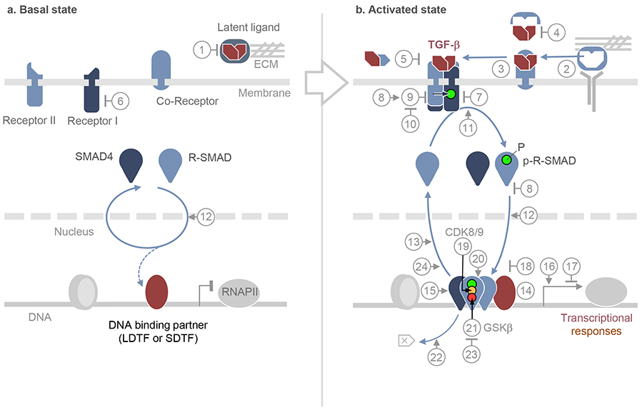
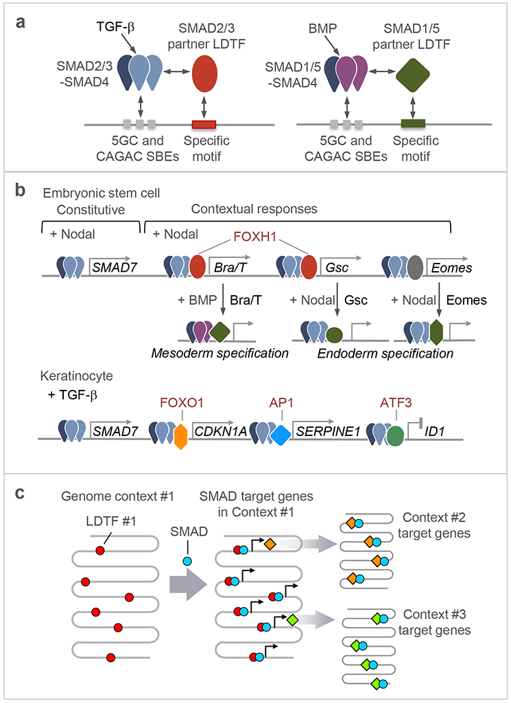
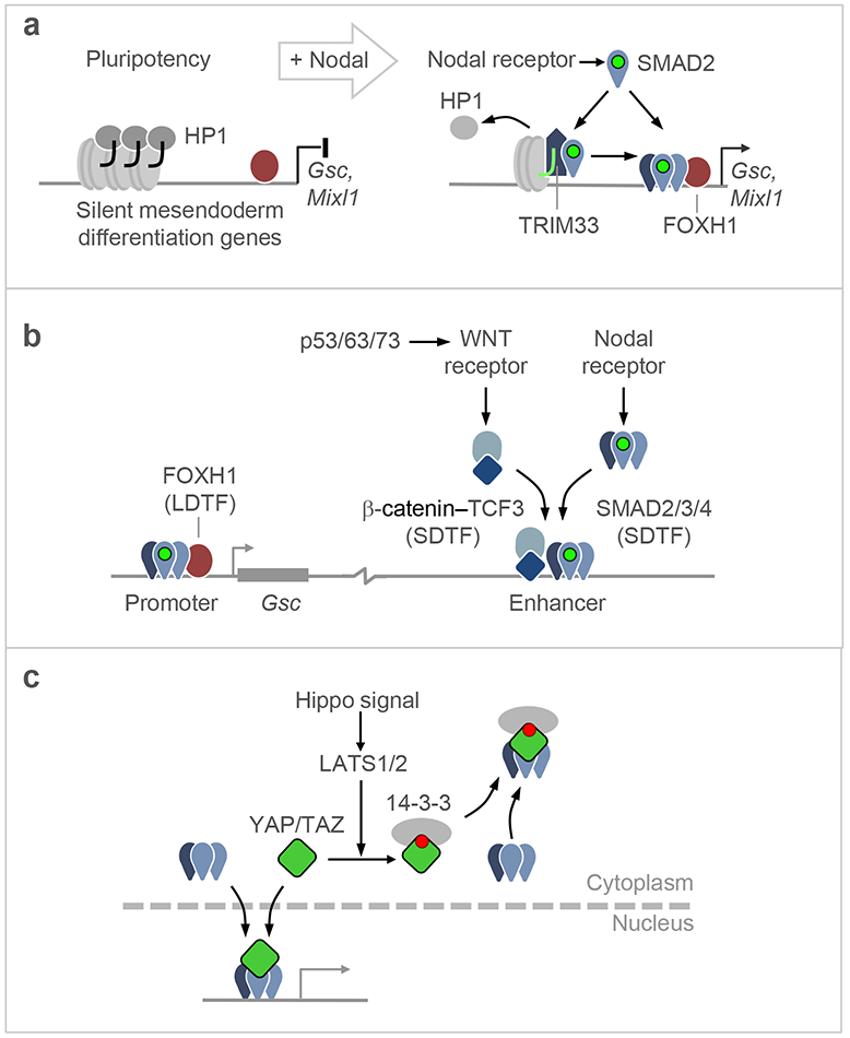
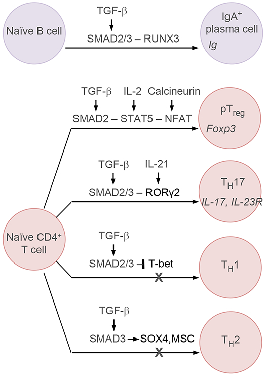
Few cell signals match the impact of the transforming growth factor-β (TGFβ) family in metazoan biology. TGFβ cytokines regulate cell fate decisions during development, tissue homeostasis and regeneration, and are major players in tumorigenesis, fibrotic disorders, immune malfunctions and various congenital diseases. The effects of the TGFβ family are mediated by a combinatorial set of ligands and receptors and by a common set of receptor-activated mothers against decapentaplegic homologue (SMAD) transcription factors, yet the effects can differ dramatically depending on the cell type and the conditions. Recent progress has illuminated a model of TGFβ action in which SMADs bind genome-wide in partnership with lineage-determining transcription factors and additionally integrate inputs from other pathways and the chromatin to trigger specific cellular responses. These new insights clarify the operating logic of the TGFβ pathway in physiology and disease.


Figures

Figure 1.
Figure 1.. The TGF-β–SMAD pathway in the basal and activated states
a. In the basal state, TGF-β family ligands are either absent or present as latent factors, sequestered by binding components of the extracellular matrix (ECM) or the membrane of other cells [1]. Allosteric inhibitors may enforce the inactive state of the receptors, all of which are transmembrane serine/threonine protein kinases [6]. SMAD proteins that serve as receptor substrates (R-SMADs), and their shared partner SMAD4, continuously transit between cytoplasm and nucleus by direct interaction with nuclear pore components [12]. SMAD partners including lineage-determining transcription factors (LDTF), other signal-driven transcription factors (SDTF), and the histone binding protein TRIM33, may already be pre-bound to the genome. Numbers in circles refer to these and other pathway regulators, as detailed in Box 1. b. In the activated state, ligand from cells in the microenvironment or released from latent complexes allosterically by integrins or enzymatically by proteases [2], binds to pairs of type I and type II receptors. Ligands can access the receptors directly or, in the case of TGF-β, Nodal and BMP9/10, with assistance of accessory co-receptors [3]. Ligand traps [4] and antagonistic ligands [5] block access to the receptors. In the ligand-induced receptor complex, the type II receptor subunits primarily phosphorylate and activate the type I receptors, which phosphorylate R-SMADs, with the assistance of adaptor proteins [11]. Receptor-phosphorylated R-SMADs form heterotrimers with SMAD4. Pseudo-receptors [7], inhibitory SMADs [8], and SKI proteins [18] inhibit the formation of receptor and SMAD complexes and provide negative feedback in the pathway. Inhibitory SMADs recruit ubiquitin ligases that target the receptor [9], which are opposed by deubiquitylases [10]. In the nucleus, activated SMAD complexes bind to hundreds of genomic loci as dictated by context-defining LDTFs and SDTFs [14]. SMADs contact DNA using a highly conserved β-hairpin structure located in the N-terminal domain (or MH1) domain. The SMAD C-terminal domain (or MH2 domain) binds not only LDTFs and SDTFs but also chromatin binding proteins [15], co-activators and co-repressors [16,17]. The nuclear kinases CDK8/9 phosphorylate the SMAD interdomain linker region [19] for recruitment of additional cofactors [20]. Subsequent phosphorylation by GSK3 targets SMADs for ubiquitin- and proteasome-dependent degradation [21,22]. Alternatively, SMADs are dephosphorylated [13, 23] and dissociated from DNA [24] for recycling. The signaling steps summarized here are reviewed in detail elsewhere,,,,,,.
Figure 2.
Figure 2.. LDTFs and SDTFs as contextual determinants of TGF-β–SMAD action
a. All R-SMADs recognize a common set of DNA elements including GGCGC and related 5bp motifs (5GC motifs), as well as the CAGAC motif. TGF-β-activated SAMAD2/3 and BMP-activated SMAD1/5 recognize different partner LDTFs, thereby achieving specificity in target gene recognition. b. Examples of interactions between SMAD complexes and DNA binding cofactors. In ES cells poised to undergo mesendodermal differentiation, the LDTF FOXH1 is pre-bound to loci throughout the genome and recruits Nodal-activated SMAD2/3:SMAD4 complexes to these loci. The target genes encode LDTFs like BRA/T and GSC. These and the related LDTF EOMES, interact in turn with Nodal-activated or BMP-activated SMADs to mediate further specification of the mesoderm and endoderm lineages (refer also to Table 2). In keratinocyte precursors, TGF-β-activated SMAD2/3:SMAD4 complexes bind the genome in partnership with different SDTFs to regulate the expression of different subsets of target genes. Although SMADs target largely different gene sets in different cell types as a function of context-specific LDTFs and SDTFs, certain SMAD-mediated gene responses appear to be constitutive to the pathway and common to many cell types, as in the case of the negative feedback regulator SMAD7. c. Schematic representation of successive waves of SMAD-LDTF driven gene expression programs. An LDTF that defines a particular phenotypic context (Context #1) within a given cell lineage recruits signal-driven SMADs to activate the expression of other LDTFs, which in turn interact with SMADs to mediate transitions into new differentiated states (Context #2, #3).
Figure 3.
Figure 3.. SMAD cooperation with different DNA binding partners
a. Certain gene responses, including the induction of LDTF-encoding genes by Nodal-driven SMAD2/3:SMAD4 in ES cells, require the participation of TRIM33. Through its PHD and Bromo domains, TRIM33 binds to histone H3 N-terminal tails that have repressive chromatin marks (H3K4me0, H3K9me3 and H3K18ac), displacing heterochromatin protein 1 (HP1) from these sites. Nodal-activated SMAD2/3 binds to TRIM33, an interaction that allows the recruitment of SMAD2/3:SMAD4 complexes by FOXH1 to induce gene expression and drive mesendodermal differentiation. TRIM33 can also act as a SMAD4 ubiquitin ligase to limit SMAD transcriptional action (not shown). b. Interactions of Nodal-activated SMAD2/3:SMAD4 with LDTFs and SDTFs on a target gene. In addition to SMAD2/3:SMAD4 recruitment by FOXH1 to the Gsc promoter, the activation of this gene requires WNT signals. A WNT-driven β-catenin:TCF3 complex and a Nodal-driven SMAD2/3:SMAD4 cooperate in binding to a distal enhancer required for activation of Gsc transcription. The WNT input is delivered by WNT3, whose expression is induced by p53 and its family members p63 and p73. The p53 family, WNT and Nodal are all essential for gastrulation, providing evidence for their involvement in SMAD-driven mesendodermal specification. c. Representation of YAP-mediated eviction of Nodal-activated SMADs from the nucleus. When YAP is phosphorylated by LATS1/2kinases in response to Hippo pathway activation. The phosphorylation sites act as binding site for 14-3-3 proteins, which drag SMADs into the nucleus and prevents TGF-β signaling.
Figure 4.
Figure 4.. Transcriptional determinants of TGF-β regulation of immune cell fate
TGF-β-activated SMADs cooperate with RUNX3 to promote class switch recombination during B-cell maturations. TGF-β-activated SMAD2/3 also regulate the specification of different cell subtypes in naïve CD4+ T cells. In this context, TGF-β-activated SMADs in Treg cells cooperate with signal-activated STAT5 and NFAT to induce Foxp3 expression for Treg differentiation. Similarly, TGF-β-activated SMAD2/3 cooperate with RORγ2 to induce a TH17 phenotype. In contrast, TGF-β-activated SMAD2/3 inhibit the specification of TH1 cell fate by repressing T-bet transcription factor, and the specification of TH2 cell fate by inducing the expression of SOX4 and MSC.
Figure 5.
Figure 5.. Determinants of TGF-β tumor suppression and its subversion in cancer
a. In mouse skin and mucosal epithelia, loss of TGF-β receptor signaling leads to hyperplasia, whereas gain of oncogenic HRAS mutations leads to TGF-β-associated apoptosis. Loss of TGF-β signaling in a HRAS-mutant background leads to the emergence of tumors. Thus, TGF-β enforces homeostasis in the wild type background, and tumor suppression in a RAS-mutant background. b. The switch of TGF-β from a signal for lineage maintenance and homeostasis to a signal for apoptosis that cancer cells must disable, as observed in pancreatic epithelial progenitors. In normal progenitors TGF-β signaling through SMAD2/3 with SMAD4 activates the expression of CDK inhibitors, SOX4, and other genes. CDK inhibitors tone down cell proliferation, whereas SOX4 associates with KLF5 to co-occupy the genome and enforce epithelial progenitor identity. Stimulation of SOX4 expression requires SMAD2/3 but not SMAD4. In premalignant pancreatic progenitors harboring oncogenic KRAS mutations, a hyperactive ERK MAPK pathway enables SMAD2/3:SMAD4 to strongly induce the expression of the EMT master regulator SNAIL. SNAIL represses KLF5 expression. In the absence of KLF5, SOX4 induces pro-apoptotic genes BIM and BMF for elimination of the cell. Nearly one half of human pancreatic ductal adenocarcinomas (PDA) progress through this bottleneck by selecting for clones that harbor SMAD4 inactivating mutations. Loss of SMAD4 does still allows TGF-β to support SOX4 expression and the SOX4:KLF5 mediated pancreatic progenitor state. The other half of PDA tumors progress with a functionally intact TGF-β signaling system but accumulate unknown alterations that prevent the pathway from triggering apoptosis.
Figure 6
Figure 6
Cancer cells can traverse the tumor suppressive TGF-β bottleneck by accumulating mutations (“X”) that disable the signal transduction pathway. In many gastrointestinal cancers, TGF-β receptor and SMAD mutations deeply disable the pathway, and this now allows stromal TGF-β to exert pro-tumorigenic effects including immune suppressive action on T and NK cells and activation of stromal paracrine loops such as the production of tumorigenic cytokines (e.g. IL-11) by cancer-associated fibroblasts (CAF). In many other types of cancer, the TGF-β receptors and SMADs remain intact but downstream alterations prevent the pathway from triggering tumor suppressive effects. Thus corrupted, the TGF-β–SMAD pathway can drive a non-lethal EMT as well the production of factors that promote metastatic extravasation, immune evasion, and colonization of target tissues.

References

    1. Massagué J How cells read TGF-beta signals. Nat Rev Mol Cell Biol 1, 169–178 (2000). - PubMed
    1. Josso N, Belville C, di Clemente N & Picard JY AMH and AMH receptor defects in persistent Mullerian duct syndrome. Hum Reprod Update 11, 351–356 (2005). - PubMed
    1. Lindsay ME & Dietz HC The genetic basis of aortic aneurysm. Cold Spring Harb Perspect Med 4, a015909 (2014). - PMC - PubMed
    1. Matzuk MM & Burns KH Genetics of mammalian reproduction: modeling the end of the germline. Annu Rev Physiol 74, 503–528 (2012). - PubMed
    1. Meng XM, Nikolic-Paterson DJ & Lan HY TGF-beta: the master regulator of fibrosis. Nat Rev Nephrol 12, 325–338 (2016). - PubMed