. 2018 Apr 11;8(1):5842.
doi: 10.1038/s41598-018-24221-5.

Pro-inflammatory cytokines activate hypoxia-inducible factor 3α via epigenetic changes in mesenchymal stromal/stem cells

Affiliations

Pro-inflammatory cytokines activate hypoxia-inducible factor 3α via epigenetic changes in mesenchymal stromal/stem cells

Francesca Cuomo et al. Sci Rep. .

Erratum in

Abstract

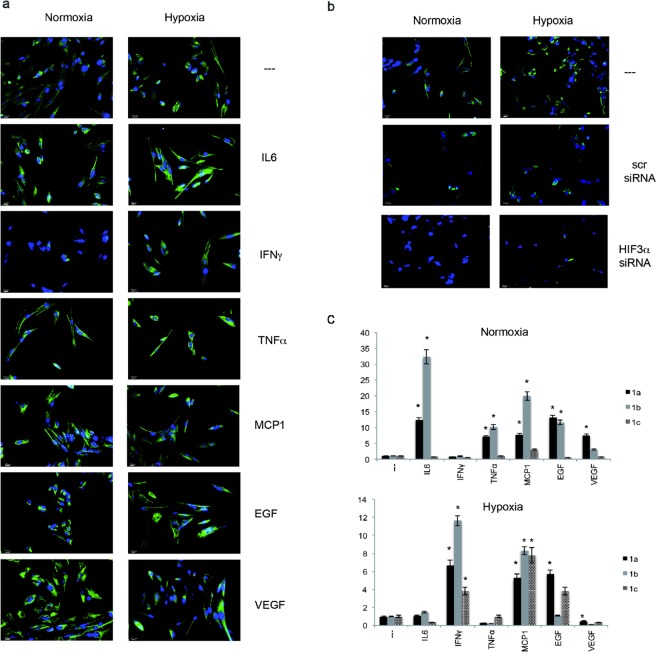
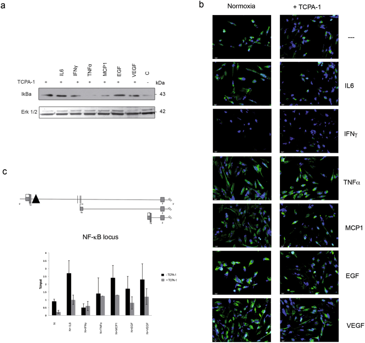
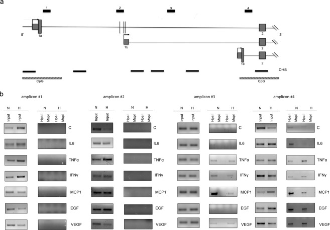
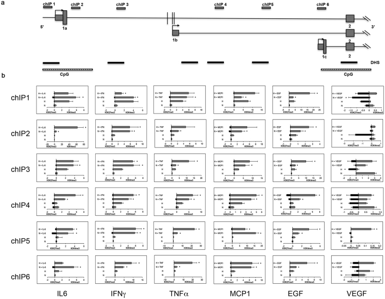
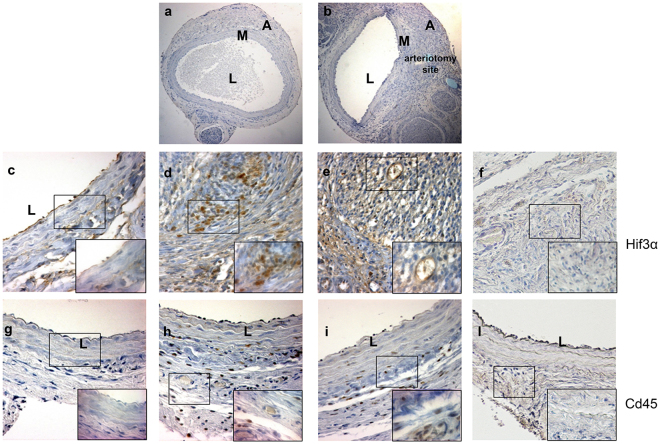
Human mesenchymal stromal/stem cells (hMSCs) emerged as a promising therapeutic tool for ischemic disorders, due to their ability to regenerate damaged tissues, promote angiogenesis and reduce inflammation, leading to encouraging, but still limited results. The outcomes in clinical trials exploring hMSC therapy are influenced by low cell retention and survival in affected tissues, partially influenced by lesion's microenvironment, where low oxygen conditions (i.e. hypoxia) and inflammation coexist. Hypoxia and inflammation are pathophysiological stresses, sharing common activators, such as hypoxia-inducible factors (HIFs) and NF-κB. HIF1α and HIF2α respond essentially to hypoxia, activating pathways involved in tissue repair. Little is known about the regulation of HIF3α. Here we investigated the role of HIF3α in vitro and in vivo. Human MSCs expressed HIF3α, differentially regulated by pro-inflammatory cytokines in an oxygen-independent manner, a novel and still uncharacterized mechanism, where NF-κB is critical for its expression. We investigated if epigenetic modifications are involved in HIF3α expression by methylation-specific PCR and histone modifications. Robust hypermethylation of histone H3 was observed across HIF3A locus driven by pro-inflammatory cytokines. Experiments in a murine model of arteriotomy highlighted the activation of Hif3α expression in infiltrated inflammatory cells, suggesting a new role for Hif3α in inflammation in vivo.


Conflict of interest statement

The authors declare no competing interests.

Figures

Figure 1
Figure 1
HIF3α expression in hMSCs. (a) Immunofluorescence analysis of HIF3α protein in hMSCs cultured in standard oxygen conditions (Normoxia), and in CoCl2-induced hypoxia (Hypoxia) for 24 h in absence and in presence of indicated cytokines and probed with antibodies against HIF3α. Scale bars: 10 μm). (b) Immunofluorescence analysis of HIF3α protein in cells grown in normoxia and hypoxia with siRNA-mediated HIF3α silencing or scrambled siRNA as control (scr). (c) Expression levels of the three alternative first exons (1a, 1b, and 1c) by qRT-PCR in hMSCs cultured in normoxia or hypoxia for 24 h in absence or in presence of indicated cytokines. Relative gene expression data are reported as 2-ΔΔCt method, normalized to housekeeping gene (b-actin mRNA) and ALU sequences. Data are expressed as means ± SEM (n = 3).*p value < 0.05.
Figure 2
Figure 2
HIF3α activation is dependent on NF-κB activation: (a) Immunoblotting analysis of IκBα: hMSCs cultured in normoxia and treated with IL6, IFNγ, TNFα, MCP1, EGF and VEGF for 24 h. Cells were pre-treated with TCPA-1 for 1 h before cytokines supplementation. (b) Immunofluorescence analysis of HIF3α protein in hMSCs cultured in standard oxygen conditions (Normoxia) in absence and in presence of indicated cytokines and probed with antibodies against HIF3α. Cells were pre-treated with TCPA-1 for 1 h before cytokines supplementation. Scale bars: 10 μm). (c) Schematic diagram of the HIF3A promoter region, where a putative NF-KB binding site is depicted (black triangle). NFκB (RelA) binding on HIF3A promoter: ChIP analysis was performed in hMSCs cultured in standard oxygen conditions and treated with IL6, IFNγ, TNFα, MCP1, EGF and VEGF for 24 h (black bars), or pre-treated with TCPA-1 for 1 h (grey bars) before adding cytokines. As control, species matched IgG were used. Data obtained by qRT-PCR are expressed as enrichment of chromatin-associated DNA fragments immunoprecipitated by NF-κB antibody compared with input (% Input) and expressed as means ± SEM of two independent experiments performed in triplicate.
Figure 3
Figure 3
DNA methylation status of human HIF3A promoter. (a) Schematic diagram of the HIF3A promoter region, CpG islands and DNAse I hypersensitive sites (DHS) surrounding the three transcription start sites. Vertical black lines represent the three methylation sites in intron 1 in association with high BMI. (b) MS-PCR analysis of genomic DNA extracted from hMSCs exposed to different pro-inflammatory cytokines in normoxic and hypoxic conditions. The locations of PCR amplicons (#1 to #4) are shown in (a). Three independent experiments were performed and a representative experiment is reported. Images derived from different part of the same gel and cropped for layout reasons and included in Suppl. Information.
Figure 4
Figure 4
ChIP-PCR analysis of histone modifications of human HIF3A promoter. (a) Schematic diagram of the HIF3A promoter region and primers used in ChIP analysis using specific antibodies to H3K4me3 and H3K27me3 histone modifications. qRT-PCR was performed on immunoprecipitated chromatin of hMSCs treated with different cytokines in normoxic and hypoxic conditions. (b) Six different regions (ChIP1-6) spanning the HIF3A gene were analysed. Results are expressed as means ± SEM of three different experiments and are reported as the ratio between treated hMSCs vs. untreated hMSCs (2-ΔΔCt method). *< 0.05 (p-value).
Figure 5
Figure 5
Immunohistochemical analysis of Hif3α and Cd45 expression in uninjured rat carotids and in carotids from Wistar male rats treated with MSCs or DMEM and harvested 7 days after arteriotomy. (a) Uninjured rat carotid; (b) arteriotomy-injured rat carotid harvested 7 days after injury, haematoxylin staining. Arrows in b indicate the injury site, where arteriotomy is followed by the application of an 8.0 polypropylene stitch (light blue). (cf) Representative immunohistochemical staining of Hif3α in uninjured rat carotid (c) and in injured carotids harvested 7 days after arteriotomy from DMEM- (d) and MSC-treated rats (e). (gl) Representative immunohistochemical staining of Cd45 in adjacent cross-sections from uninjured rat carotid (g) and in injured carotids harvested 7 days after arteriotomy from DMEM- (h) and MSC-treated rats (i). (f,l) Immunohistochemical staining of serial cross-sections of rat carotids used in d and h without primary antibody as negative control. (a,b) 10x magnification; (cl) 20x magnification; small insets: 40x magnification of selected areas enclosed in black rectangles, representative of nuclei positive to Hif3α and Cd45 in adventitia, vasa vasorum and perivascular tissue. Brown staining corresponds to target protein expression. Nuclei were counterstained with haematoxylin. L: lumen; M: media; A: adventitia.

Similar articles

Cited by

References

    1. Kean TJ, Lin P, Caplan AI, Dennis JE. MSCs: Delivery Routes and Engraftment, Cell-Targeting Strategies, and Immune Modulation. Stem Cells Int. 2013;2013:732742. doi: 10.1155/2013/732742. - DOI - PMC - PubMed
    1. Kfouri Y, Scadden D. Mesenchymal cell contributions to the stem cell niche. Cell Stem Cell. 2015;16:239–253. doi: 10.1016/j.stem.2015.02.019. - DOI - PubMed
    1. Schiavetta A, et al. A phase II trial of autologous transplantation of bone marrow stem cells for critical limb ischemia: results of the Naples and Pietra Ligure Evaluation of Stem Cells study. Stem Cells Transl Med. 2012;1:572–578. doi: 10.5966/sctm.2012-0021. - DOI - PMC - PubMed
    1. Madrigal M, Rao KS, Riordan NH. A review of therapeutic effects of mesenchymal stem cell secretions and induction of secretory modification by different culture methods. J Transl Med. 2014;12:260. doi: 10.1186/s12967-014-0260-8. - DOI - PMC - PubMed
    1. Forte A, et al. Stem cell therapy for arterial restenosis: potential parameters contributing to the success of bone marrow-derived mesenchymal stromal cells. Cardiovasc Drugs Ther. 2012;26:9–21. doi: 10.1007/s10557-011-6359-8. - DOI - PubMed

Publication types

MeSH terms

LinkOut - more resources