. 2018 Mar 28:12:200.
doi: 10.3389/fnins.2018.00200. eCollection 2018.

Persistent Adult Neuroimmune Activation and Loss of Hippocampal Neurogenesis Following Adolescent Ethanol Exposure: Blockade by Exercise and the Anti-inflammatory Drug Indomethacin

Affiliations

Persistent Adult Neuroimmune Activation and Loss of Hippocampal Neurogenesis Following Adolescent Ethanol Exposure: Blockade by Exercise and the Anti-inflammatory Drug Indomethacin

Ryan P Vetreno et al. Front Neurosci. .

Abstract

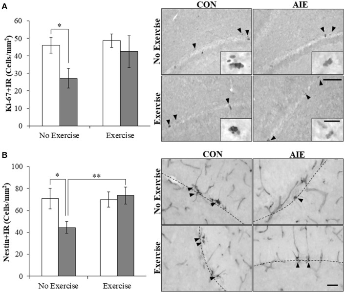
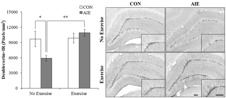
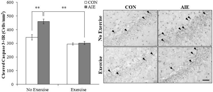
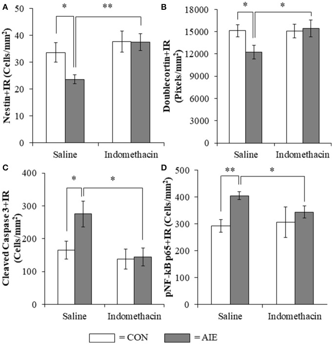
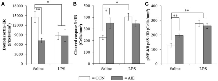
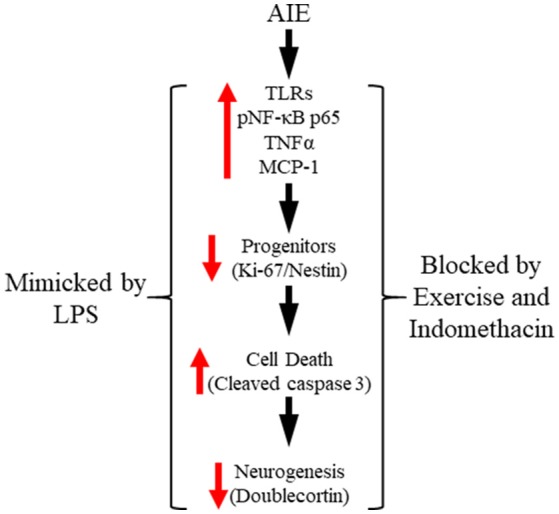
Alcohol abuse and binge drinking are common during adolescence, a developmental period characterized by heightened neuroplasticity. Animal studies reveal that adolescent ethanol exposure decreases hippocampal neurogenesis that persists into adulthood, but the mechanism remains to be fully elucidated. Using a rodent model of adolescent intermittent ethanol (AIE; 5.0 g/kg, i.g., 2-days on/2-days off from postnatal day [P]25 to P55), we tested the hypothesis that AIE-induced upregulation of neuroimmune signaling contributes to the loss of hippocampal neurogenesis in adulthood. We found that AIE caused upregulation of multiple proinflammatory Toll-like receptors (TLRs), increased expression of phosphorylated NF-κB p65 (pNF-κB p65) and the cell death marker cleaved caspase 3, and reduced markers of neurogenesis in the adult (P80) hippocampus, which is consistent with persistently increased neuroimmune signaling reducing neurogenesis. We observed a similar increase of pNF-κB p65-immunoreactive cells in the post-mortem human alcoholic hippocampus, an effect that was negatively correlated with age of drinking onset. Voluntary wheel running from P24 to P80 prevented the AIE-induced loss of neurogenesis markers (i.e., nestin and doublecortin) in the adult hippocampus that was paralleled by blockade of increased expression of the cell death marker cleaved caspase 3. Wheel running also prevented the AIE-induced increase of hippocampal pNF-κB p65 and induction of neuroimmune NF-κB target genes, including TNFα and IκBα in the adult brain. Administration of the anti-inflammatory drug indomethacin during AIE prevented the loss of neurogenesis markers (i.e., nestin and doublecortin) and the concomitant increase of cleaved caspase 3, an effect that was accompanied by blockade of the increase of pNF-κB p65. Similarly, administration of the proinflammatory TLR4 activator lipopolysaccharide resulted in a loss of doublecortin that was paralleled by increased expression of cleaved caspase 3 and pNF-κB p65 in the hippocampal dentate gyrus of CON animals that mimicked the AIE-induced loss of neurogenesis. Taken together, these data suggest that exercise and anti-inflammatory drugs protect against adolescent binge ethanol-induced brain neuroimmune signaling and the loss of neurogenesis in the adult hippocampus.

Keywords: alcohol; brain development; cytokines; inflammation; neuroprogenitors.


Figures

Figure 1
Figure 1
Adolescent intermittent ethanol (AIE) treatment upregulated expression of Toll-like receptor (TLR) genes in the adult hippocampus. Quantitative PCR assessment of TLR mRNA in adult (P80) hippocampal tissue samples revealed an approximate 3-fold increase of TLR1, TLR5, and TLR8, an approximate 2.5-fold increase of TLR4 and TLR6, an approximate 2-fold increase of TLR2, and an approximate 50% increase of TLR7, relative to CONs. We did not observe an effect of AIE on mRNA levels of TLR3 or TLR9. qPCR analyses were run in triplicate. Data are presented as mean ± SEM. *p < 0.05, **p < 0.01 relative to CON.
Figure 2
Figure 2
Increased expression of the proinflammatory transcription factor nuclear factor kappa-light-chain-enhancer of activated B cells p65 (phosphorylated [pNF-κB p65]) in the hippocampal dentate gyrus of adolescent intermittent ethanol (AIE)-treated adult rats and post-mortem human alcoholics. (A) Modified unbiased stereological quantification of pNF-κB p65+IR cells revealed a 40% (±12%) increase in the adult (P80) hippocampal dentate gyrus of AIE-treated animals, relative to CONs. (B) High magnification photomicrographs of pNF-κB p65 (green) colocalization with the immature neuron marker doublecortin and the microglial marker Iba-1 (red) in the hippocampal granule cell layer of an AIE-treated rat. White arrowheads indicate pNF-κB p65+IR cells that colocalized with a cellular marker. Scale bar = 10 μm. (C) pNF-κB p65+IR was increased in the post-mortem human alcoholic (ALC) hippocampal dentate gyrus (n = 8), relative to moderate drinking controls (CON; n = 8). Representative photomicrographs of pNF-κB p65+IR in a moderate drinking control and alcoholic subject. Dashed lines indicate outline of the granule cell layer of the hippocampal dentate gyrus. Scale bar = 25 μm (Insert: 10 μm). (D) Across subjects, age of drinking onset was negatively correlated with expression of pNF-κB p65+IR cells in the hippocampal dentate gyrus (r = −0.57, N = 16, p < 0.05). Moderate alcohol drinking controls tended to report a later age of drinking onset (25 ± 1 years of age) relative to individuals that met criteria for alcoholism (17 ± 1 years of age). *p < 0.05, **p < 0.01, relative to CONs.
Figure 3
Figure 3
Voluntary wheel running prevented the adolescent intermittent ethanol (AIE)-induced loss of neuroprogenitor marker expression in the adult hippocampal dentate gyrus. (A) Modified unbiased stereological assessment revealed a 41% (±12%) reduction of Ki-67+IR cells in the adult (P80) hippocampal dentate gyrus of AIE-treated animals, relative to CONs. Running wheel exposure from P24 to P80 blunted the AIE-induced loss of Ki-67+IR cells, relative to the AIE animals. Representative photomicrographs of Ki-67+IR cells in the adult hippocampal dentate gyrus from CON- and AIE-treated animals across exercise conditions. Scale bar = 100 μm (Insert: 10 μm). (B) Modified unbiased stereological assessment revealed a 37% (±8%) reduction of nestin+IR cells in the adult hippocampal dentate gyrus of AIE-treated animals, relative to CONs. Subjects in the AIE treatment group that were exposed to running wheels from P24 to P80 did not evidence the observed loss of nestin+IR cells, relative to the AIE animals. Representative photomicrographs of nestin+IR cells in the adult hippocampal dentate gyrus from CON- and AIE-treated animals across exercise conditions. Scale bar = 50 μm. Dashed lines indicate outline of the granule cell layer of the hippocampal dentate gyrus. Data are presented as mean ± SEM. *p < 0.05, **p < 0.01.
Figure 4
Figure 4
Voluntary exercise prevented the adolescent intermittent ethanol (AIE)-induced loss of the immature neuron marker doublecortin in the adult hippocampal dentate gyrus. Pixel density quantification revealed a 36% (±6%) reduction of doublecortin+IR in the adult (P80) hippocampal dentate gyrus of AIE-treated animals, relative to CONs. Subjects in the AIE treatment group that were exposed to running wheels from P24 to P80 did not evidence the observed loss of doublecortin+IR, relative to standard housed AIE animals. Representative photomicrographs of doublecortin+IR in the adult hippocampal dentate gyrus from CON- and AIE-treated animals across exercise conditions. Scale bar = 100 μm (Insert: 100 μm). Data are presented as mean ± SEM. *p < 0.05, **p < 0.01.
Figure 5
Figure 5
Running wheel exposure prevented the adolescent intermittent ethanol (AIE)-induced increase of the cell death marker cleaved caspase 3 in the adult hippocampal dentate gyrus. Modified unbiased stereological quantification revealed a 34% (±5%) increase of cleaved caspase 3+IR cells in the adult (P80) hippocampal dentate gyrus of AIE-treated animals, relative to CONs. Voluntary wheel running from P24 to P80 prevented the AIE-induced increase of cleaved caspase 3+IR cells, relative to AIE animals. Representative photomicrographs of cleaved caspase 3+IR cells in the adult hippocampal dentate gyrus from CON- and AIE-treated animals across exercise conditions. Scale bar = 50 μm. Data are presented as mean ± SEM. **p < 0.01.
Figure 6
Figure 6
Voluntary wheel running prevented the adolescent intermittent ethanol (AIE)-induced phosphorylation of the proinflammatory transcription factor nuclear factor kappa-light-chain-enhancer of activated B cells p65 (phosphorylated [pNF-κB p65]) in the adult hippocampal dentate gyrus. Modified unbiased stereological quantification of pNF-κB p65+IR cells revealed a 40% (±12%) increase in the adult (P80) hippocampal dentate gyrus of AIE-treated animals, relative to CONs. Interestingly, subjects in the AIE treatment group that were exposed to voluntary wheel running from P24 to P80 did not evidence the observed increase of pNF-κB p65+IR cells, relative to AIE animals. Representative photomicrographs of pNF-κB p65+IR cells in the adult hippocampal dentate gyrus from CON- and AIE-treated animals across exercise conditions. Scale bar = 100 μm. Data are presented as mean ± SEM. **p < 0.01.
Figure 7
Figure 7
Indomethacin treatment prevented the adolescent intermittent ethanol (AIE)-induced loss of neurogenesis markers, the increase of the cell death marker cleaved caspase 3, and phosphorylation of the proinflammatory transcription factor nuclear factor kappa-light-chain-enhancer of activated B cells p65 (phosphorylated [pNF-κB p65]) in the hippocampal dentate gyrus. (A) Modified unbiased stereological assessment revealed a 30% (±5%) reduction of the neural progenitor marker nestin in the hippocampal dentate gyrus of late adolescent (P56) AIE-treated animals, relative to CONs. Treatment with indomethacin prevented the AIE-induced loss of nestin+ cells. (B) Quantification of doublecortin pixel density revealed a 21% (±6%) reduction in the hippocampal dentate gyrus of AIE-treated animals, relative to CONs. Subjects in the AIE treatment group that received indomethacin treatment did not evidence the AIE-induced loss of doublecortin+IR, relative to AIE animals. (C) Modified unbiased stereological quantification revealed a 66% (±24%) increase of the cell death marker cleaved caspase 3 in the hippocampal dentate gyrus of AIE-treated animals, relative to CONs. Subjects in the AIE treatment group that received indomethacin treatment did not evidence the observed increase of cleaved caspase 3+IR cells, relative to AIE animals. (D) Modified unbiased stereological quantification of pNF-κB p65+IR cells revealed a 38% (±5%) increase in the hippocampal dentate gyrus of AIE-treated animals, relative to CONs. Furthermore, subjects in the AIE treatment group that received indomethacin during AIE did not evidence the increase of pNF-κB p65+IR, relative to AIE animals. Data are presented as mean ± SEM. *p < 0.05, **p < 0.01.
Figure 8
Figure 8
Lipopolysaccharide (LPS) treatment mimicked the AIE-induced loss of doublecortin, and increased expression of cleaved caspase 3 and phosphorylation of the proinflammatory transcription factor nuclear factor kappa-light-chain-enhancer of activated B cells p65 (phosphorylated [pNF-κB p65]) in the adult hippocampal dentate gyrus. (A) Quantification of doublecortin pixel density in the hippocampal dentate gyrus of adult rats (P80) revealed a significant reduction in AIE- (49% [±5%]), CON+LPS- (41% [±8%]), and AIE+LPS-treated animals (41% [±12%]), relative to CONs. (B) Modified unbiased stereological quantification of cleaved caspase 3+IR cells in the hippocampal dentate gyrus of adult rats revealed a significant increase in AIE- (54% [±20%]), CON+LPS-treated animals (77% [±13%]), and a tread toward an increase in AIE+LPS-treated animals (51% [±11%]), relative to CONs. (C) Modified unbiased stereological quantification of pNF-κB p65+IR cells in the hippocampal dentate gyrus of adult rats revealed a significant increase in AIE- (52% [±14%]), CON+LPS- (116% [±9%]), and AIE+LPS-treated animals (104% [±14%]), relative to CONs. Data are presented as mean ± SEM. *p < 0.05, **p < 0.01.
Figure 9
Figure 9
Proposed mechanism of adolescent intermittent ethanol (AIE)-induced loss of hippocampal neurogenesis. Adolescent binge ethanol exposure upregulations Toll-like receptor (TLR) expression in the hippocampus resulting in increased phosphorylation of the proinflammatory transcription factor nuclear factor kappa-light-chain-enhancer of activated B cells p65 (phosphorylated [pNF-κB p65]). This in turn results in the induction of neuroimmune signaling molecules, including tumor necrosis factor alpha (TNFα) that activate positive loops of amplification that persist into adulthood (Crews and Vetreno, 2016). The persistent proinflammatory neuroimmune induction results in diminished neurogenesis and increased cell death that persists into adulthood. Importantly, exposure to either voluntary wheel running, which conveys immune-modulatory effects, or treatment with the anti-inflammatory drug indomethacin restores hippocampal neurogenesis by preventing the AIE-induced increase of cell death (i.e., cleaved caspase 3) as well as increased phosphorylation of pNF-κB p65. Moreover, treatment with the endotoxin lipopolysaccharide (LPS) mimicked the AIE-induced loss of neurogenesis while increasing expression of cleaved caspase 3 and pNF-κB p65. Together, these data suggest that increased expression of neuroimmune molecules increases cell death in the hippocampus resulting in the persistent AIE-induced loss of neurogenesis.

Similar articles

Cited by

References

    1. Ang E. T., Wong P. T., Moochhala S., Ng Y. K. (2003). Neuroprotection associated with running: is it a result of increased endogenous neurotrophic factors? Neuroscience 118, 335–345. 10.1016/S0306-4522(02)00989-2 - DOI - PubMed
    1. Ang E. T., Wong P. T., Moochhala S., Ng Y. K. (2004). Cytokine changes in the horizontal diagonal band of Broca in the septum after running and stroke: a correlation to glial activation. Neuroscience 129, 337–347. 10.1016/j.neuroscience.2004.06.087 - DOI - PubMed
    1. Bernardi A., Braganhol E., Jager E., Figueiro F., Edelweiss M. I., Pohlmann A. R., et al. . (2009). Indomethacin-loaded nanocapsules treatment reduces in vivo glioblastoma growth in a rat glioma model. Cancer Lett. 281, 53–63. 10.1016/j.canlet.2009.02.018 - DOI - PubMed
    1. Beynon S. B., Walker F. R. (2012). Microglial activation in the injured and healthy brain: what are we really talking about? Practical and theoretical issues associated with the measurement of changes in microglial morphology. Neuroscience 225, 162–171. 10.1016/j.neuroscience.2012.07.029 - DOI - PubMed
    1. Bianchi M. E. (2007). DAMPs, PAMPs and alarmins: all we need to know about danger. J. Leukoc. Biol. 81, 1–5. 10.1189/jlb.0306164 - DOI - PubMed