Review
. 2018 Mar 28:9:133.
doi: 10.3389/fendo.2018.00133. eCollection 2018.

Tau Phosphorylation in Female Neurodegeneration: Role of Estrogens, Progesterone, and Prolactin

Affiliations
Review

Tau Phosphorylation in Female Neurodegeneration: Role of Estrogens, Progesterone, and Prolactin

Daniel Muñoz-Mayorga et al. Front Endocrinol (Lausanne). .

Abstract

Sex differences are important to consider when studying different psychiatric, neurodevelopmental, and neurodegenerative disorders, including Alzheimer's disease (AD). These disorders can be affected by dimorphic changes in the central nervous system and be influenced by sex-specific hormones and neuroactive steroids. In fact, AD is more prevalent in women than in men. One of the main characteristics of AD is the formation of neurofibrillary tangles, composed of the phosphoprotein Tau, and neuronal loss in specific brain regions. The scope of this work is to review the existing evidence on how a set of hormones (estrogen, progesterone, and prolactin) affect tau phosphorylation in the brain of females under both physiological and pathological conditions.

Keywords: estrogen; hippocampus; neurodegenerative disease; neuroprotection; progesterone; prolactin; reproduction; tau phosphorylation.


Figures

Figure 1
Figure 1
Regulation of tau phosphorylation. Tau is a native phosphoprotein that relies on an equilibrium of phosphorylation and dephosphorylation to perform physiological and neuroplasticity processes. Tau most studied kinases and phosphatase are shown; kinases such as cdk5 and GSK3 have been related to the hallmark lesions in Alzheimer’s disease.
Figure 2
Figure 2
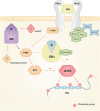
Actions of estrogen, progesterone, and prolactin on tau phosphorylation. Interacting mechanisms of hormone action that affect the dynamics of tau phosphorylation. The three hormones have documented activity in the activation of Akt that makes GSK3 inactive, which results in the inhibition of tau phosphorylation for this particular kinase. Since GSK3 has been linked in the pathological development of Alzheimer’s disease (AD), the interplay between these hormones, their pathways, and GSK3 might explain why the absence of hormones could account for the higher risk of developing AD. On the other hand, the presence of hormones like prolactin might prove neuroprotective for the neurodegeneration of the female brain.

References

    1. Weingarten MD, Lockwood AH, Hwo SY, Kirschner MW. A protein factor essential for microtubule assembly. Proc Natl Acad Sci U S A (1975) 72(5):1858–62.10.1073/pnas.72.5.1858 - DOI - PMC - PubMed
    1. Kolarova M, García-Sierra F, Bartos A, Ricny J, Ripova D. Structure and pathology of tau protein in Alzheimer disease. Int J Alzheimers Dis (2012) 2012:731526.10.1155/2012/731526 - DOI - PMC - PubMed
    1. Binder L, Frankfurter A, Rebhuhn L. The distribution of tau in the mammalian central nervous system. J Cell Biol (1985) 101:1371–8.10.1083/jcb.101.4.1371 - DOI - PMC - PubMed
    1. Trojanowsky JQ, Schuck T, Schmidt ML, Lee VM-Y. Distribution of tau proteins in the normal human central and peripheral nervous system. J Histochem Cytochem (1989) 37(2):209–15.10.1177/37.2.2492045 - DOI - PubMed
    1. Kosik KS, Finch EA. MAP2 and tau segregate into dendritic and axonal domains after the elaboration of morphologically distinct neurites: an immunocytochemical study of cultured rat cerebrum. J Neurosci (1987) 7(10):3142–53. - PMC - PubMed

LinkOut - more resources