

IL-8 associates with a pro-angiogenic and mesenchymal subtype in glioblastoma
- PMID: 29644004
- PMCID: PMC5884659
- DOI: 10.18632/oncotarget.24595
IL-8 associates with a pro-angiogenic and mesenchymal subtype in glioblastoma
Abstract
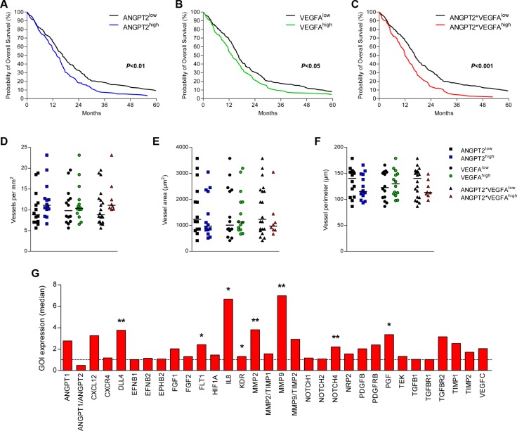
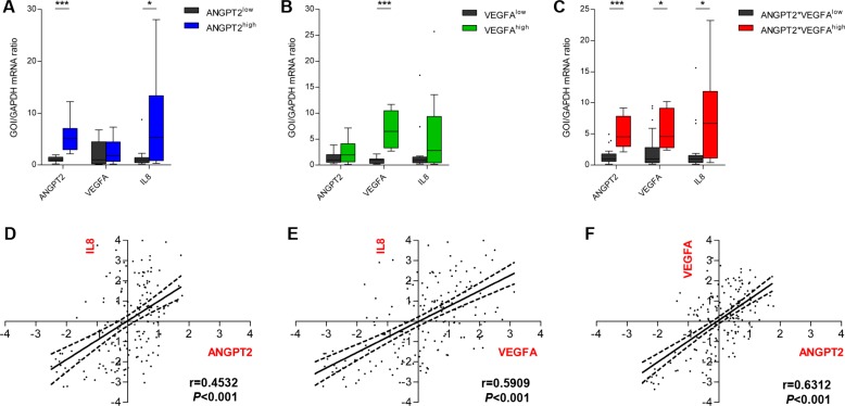
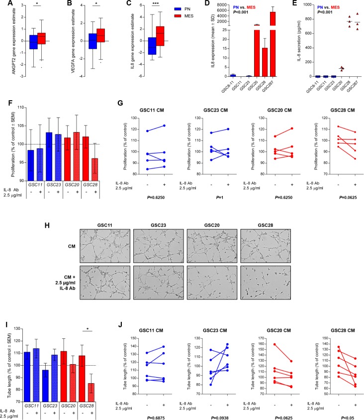
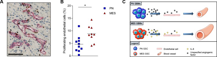
Glioblastoma (GBM) is a highly aggressive brain tumor characterized by a high rate of vascularization. However, therapeutic targeting of the vasculature through anti-vascular endothelial growth factor (VEGF) treatment has been disappointing, for which Angiopoietin-2 (Ang-2) upregulation has partly been held accountable. In this study we therefore explored the interplay of Ang-2 and VEGFA and their effect on angiogenesis in GBM, especially in the context of molecular subclasses. In a large patient cohort we identified that especially combined high expression of Ang-2 and VEGFA predicted poor overall survival of GBM patients. The high expression of both factors was also associated with increased IL-8 expression in GBM tissues, but in vitro stimulation with Ang-2 and/or VEGFA did not indicate tumor or endothelial cell-specific IL-8 responses. Glioblastoma stem cells (GSCs) of the mesenchymal (MES) subtype showed dramatically higher expression of IL8 when compared to proneural (PN) GSCs. Secreted IL-8 derived from MES GSCs induced endothelial proliferation and tube formation, and the MES GBMs had increased counts of proliferating endothelial cells. Our results highlight a critical pro-angiogenic role of IL-8 in MES GBMs.
Keywords: IL-8; angiogenesis; glioblastoma; subclasses.
Conflict of interest statement
CONFLICTS OF INTEREST The authors declare no conflicts of interest.
Figures

Similar articles
-
Molecular subtypes and differentiation programmes of glioma stem cells as determinants of extracellular vesicle profiles and endothelial cell-stimulating activities.J Extracell Vesicles. 2018 Jul 17;7(1):1490144. doi: 10.1080/20013078.2018.1490144. eCollection 2018. J Extracell Vesicles. 2018. PMID: 30034643 Free PMC article.
-
Peroxisome proliferator-activated receptor gamma as a theragnostic target for mesenchymal-type glioblastoma patients.Exp Mol Med. 2020 Apr;52(4):629-642. doi: 10.1038/s12276-020-0413-1. Epub 2020 Apr 13. Exp Mol Med. 2020. PMID: 32280134 Free PMC article.
-
Stem cell signature in glioblastoma: therapeutic development for a moving target.J Neurosurg. 2015 Feb;122(2):324-30. doi: 10.3171/2014.9.JNS132253. Epub 2014 Nov 14. J Neurosurg. 2015. PMID: 25397368 Review.
-
Novel insights into vascularization patterns and angiogenic factors in glioblastoma subclasses.J Neurooncol. 2017 Jan;131(1):11-20. doi: 10.1007/s11060-016-2269-8. Epub 2016 Sep 15. J Neurooncol. 2017. PMID: 27633774 Free PMC article.
-
Vascular heterogeneity and targeting: the role of YKL-40 in glioblastoma vascularization.Oncotarget. 2015 Dec 1;6(38):40507-18. doi: 10.18632/oncotarget.5943. Oncotarget. 2015. PMID: 26439689 Free PMC article. Review.
Cited by
-
Patient-Derived Glioma Models: From Patients to Dish to Animals.Cells. 2019 Sep 30;8(10):1177. doi: 10.3390/cells8101177. Cells. 2019. PMID: 31574953 Free PMC article. Review.
-
STAT3 activation confers trastuzumab-emtansine (T-DM1) resistance in HER2-positive breast cancer.Cancer Sci. 2018 Oct;109(10):3305-3315. doi: 10.1111/cas.13761. Epub 2018 Aug 31. Cancer Sci. 2018. PMID: 30076657 Free PMC article.
-
Endometriosis-Associated Angiogenesis and Anti-angiogenic Therapy for Endometriosis.Front Glob Womens Health. 2022 Apr 5;3:856316. doi: 10.3389/fgwh.2022.856316. eCollection 2022. Front Glob Womens Health. 2022. PMID: 35449709 Free PMC article. Review.
-
Plasma IL-8 and ICOSLG as prognostic biomarkers in glioblastoma.Neurooncol Adv. 2021 Jun 1;3(1):vdab072. doi: 10.1093/noajnl/vdab072. eCollection 2021 Jan-Dec. Neurooncol Adv. 2021. PMID: 34286278 Free PMC article.
-
Therapeutic Targets and Emerging Treatments in Advanced Chondrosarcoma.Int J Mol Sci. 2022 Jan 20;23(3):1096. doi: 10.3390/ijms23031096. Int J Mol Sci. 2022. PMID: 35163019 Free PMC article. Review.
References
-
- Huse JT, Holland EC. Targeting brain cancer: Advances in the molecular pathology of malignant glioma and medulloblastoma. Nat Rev Cancer. 2010;10:319–331. - PubMed
-
- Preusser M, de Ribaupierre S, Wohrer A, Erridge SC, Hegi M, Weller M, Stupp R. Current concepts and management of glioblastoma. Ann Neurol. 2011;70:9–21. - PubMed
-
- Stupp R, Mason WP, van den Bent MJ, Weller M, Fisher B, Taphoorn MJ, Belanger K, Brandes AA, Marosi C, Bogdahn U, Curschmann J, Janzer RC, Ludwin SK, et al. European Organisation for Research and Treatment of Cancer Brain Tumor and Radiotherapy Groups, and National Cancer Institute of Canada Clinical Trials Group. Radiotherapy plus concomitant and adjuvant temozolomide for glioblastoma. N Engl J Med. 2005;352:987–996. - PubMed
-
- Stupp R, Hegi ME, Mason WP, van den Bent MJ, Taphoorn MJ, Janzer RC, Ludwin SK, Allgeier A, Fisher B, Belanger K, Hau P, Brandes AA, Gijtenbeek J, et al. European Organisation for Research and Treatment of Cancer Brain Tumour and Radiation Oncology Groups, and National Cancer Institute of Canada Clinical Trials Group. Effects of radiotherapy with concomitant and adjuvant temozolomide versus radiotherapy alone on survival in glioblastoma in a randomised phase III study: 5-year analysis of the EORTC-NCIC trial. Lancet Oncol. 2009;10:459–466. - PubMed
-
- Chaudhry IH, O’Donovan DG, Brenchley PE, Reid H, Roberts IS. Vascular endothelial growth factor expression correlates with tumour grade and vascularity in gliomas. Histopathology. 2001;39:409–415. - PubMed
LinkOut - more resources
Full Text Sources
Other Literature Sources
Miscellaneous
