. 2018 Mar 2;9(21):15766-15779.
doi: 10.18632/oncotarget.24607. eCollection 2018 Mar 20.

Calcium-sensing receptor (CaSR) promotes development of bone metastasis in renal cell carcinoma

Affiliations

Calcium-sensing receptor (CaSR) promotes development of bone metastasis in renal cell carcinoma

Sebastian Frees et al. Oncotarget. .

Abstract

Bone metastasis is an important prognostic factor in renal cell carcinoma (RCC). The calcium-sensing receptor (CaSR) has been associated with bone metastasis in several different malignancies. We analyzed the impact of CaSR in bone metastasis in RCC in vitro and in vivo. The RCC cell line 786-O was stably transfected with the CaSR gene and treated with calcium alone or in combination with the CaSR antagonist NPS2143. Afterwards migration, adhesion, proliferation and prominent signaling molecules were analyzed. Calcium treated CaSR-transfected 768-O cells showed an increased adhesion to endothelial cells and the extracellular matrix components fibronectin and collagen I, but not to collagen IV. The chemotactic cell migration and proliferation was also induced by calcium. The activity of SHC, AKT, ERK, P90RSK and JNK were enhanced after calcium treatment of CaSR-transfected cells. These effects were abolished by NPS2143. Development of bone metastasis was evaluated in vivo in a mouse model. Intracardiac injection of CaSR-transfected 768-O cells showed an increased rate of bone metastasis. The results indicate CaSR as an important component in the mechanism of bone metastasis in RCC. Therefore, targeting CaSR might be beneficial in patients with bone metastatic RCC with a high CaSR expression.

Keywords: bone metastases, metastasis; calcium-sensing receptor; kidney cancer; renal cell carcinoma.


Conflict of interest statement

CONFLICTS OF INTEREST No conflict of interest.

Figures

Figure 1
Figure 1. Cell adhesion of CaSR-transfected 786-O cells on endothelial cells (HUVEC)
Cells were treated with calcium (5 mM) or a combination of calcium (5 mM) and NPS2143 (10 µM). (A) The adhesion value is shown as percentage of the adhesion of untreated vector-transfected cells. (B) Microscopic images of cell adhesion on HUVEC. Calcium triggered cell adhesion on HUVEC in CaSR-transfected cells significantly. Significance was calculated by Student’s T-test, p < 0.05.
Figure 2
Figure 2
Cell adhesion of CaSR-transfected 786-O cells on extracellular matrix components fibronectin (A), collagen I (B), collagen IV (C) and BSA (D). Cells were treated with calcium (5 mM) or a combination of calcium (5 mM) and NPS2143 (10 µM). The adhesion value is shown as percentage of the adhesion of untreated vector-transfected cells. BSA was used as control. Calcium triggered cell adhesion on fibronectin and collagen I in CaSR-transfected cells significantly. Significance was calculated by Student’s T-test, p < 0.05.
Figure 3
Figure 3. Chemotactical cell migration of CaSR-transfected 786-O cells using calcium as chemotaxin
Cells were treated with NPS2143 (10 µM). Migration was determined in a Boyden chamber using serum-free medium as control or calcium (5 mM) as chemotaxin. (A) The migration value is shown as percentage of the migration of untreated vector-transfected cells. (B) Microscopic images of migrated cells. CaSR-transfected cells showed a significant increased migration. Significance was calculated by Student’s T-test, p < 0.05.
Figure 4
Figure 4. Cell proliferation of CaSR-transfected 786-O
Cells were treated with calcium (5 mM) or a combination of calcium (5 mM) and NPS2143 (10 µM). The proliferation value is shown as percentage of the proliferation of untreated vector-transfected cells. Calcium triggered cell proliferation in CaSR-transfected cells significantly. Significance was calculated by Student’s T-test, p < 0.05.
Figure 5
Figure 5
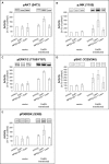
Activity of (A) AKT, (B) JNK, (C) ERK1/2, (D) SHC, and (E) P90RSK of CaSR-transfected 786-O. Cells were treated with calcium (5 mM) or a combination of calcium (5 mM) and NPS2143 (10 µM). The activity value is shown as percentage of untreated vector-transfected cells. Exemplary Western blot bands are shown above the diagram. Calcium triggered activity of AKT, JNK, ERK1/2, SHC and P90RSK in CaSR-transfected cells.
Figure 6
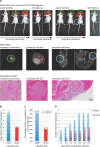
Figure 6. Development of bone metastases after intracardiac injection of CaSR overexpressing cells into a xenograft mouse model
Detection of bone metastases using bioluminescence (IVIS), representative MRI-images and histopathology (A) (representative images shown - each single lesion is represented by one color) confirmed a higher number of total bone metastases (B) as well as a higher number of bone metastases per total animals (C) (Students T-Test, p < 0.05) and an earlier development of bone metastases (D) for the CaSR overexpressing cells.
Figure 7
Figure 7. Signaling pathways influenced by CaSR
CaSR, stimulated by calcium, triggers AKT activity as well as MAPK activity of JNK, ERK1/2, p90RSK and SHC activity. Both signaling pathways lead downstream to an increase in cellular processes like cell migration, proliferation and adhesion. Red arrows show increased activity in CaSR overexpressing cells after Calcium stimulation.

Similar articles

Cited by

References

    1. Society AC . Cancer Facts & Figures 2016. Atlanta: American Cancer Society; 2016.
    1. Chow WH, Devesa SS, Warren JL, Fraumeni JF., Jr Rising incidence of renal cell cancer in the United States. JAMA. 1999;281:1628–31. - PubMed
    1. Decastro GJ, McKiernan JM. Epidemiology, clinical staging, and presentation of renal cell carcinoma. Urol Clin North Am. 2008;35:581–92. https://doi.org/10.1016/j.ucl.2008.07.005. - DOI - PubMed
    1. Sun M, Choueiri TK. Kidney cancer: Recurrence in renal cell carcinoma: the work is not done. Nat Rev Urol. 2016;13:246–7. https://doi.org/10.1038/nrurol.2016.57. - DOI - PubMed
    1. Mikami S, Oya M, Mizuno R, Kosaka T, Katsube KI, Okada Y. Invasion and metastasis of renal cell carcinoma. Medical molecular morphology. 2014;47:63–7. - PubMed