. 2018 Apr 10:9:9.
doi: 10.1186/s13227-018-0096-z. eCollection 2018.

Molecular, phylogenetic and developmental analyses of Sall proteins in bilaterians

Affiliations

Molecular, phylogenetic and developmental analyses of Sall proteins in bilaterians

José Lorente-Sorolla et al. Evodevo. .

Abstract

Background: Sall (Spalt-like) proteins are zinc-finger transcription factors involved in a number of biological processes. They have only been studied in a few model organisms, such as Drosophila melanogaster, Caenorhabditis elegans, Schmidtea mediterranea and some vertebrates. Further taxon sampling is critical to understand the evolution and diversification of this protein and its functional roles in animals.

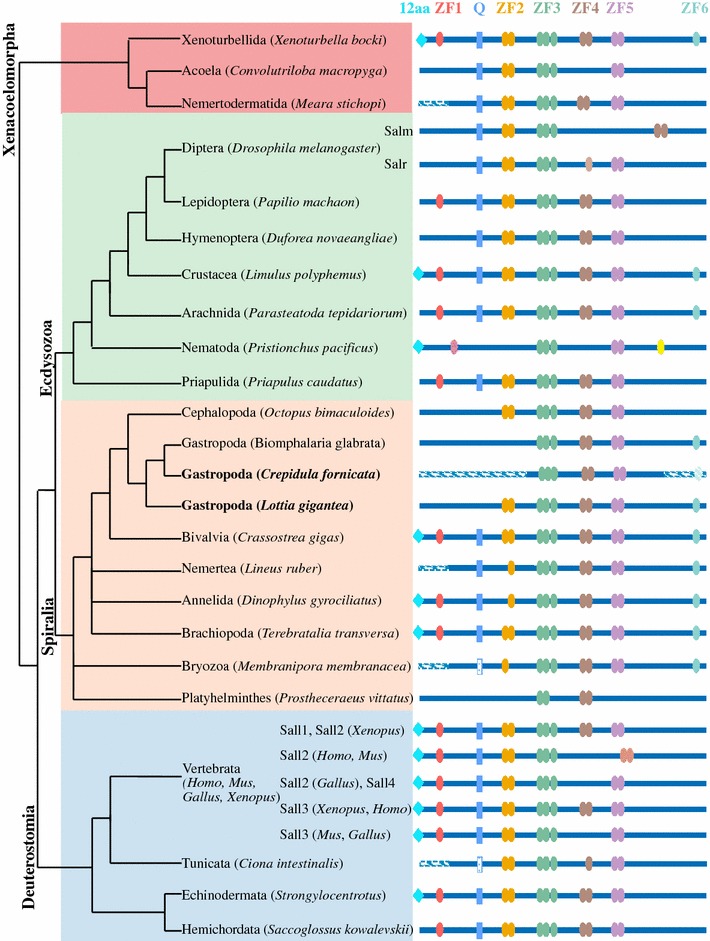
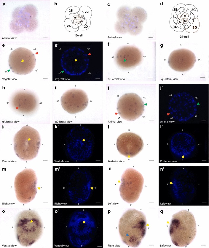
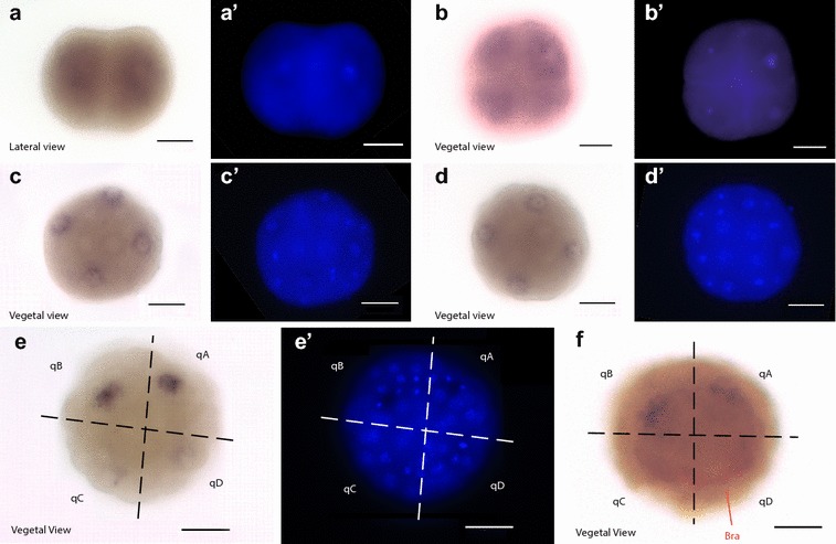
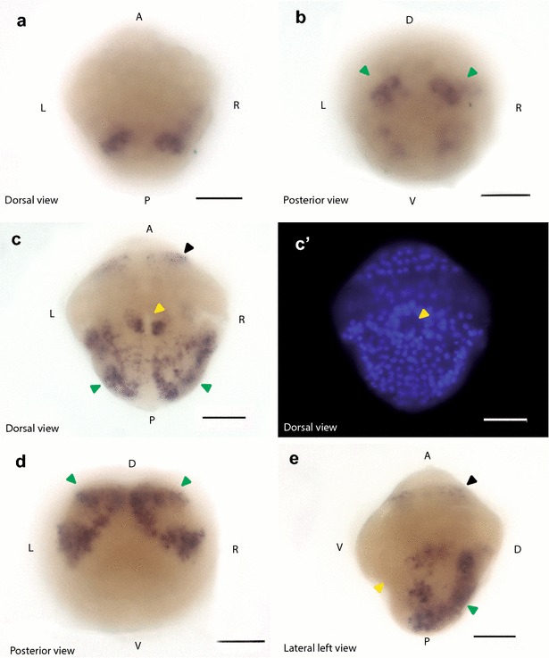
Results: Using genome and transcriptome mining, we confirmed the presence of sall genes in a range of additional animal taxa, for which their presence had not yet been described. We show that sall genes are broadly conserved across the Bilateria, and likely appeared in the bilaterian stem lineage. Our analysis of the protein domains shows that the characteristic arrangement of the multiple zinc-finger domains is conserved in bilaterians and may represent the ancient arrangement of this family of transcription factors. We also show the existence of a previously unknown zinc-finger domain. In situ hybridization was used to describe the gene expression patterns in embryonic and larval stages in two species of snails: Crepidula fornicata and Lottia gigantea. In L. gigantea, sall presents maternal expression, although later on the expression is restricted to the A and B quadrants during gastrulation and larval stage. In C. fornicata, sall has no maternal expression and it is expressed mainly in the A, C and D quadrants during blastula stages and in an asymmetric fashion during the larval stage.

Discussion: Our results suggest that the bilaterian common ancestor had a Sall protein with at least six zinc-finger domains. The evolution of Sall proteins in bilaterians might have occurred mostly as a result of the loss of protein domains and gene duplications leading to diversification. The new evidence complements previous studies in highlighting an important role of Sall proteins in bilaterian development. Our results show maternal expression of sall in the snail L. gigantea, but not C. fornicata. The asymmetric expression shown in the ectoderm of the trochophore larva of snails is probably related to shell/mantle development. The observed sall expression in cephalic tissue in snails and some other bilaterians suggests a possible ancestral role of sall in neural development in bilaterians.

Keywords: Crepidula fornicata; Gastropoda; Gene evolution; Lottia gigantea; Protein domains; Sall; Spalt; Spiralia.


Figures

Fig. 1
Fig. 1
Organization of conserved domains in “Sal-box” containing proteins. Colored ovals represent the zinc-finger motifs. The blue rectangle represents the poly-Q region. The turquoise diamond represents the 12 conserved amino acids at the N-terminal end of the Sall proteins that interact with the HDC NuRD [14]. Protein length range is indicated at the right
Fig. 2
Fig. 2
Organization of conserved domains in Sall proteins in Bilateria. The light blue diamond represents the 12 conserved amino acids at the N-terminal end that interact with the HDC NuRD [14]. Colored ovals represent the zinc-finger motifs from ZF1 to ZF6. The blue rectangle represents the poly-Q region. The striped lines represent missing sequence. Phylogenetic tree based on Cannon et al. [63] and Peters et al. [64]. The blue box on the tree highlights the deuterostomes; the orange box, the spiralians; the green box, the ecdysozoans; and the red box, the xenacoelomorphs. Bold names highlight the snail species studied in more detail in this work
Fig. 3
Fig. 3
Sall expression in C. fornicata. In situ hybridization of sall mRNA in embryos from 16-cell stage to organogenesis. The distribution of labeled mRNA is shown by the dark blue staining. In all images orientation is indicated at the bottom left corner of each panel. Scale bar at the bottom right corner equals 40 µm. Yellow arrowhead indicates the blastopore/stomodeum. a, c 16- and 24-cell stage embryos, respectively. mRNA is expressed in the macromeres. The lighter colored nuclei are labeled with Hoechst (blue). b, d Schematic representation of 16- and 24-cell embryos, respectively, in animal view (modified from Henry et al., [47]). Capital letters indicate the macromeres and lowercase letters the micromeres. ej′ Localization of sall mRNA in blastula stage. ee′, jj′ Bright-field and fluorescent Hoechst-labeled corresponding images. fi Lateral view of each quadrant. Green and red arrowheads indicate stained reference cells in quadrants C and A, respectively, in different views. q quadrant, A animal pole, V vegetal pole. kn′ Localization of sall mRNA in gastrula stage. Each bright-field image has a corresponding fluorescent Hoechst-labeled image on the right. oq′ Localization of sall mRNA in organogenesis. o, o′ Bright-field and fluorescent Hoechst-labeled corresponding images. p Bright-field right view. The blue arrow indicates the asymmetric patch of expression present in this side. q Bright-field left view. A: anterior; P: posterior; V: ventral; D: dorsal; R: right; L: left
Fig. 4
Fig. 4
Expression of sall during early cleavage in L. gigantea. In situ hybridization of sall mRNA in embryos from 2-cell stage to blastula stage. Each bright-field image has its corresponding fluorescent Hoechst-labeled image at the right. The distribution of labeled mRNA is shown by the dark blue staining. Orientation is indicated at the bottom left corner of each panel. Scale bar at the bottom right corner equals 40 µm. aa′ Expression in all cells at 2-cell stage. bb′ Expression in all cells at 4-cell stage. cc Expression in the 2 m micromeres at 16-cell stage. dd′ Expression in the 2 m micromeres at 24-cell stage. ee′ Expression of sall mRNA at blastula stage. f Double in situ hybridization of sall (dark blue) and brachyury (red). Indicated in red, labeled brachyury mRNA in the D quadrant. Note that sall expression is restricted to the A and B quadrants. q: quadrant; bra: brachyury. Quadrants are separated by dashed lines in e, f
Fig. 5
Fig. 5
Expression of sall during trochophore stages in L. gigantea. The distribution of the labeled mRNA is shown by the dark blue staining. Orientation is indicated at the bottom left corner of each panel. Scale bar at the bottom right corner equals 40 µm. A: anterior; P: posterior; V: ventral; D: dorsal; R: right; L: left. a, b Localization of sall mRNA in pre-trochophore stage. Expression starts to extend from the progeny of 2 m micromeres. ce In situ hybridization of sall mRNA at trochophore stage. Black arrowhead indicates the stained ring in the cephalic region. Green arrowhead indicates the dorsolateral ectodermic strip. Yellow arrowhead indicates the stomodeum

References

    1. Kuhnlein RP, Frommer G, Friedrich M, Gonzalez-Gaitan M, Weber A, Wagner-Bernholz JF, Gehring WJ, Jackle H, Schuh R. Spalt encodes an evolutionarily conserved zinc finger protein of novel structure which provides homeotic gene function in the head and tail region of the Drosophila embryo. EMBO J. 1994;13(1):168–179. - PMC - PubMed
    1. Frei E, Schuh R, Baumgartner S, Burri M, Noll M, Jurgens G, Seifert E, Nauber U, Jackle H. Molecular characterization of spalt, a homeotic gene required for head and tail development in the Drosophila embryo. EMBO J. 1988;7(1):197–204. - PMC - PubMed
    1. Schuh R, Aicher W, Gaul U, Cote S, Preiss A, Maier D, Seifert E, Nauber U, Schroder C, Kemler R. A conserved family of nuclear proteins containing structural elements of the finger protein encoded by Kruppel, a Drosophila segmentation gene. Cell. 1986;47:1025–1032. doi: 10.1016/0092-8674(86)90817-2. - DOI - PubMed
    1. Arora K, Dai H, Kazuko SG, Jamal J, O’Connor MB, Letsou A, Warrior R. The Drosophila schnurri gene acts in the Dpp/TGF signaling pathway and encodes a transcription factor homologous to the human MBP family. Cell. 1995;81(5):781–790. doi: 10.1016/0092-8674(95)90539-1. - DOI - PubMed
    1. Grieder NC, Nellen D, Burke R, Basler K, Affolter M. Schnurri is required for Drosophila Dpp signaling and encodes a zinc finger protein similar to the mammalian transcription factor PRDII-BF1. Cell. 1995;81(5):791–800. doi: 10.1016/0092-8674(95)90540-5. - DOI - PubMed